Show PDB file:   
         Plain Text   HTML   (compressed file size)
QuickSearch:   
by PDB,NDB,UniProt,PROSITE Code or Search Term(s)  
(-)Asym./Biol. Unit
collapse expand < >
Image Asym./Biol. Unit
Asym./Biol. Unit  (Jmol Viewer)

(-) Description

Title :  CRYSTAL STRUCTURE OF THE NEUTRALIZING HIV ANTIBODY 2F5 FAB FRAGMENT (RECOMBINANTLY PRODUCED IGG) WITH 11 AA GP41 MPER-DERIVED PEPTIDE
 
Authors :  N. I. Nicely, S. M. Dennison, G. Kelsoe, H. -X. Liao, S. M. Alam, B. F. Hayn
Date :  22 Apr 10  (Deposition) - 17 Nov 10  (Release) - 05 Feb 14  (Revision)
Method :  X-RAY DIFFRACTION
Resolution :  2.20
Chains :  Asym./Biol. Unit :  H,L,P
Keywords :  Hiv-1, Hiv Gp41 Mper, 13H11, 2F5, Z13, 4E10, Fab Antibody, Immune System (Keyword Search: [Gene Ontology, PubMed, Web (Google))
 
Reference :  N. I. Nicely, S. M. Dennison, G. Kelsoe, Y. Ueda, H. -X. Liao, S. M. Alam, B. F. Haynes
Crystal Structure Of A Non-Neutralizing Hiv-1 Gp41 Envelope Antibody Demonstrates Neutralization Mechanism Of Gp41 Antibodies
To Be Published
PubMed: search

(-) Compounds

Molecule 1 - GP41 MPER-DERIVED PEPTIDE
    ChainsP
    EngineeredYES
    Organism ScientificHUMAN IMMUNODEFICIENCY VIRUS 1
    Organism Taxid11676
    Other DetailsCHAIN P IS A CHEMICALLY SYNTHESIZED PEPTIDE OF SEQUENCE DERIVED FROM THE MEMBRANE PROXIMAL EXTERNAL REGION OF GP41
    SyntheticYES
 
Molecule 2 - ANTI-HIV-1 ANTIBODY 2F5 LIGHT CHAIN
    ChainsL
    EngineeredYES
    Expression SystemHOMO SAPIENS
    Expression System Cell LineHEK293T CELL
    Expression System CommonHUMAN
    Expression System OrganKIDNEY
    Expression System Taxid9606
    Expression System Vector TypePLASMID
    Organism CommonHUMAN
    Organism ScientificHOMO SAPIENS
    Organism Taxid9606
 
Molecule 3 - ANTI-HIV-1 ANTIBODY 2F5 HEAVY CHAIN
    ChainsH
    EngineeredYES
    Expression SystemHOMO SAPIENS
    Expression System Cell LineHEK293T CELL
    Expression System CommonHUMAN
    Expression System OrganKIDNEY
    Expression System Taxid9606
    Expression System Vector TypePLASMID
    Organism CommonHUMAN
    Organism ScientificHOMO SAPIENS
    Organism Taxid9606

 Structural Features

(-) Chains, Units

  123
Asymmetric/Biological Unit HLP

Summary Information (see also Sequences/Alignments below)

(-) Ligands, Modified Residues, Ions  (1, 1)

Asymmetric/Biological Unit (1, 1)
No.NameCountTypeFull Name
1NH21Mod. Amino AcidAMINO GROUP

(-) Sites  (0, 0)

(no "Site" information available for 3MOD)

(-) SS Bonds  (4, 4)

Asymmetric/Biological Unit
No.Residues
1H:22 -H:92
2H:140 -H:196
3L:23 -L:88
4L:134 -L:194

(-) Cis Peptide Bonds  (5, 5)

Asymmetric/Biological Unit
No.Residues
1Ser L:7 -Pro L:8
2Tyr L:94 -Pro L:95
3Tyr L:140 -Pro L:141
4Phe H:146 -Pro H:147
5Glu H:148 -Pro H:149

 Sequence-Structure Mapping

(-) SAPs(SNPs)/Variants  (0, 0)

(no "SAP(SNP)/Variant" information available for 3MOD)

(-) PROSITE Motifs  (0, 0)

(no "PROSITE Motif" information available for 3MOD)

(-) Exons   (0, 0)

(no "Exon" information available for 3MOD)

(-) Sequences/Alignments

Asymmetric/Biological Unit
   Reformat: Number of residues per line =  ('0' or empty: single-line sequence representation)
  Number of residues per labelling interval =   
  UniProt sequence: complete  aligned part    
   Show mapping: SCOP domains CATH domains Pfam domains Secondary structure (by author)
SAPs(SNPs) PROSITE motifs Exons
(details for a mapped element are shown in a popup box when the mouse pointer rests over it)
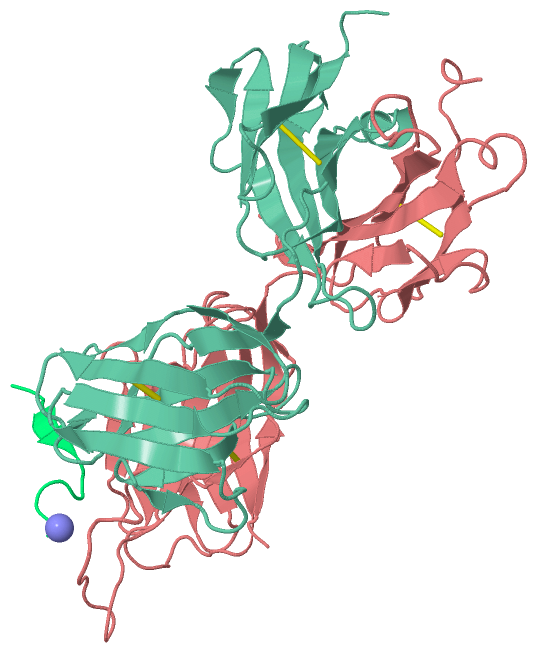
Chain H from PDB  Type:PROTEIN  Length:237
                                                                                                                                                                                                                                                                              
               SCOP domains --------------------------------------------------------------------------------------------------------------------------------------------------------------------------------------------------------------------------------------------- SCOP domains
               CATH domains --------------------------------------------------------------------------------------------------------------------------------------------------------------------------------------------------------------------------------------------- CATH domains
               Pfam domains --------------------------------------------------------------------------------------------------------------------------------------------------------------------------------------------------------------------------------------------- Pfam domains
         Sec.struct. author ..eeeee...ee.....eeeeeeee.........eeeeeee......eeeeeee....eee.......eeeeee....eeeeee........eeeeeeee....................ee...eeeee........eeeee..........eeeeeeeeeee.....eeee........eee...ee.....eeeeeeeeee.........eeeeee....eeeeee........ Sec.struct. author
                 SAPs(SNPs) --------------------------------------------------------------------------------------------------------------------------------------------------------------------------------------------------------------------------------------------- SAPs(SNPs)
                    PROSITE --------------------------------------------------------------------------------------------------------------------------------------------------------------------------------------------------------------------------------------------- PROSITE
                 Transcript --------------------------------------------------------------------------------------------------------------------------------------------------------------------------------------------------------------------------------------------- Transcript
                3mod H    1 RITLKESGPPLVKPTQTLTLTCSFSGFSLSDFGVGVGWIRQPPGKALEWLAIIYSDDDKRYSPSLNTRLTITKDTSKNQVVLVMTRVSPVDTATYFCAHRRGPTTLFGVPIARGPVNAMDVWGQGITVTISSTSTKGPSVFPLAPSSKSTSGGTAALGCLVKDYFPEPVTVSWNSGALTSGVHTFPAVLQSSGLYSLSSVVTVPSSSLGTQTYICNVNHKPSNTKVDKRVEPKSCDK  218
                                    10        20        30     || 38        48        58        68        78    ||| 85        95     |100E|||||||101       111       121       131       141       151       161       171       181       191       201       211       
                                                             35A|                                             82A||               100A|||||100J||||                                                                                                                      
                                                              35B                                              82B|                100B|||||100K|||                                                                                                                      
                                                                                                                82C                 100C|||||100L||                                                                                                                      
                                                                                                                                     100D|||||100M|                                                                                                                      
                                                                                                                                      100E|||| 100N                                                                                                                      
                                                                                                                                       100F|||                                                                                                                           
                                                                                                                                        100G||                                                                                                                           
                                                                                                                                         100H|                                                                                                                           
                                                                                                                                          100I                                                                                                                           

Chain L from PDB  Type:PROTEIN  Length:214
                                                                                                                                                                                                                                                       
               SCOP domains ---------------------------------------------------------------------------------------------------------------------------------------------------------------------------------------------------------------------- SCOP domains
               CATH domains ---------------------------------------------------------------------------------------------------------------------------------------------------------------------------------------------------------------------- CATH domains
               Pfam domains ---------------------------------------------------------------------------------------------------------------------------------------------------------------------------------------------------------------------- Pfam domains
         Sec.struct. author ...eeee..eeee.....eeeeeee.......eeeeee......eeeee...ee.......eeeeee..eeeeee........eeeeeee..ee..ee...eeeee.......eeeee...hhhhh..eeeeeeeeeee.....eeeeee..ee....eeeee.........eeeeeeeeeehhhhh...eeeeeee.......eeeeee.... Sec.struct. author
                 SAPs(SNPs) ---------------------------------------------------------------------------------------------------------------------------------------------------------------------------------------------------------------------- SAPs(SNPs)
                    PROSITE ---------------------------------------------------------------------------------------------------------------------------------------------------------------------------------------------------------------------- PROSITE
                 Transcript ---------------------------------------------------------------------------------------------------------------------------------------------------------------------------------------------------------------------- Transcript
                3mod L    1 ALQLTQSPSSLSASVGDRITITCRASQGVTSALAWYRQKPGSPPQLLIYDASSLESGVPSRFSGSGSGTEFTLTISTLRPEDFATYYCQQLHFYPHTFGGGTRVDVRRTVAAPSVFIFPPSDEQLKSGTASVVCLLNNFYPREAKVQWKVDNALQSGNSQESVTEQDSKDSTYSLSSTLTLSKADYEKHKVYACEVTHQGLSSPVTKSFNRGEC  214
                                    10        20        30        40        50        60        70        80        90       100       110       120       130       140       150       160       170       180       190       200       210    

Chain P from PDB  Type:PROTEIN  Length:12
 aligned with ENV_HV1Z6 | P04580 from UniProtKB/Swiss-Prot  Length:855

    Alignment length:12
                                   668  
           ENV_HV1Z6    659 LLELDKWASLWN  670
               SCOP domains ------------ SCOP domains
               CATH domains ------------ CATH domains
               Pfam domains ------------ Pfam domains
         Sec.struct. author ..ee........ Sec.struct. author
                 SAPs(SNPs) ------------ SAPs(SNPs)
                    PROSITE ------------ PROSITE
                 Transcript ------------ Transcript
                3mod P  660 LLELDKWASLWx  671
                                   669 |
                                     671-NH2

   Legend:   → Mismatch (orange background)
  - → Gap (green background, '-', border residues have a numbering label)
    → Modified Residue (blue background, lower-case, 'x' indicates undefined single-letter code, labelled with number + name)
  x → Chemical Group (purple background, 'x', labelled with number + name, e.g. ACE or NH2)
  extra numbering lines below/above indicate numbering irregularities and modified residue names etc., number ends below/above '|'

 Classification and Annotation

(-) SCOP Domains  (0, 0)

(no "SCOP Domain" information available for 3MOD)

(-) CATH Domains  (0, 0)

(no "CATH Domain" information available for 3MOD)

(-) Pfam Domains  (0, 0)

(no "Pfam Domain" information available for 3MOD)

(-) Gene Ontology  (21, 21)

Asymmetric/Biological Unit(hide GO term definitions)
Chain P   (ENV_HV1Z6 | P04580)
molecular function
    GO:0005198    structural molecule activity    The action of a molecule that contributes to the structural integrity of a complex or its assembly within or outside a cell.
biological process
    GO:0075512    clathrin-dependent endocytosis of virus by host cell    Any clathrin-mediated endocytosis that is involved in the uptake of a virus into a host cell. Begins by invagination of a specific region of the host cell plasma membrane around the bound virus to form a clathrin-coated pit, which then pinches off to form a clathrin-coated endocytic vesicle containing the virus.
    GO:0075509    endocytosis involved in viral entry into host cell    Any endocytosis that is involved in the uptake of a virus into a host cell.
    GO:0030683    evasion or tolerance by virus of host immune response    Any process, either active or passive, by which a virus avoids the effects of the host organism's immune response. The host is defined as the larger of the organisms involved in a symbiotic interaction.
    GO:0039654    fusion of virus membrane with host endosome membrane    Fusion of a virus membrane with a host endosome membrane. Occurs after internalization of the virus through the endosomal pathway, and results in release of the virus contents into the cell.
    GO:0019064    fusion of virus membrane with host plasma membrane    Fusion of a viral membrane with the host cell membrane during viral entry. Results in release of the virion contents into the cytoplasm.
    GO:0039663    membrane fusion involved in viral entry into host cell    Merging of the virion membrane and a host membrane (host plasma membrane or host organelle membrane) that is involved in the uptake of a virus into a host cell.
    GO:0002223    stimulatory C-type lectin receptor signaling pathway    Any series of molecular signals generated as a consequence of binding to a C-type lectin receptor capable of cellular activation.
    GO:0046718    viral entry into host cell    The process that occurs after viral attachment by which a virus, or viral nucleic acid, breaches the plasma membrane or cell envelope and enters the host cell. The process ends when the viral nucleic acid is released into the host cell cytoplasm.
    GO:0016032    viral process    A multi-organism process in which a virus is a participant. The other participant is the host. Includes infection of a host cell, replication of the viral genome, and assembly of progeny virus particles. In some cases the viral genetic material may integrate into the host genome and only subsequently, under particular circumstances, 'complete' its life cycle.
    GO:0019082    viral protein processing    Any protein maturation process achieved by the cleavage of a peptide bond or bonds within a viral protein.
    GO:0019062    virion attachment to host cell    The process by which a virion protein binds to molecules on the host cellular surface or host cell surface projection.
cellular component
    GO:0044174    host cell endosome    A membrane-bounded organelle that carries materials newly ingested by endocytosis. It passes many of the materials to host cell lysosomes for degradation.
    GO:0044175    host cell endosome membrane    The lipid bilayer surrounding a host cell endosome.
    GO:0033644    host cell membrane    Double layer of lipid molecules as it encloses host cells, and, in eukaryotes, many organelles; may be a single or double lipid bilayer; also includes associated proteins. The host is defined as the larger of the organisms involved in a symbiotic interaction.
    GO:0020002    host cell plasma membrane    The plasma membrane surrounding a host cell.
    GO:0016021    integral component of membrane    The component of a membrane consisting of the gene products and protein complexes having at least some part of their peptide sequence embedded in the hydrophobic region of the membrane.
    GO:0016020    membrane    A lipid bilayer along with all the proteins and protein complexes embedded in it an attached to it.
    GO:0019031    viral envelope    The lipid bilayer of a virion that surrounds the protein capsid. May also contain glycoproteins.
    GO:0019012    virion    The complete fully infectious extracellular virus particle.
    GO:0055036    virion membrane    The lipid bilayer surrounding a virion.

 Visualization

(-) Interactive Views

Asymmetric/Biological Unit
  Complete Structure
    Jena3D(integrated viewing of ligand, site, SAP, PROSITE, SCOP information)
    WebMol | AstexViewer[tm]@PDBe
(Java Applets, require no local installation except for Java; loading may be slow)
    STRAP
(Java WebStart application, automatic local installation, requires Java; full application with system access!)
    RasMol
(require local installation)
    Molscript (VRML)
(requires installation of a VRML viewer; select preferred view via VRML and generate a mono or stereo PDF format file)
 
  Ligands, Modified Residues, Ions
    NH2  [ RasMol | Jena3D ]  +environment [ RasMol | Jena3D ]
 
  Sites
(no "Sites" information available for 3mod)
 
  Cis Peptide Bonds
    Glu H:148 - Pro H:149   [ RasMol ]  
    Phe H:146 - Pro H:147   [ RasMol ]  
    Ser L:7 - Pro L:8   [ RasMol ]  
    Tyr L:140 - Pro L:141   [ RasMol ]  
    Tyr L:94 - Pro L:95   [ RasMol ]  
 

(-) Still Images

Jmol
  protein: cartoon or spacefill or dots and stick; nucleic acid: cartoon and stick; ligands: spacefill; active site: stick
Molscript
  protein, nucleic acid: cartoon; ligands: spacefill; active site: ball and stick

 Databases and Analysis Tools

(-) Databases

Access by PDB/NDB ID
  3mod
    Family and Domain InformationProDom | SYSTERS
    General Structural InformationGlycoscienceDB | MMDB | NDB | OCA | PDB | PDBe | PDBj | PDBsum | PDBWiki | PQS | PROTEOPEDIA
    Orientation in MembranesOPM
    Protein SurfaceSURFACE
    Secondary StructureDSSP (structure derived) | HSSP (homology derived)
    Structural GenomicsGeneCensus
    Structural NeighboursCE | VAST
    Structure ClassificationCATH | Dali | SCOP
    Validation and Original DataBMRB Data View | BMRB Restraints Grid | EDS | PROCHECK | RECOORD | WHAT_CHECK
 
Access by UniProt ID/Accession number
  ENV_HV1Z6 | P04580
    Comparative Protein Structure ModelsModBase
    Genomic InformationEnsembl
    Protein-protein InteractionDIP
    Sequence, Family and Domain InformationInterPro | Pfam | SMART | UniProtKB/SwissProt
 
Access by Enzyme Classificator   (EC Number)
  (no 'Enzyme Classificator' available)
    General Enzyme InformationBRENDA | EC-PDB | Enzyme | IntEnz
    PathwayKEGG | MetaCyc
 
Access by Disease Identifier   (MIM ID)
  (no 'MIM ID' available)
    Disease InformationOMIM
 
Access by GenAge ID
  (no 'GenAge ID' available)
    Age Related InformationGenAge

(-) Analysis Tools

Access by PDB/NDB ID
    Domain InformationXDom
    Interatomic Contacts of Structural UnitsCSU
    Ligand-protein ContactsLPC
    Protein CavitiescastP
    Sequence and Secondary StructurePDBCartoon
    Structure AlignmentSTRAP(Java WebStart application, automatic local installation, requires Java; full application with system access!)
    Structure and Sequence BrowserSTING
 
Access by UniProt ID/Accession number
  ENV_HV1Z6 | P04580
    Protein Disorder PredictionDisEMBL | FoldIndex | GLOBPLOT (for more information see DisProt)

 Related Entries

(-) Entries Sharing at Least One Protein Chain (UniProt ID)

UniProtKB/Swiss-Prot
        ENV_HV1Z6 | P045801tjh 1tji 2zzo 3f4y 3f50 3g7a 3moa 3mob 4i2l

(-) Related Entries Specified in the PDB File

1tjh CRYSTAL STRUCTURE OF THE BROADLY NEUTRALIZING ANTI-HIV-1 ANTIBODY 2F5 IN COMPLEX WITH A GP41 11MER EPITOPE
3mnv CRYSTAL STRUCTURE OF THE NON-NEUTRALIZING HIV ANTIBODY 13H11 FAB FRAGMENT
3mnw CRYSTAL STRUCTURE OF THE NON-NEUTRALIZING HIV ANTIBODY 13H11 FAB FRAGMENT WITH A GP41 MPER-DERIVED PEPTIDE IN A HELICAL CONFORMATION
3mnz CRYSTAL STRUCTURE OF THE NON-NEUTRALIZING HIV ANTIBODY 13H11 FAB FRAGMENT WITH A GP41 MPER-DERIVED PEPTIDE BEARING ALA SUBSTITUTIONS IN A HELICAL CONFORMATION
3mo1 CRYSTAL STRUCTURE OF THE NON-NEUTRALIZING HIV ANTIBODY 13H11 FAB FRAGMENT
3moa CRYSTAL STRUCTURE OF THE NEUTRALIZING HIV ANTIBODY 2F5 FAB FRAGMENT (RECOMBINANTLY PRODUCED FAB) WITH 17 AA GP41 MPER- DERIVED PEPTIDE
3mob CRYSTAL STRUCTURE OF THE NEUTRALIZING HIV ANTIBODY 2F5 FAB FRAGMENT (RECOMBINANTLY PRODUCED FAB) WITH 11 AA GP41 MPER- DERIVED PEPTIDE