|
|
|
|
 Description
|
|
Compounds
|
||||||||||||||||||||||||||||||||||||
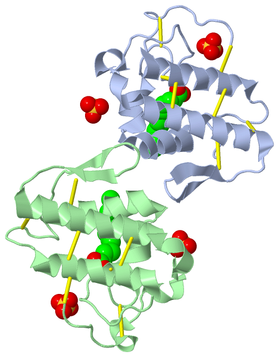
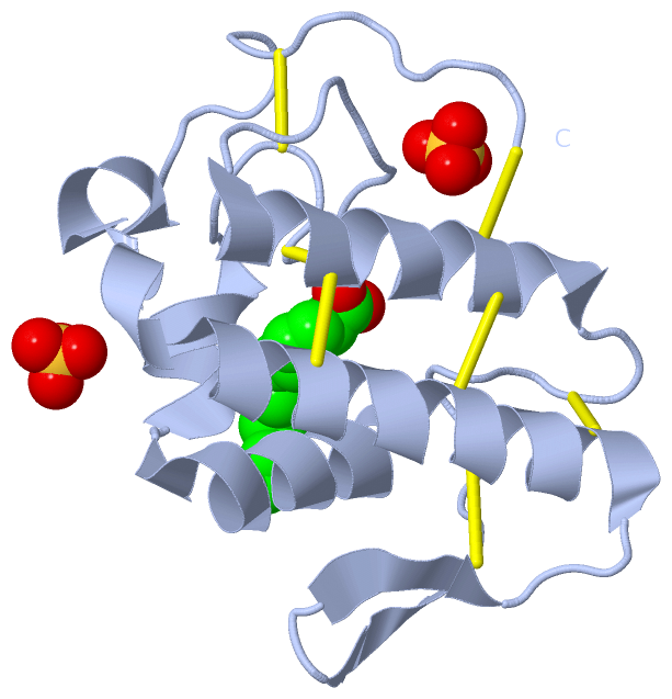
Chains, Units
Summary Information (see also Sequences/Alignments below) |
Ligands, Modified Residues, Ions (2, 6)| Asymmetric Unit (2, 6) Biological Unit 1 (2, 3) Biological Unit 2 (2, 3) |
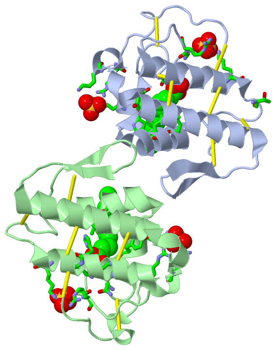
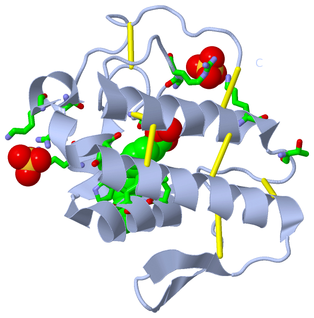
Sites (6, 6)
Asymmetric Unit (6, 6)
|
SS Bonds (14, 14)
Asymmetric Unit
|
||||||||||||||||||||||||||||||||||||||||||||||||||||||||||||
Cis Peptide Bonds (1, 1)
Asymmetric Unit
|
||||||||
SAPs(SNPs)/Variants (1, 2)
Asymmetric Unit (1, 2)
|
||||||||||||||||||||||||||||||||||||||||||||||||||||||||||||||||||||||||||||||||||||||||||||||||||||||||||||||||||||||||||||||||||||||||||||||||||||||||||||||||||||||||||||||
PROSITE Motifs (2, 4)
Asymmetric Unit (2, 4)
|
||||||||||||||||||||||||||||||||||||||||||||||||||||||||||||||||||||||||||||||||||||||||||||||||
Exons (0, 0)| (no "Exon" information available for 3MLM) |
Sequences/Alignments
Asymmetric UnitChain A from PDB Type:PROTEIN Length:121 aligned with PA2H_BOTPA | Q9IAT9 from UniProtKB/Swiss-Prot Length:120 Alignment length:121 1 | 9 19 29 39 49 59 69 79 89 99 109 119 PA2H_BOTPA - -SFELGKMILQETGKNPAKSYGAYGCNCGVLGRGQPKDATDRCCYVHKCCYKKLTGCDPKKDRYSYSWKDKTIVCGENNPCLKELCECDKAVAICLRENLGTYNKKYRYHLKPFCKKADPC 120 SCOP domains d3mlma_ A: Snake phospholipase A2 SCOP domains CATH domains ------------------------------------------------------------------------------------------------------------------------- CATH domains Pfam domains ------------------------------------------------------------------------------------------------------------------------- Pfam domains SAPs(SNPs) ----------------------------------G-------------------------------------------------------------------------------------- SAPs(SNPs) PROSITE ------------------------------------------PA2_HIS ----------------------------------PA2_ASP -------------------------- PROSITE Transcript ------------------------------------------------------------------------------------------------------------------------- Transcript 3mlm A 1 SLFELGKMILQETGKNPAKSYGAYGCNCGVLGRGGPKDATDRCCYVHKCCYKKITGCDPKKDRYSYSWKDKTIVCGENNPCLKELCECDKAVAICLRENLGTYNKKYRYHLKPFCKKADPC 121 10 20 30 40 50 60 70 80 90 100 110 120 Chain B from PDB Type:PROTEIN Length:121 aligned with PA2H_BOTPA | Q9IAT9 from UniProtKB/Swiss-Prot Length:120 Alignment length:121 1 | 9 19 29 39 49 59 69 79 89 99 109 119 PA2H_BOTPA - -SFELGKMILQETGKNPAKSYGAYGCNCGVLGRGQPKDATDRCCYVHKCCYKKLTGCDPKKDRYSYSWKDKTIVCGENNPCLKELCECDKAVAICLRENLGTYNKKYRYHLKPFCKKADPC 120 SCOP domains d3mlmb_ B: Snake phospholipase A2 SCOP domains CATH domains ------------------------------------------------------------------------------------------------------------------------- CATH domains Pfam domains ------------------------------------------------------------------------------------------------------------------------- Pfam domains SAPs(SNPs) ----------------------------------G-------------------------------------------------------------------------------------- SAPs(SNPs) PROSITE ------------------------------------------PA2_HIS ----------------------------------PA2_ASP -------------------------- PROSITE Transcript ------------------------------------------------------------------------------------------------------------------------- Transcript 3mlm B 1 SLFELGKMILQETGKNPAKSYGAYGCNCGVLGRGGPKDATDRCCYVHKCCYKKITGCDPKKDRYSYSWKDKTIVCGENNPCLKELCECDKAVAICLRENLGTYNKKYRYHLKPFCKKADPC 121 10 20 30 40 50 60 70 80 90 100 110 120
|
||||||||||||||||||||
SCOP Domains (1, 2)
Asymmetric Unit
 |
CATH Domains (0, 0)| (no "CATH Domain" information available for 3MLM) |
Pfam Domains (0, 0)| (no "Pfam Domain" information available for 3MLM) |
Gene Ontology (5, 5)|
Asymmetric Unit(hide GO term definitions) Chain A,B (PA2H_BOTPA | Q9IAT9)
|
||||||||||||||||||||||||||||||||||||||||||||||||
Interactive Views
|
||||||||||||||||||||||||||||||||||||||||||||||||||||||||||||||||||||||||||||||||||||||||||||||||||||||||||||||||||||||||||||||||||||||||||||||||||||||||||||||||||||||||||||||||||||||||
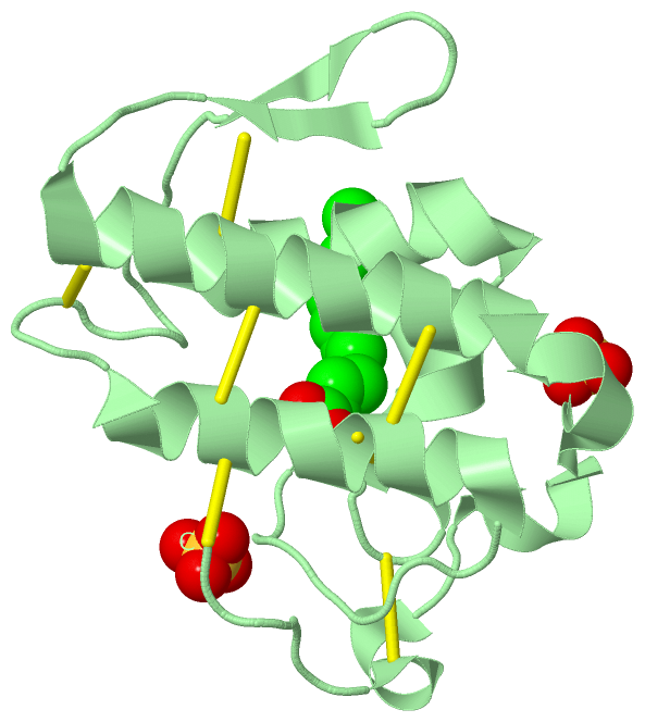
Still Images
|
||||||||||||||||
Databases
|
||||||||||||||||||||||||||||||||||||||||||||||||||||||||||||||||||||||||||||||||||||||||||||||||||||||||||||||||||||||||||||||||||||||||||||||||||||||||||||||||
Analysis Tools
|
|||||||||||||||||||||||||||||||||||||||||||||||||||||||||||||
Entries Sharing at Least One Protein Chain (UniProt ID)
Related Entries Specified in the PDB File
|
|