Show PDB file:   
         Plain Text   HTML   (compressed file size)
QuickSearch:   
by PDB,NDB,UniProt,PROSITE Code or Search Term(s)  
(-)Asym./Biol. Unit
(-)Asym./Biol. Unit - sites
collapse expand < >
Image Asym./Biol. Unit
Asym./Biol. Unit  (Jmol Viewer)
Image Asym./Biol. Unit - sites
Asym./Biol. Unit - sites  (Jmol Viewer)

(-) Description

Title :  MONOMERIC GRIFFITHSIN IN COMPLEX WITH A HIGH-MANNOSE BRANCHED CARBOHYDRATE
 
Authors :  T. Moulaei, A. Wlodawer
Date :  28 Jan 10  (Deposition) - 06 Oct 10  (Release) - 06 Oct 10  (Revision)
Method :  X-RAY DIFFRACTION
Resolution :  0.97
Chains :  Asym./Biol. Unit :  A
Keywords :  Lectin, Sugar-Binding, Anti-Hiv, High Mannose, Man9, Gp120, Gp41, Jacalin-Related, Mannose-Binding, Sugar Binding Protein (Keyword Search: [Gene Ontology, PubMed, Web (Google))
 
Reference :  T. Moulaei, S. R. Shenoy, B. Giomarelli, C. Thomas, J. B. Mcmahon, Z. Dauter, B. R. O'Keefe, A. Wlodawer
Monomerization Of Viral Entry Inhibitor Griffithsin Elucidates The Relationship Between Multivalent Binding To Carbohydrates And Anti-Hiv Activity.
Structure V. 18 1104 2010
PubMed-ID: 20826337  |  Reference-DOI: 10.1016/J.STR.2010.05.016

(-) Compounds

Molecule 1 - GRIFFITHSIN
    ChainsA
    EngineeredYES
    Expression SystemESCHERICHIA COLI
    Expression System PlasmidPET-15B
    Expression System StrainBL21(DE3)
    Expression System Taxid562
    Expression System Vector TypePLASMID
    MutationYES
    Organism CommonRED ALGA
    Organism ScientificGRIFFITHSIA
    Organism Taxid373036
    StrainQ66D336
    SynonymGRFT

 Structural Features

(-) Chains, Units

  1
Asymmetric/Biological Unit A

Summary Information (see also Sequences/Alignments below)

(-) Ligands, Modified Residues, Ions  (3, 16)

Asymmetric/Biological Unit (3, 16)
No.NameCountTypeFull Name
1EDO7Ligand/Ion1,2-ETHANEDIOL
2MAN8Ligand/IonALPHA-D-MANNOSE
3PEG1Ligand/IonDI(HYDROXYETHYL)ETHER

(-) Sites  (16, 16)

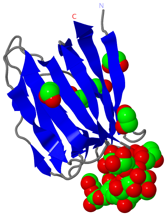
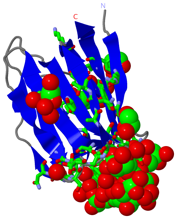
Asymmetric Unit (16, 16)
No.NameEvidenceResiduesDescription
01AC1SOFTWARESER A:27 , TYR A:68 , MAN A:123 , MAN A:127 , HOH A:207 , HOH A:222BINDING SITE FOR RESIDUE MAN A 122
02AC2SOFTWAREASP A:109 , MAN A:122 , MAN A:124 , MAN A:125 , HOH A:226 , HOH A:242BINDING SITE FOR RESIDUE MAN A 123
03AC3SOFTWAREMAN A:123 , EDO A:137 , HOH A:166 , HOH A:170 , HOH A:218 , HOH A:243 , HOH A:273BINDING SITE FOR RESIDUE MAN A 124
04AC4SOFTWAREGLY A:26 , SER A:27 , TYR A:28 , ASP A:30 , GLY A:43 , GLY A:44 , ASP A:109 , MAN A:123 , MAN A:126 , EDO A:137 , HOH A:170BINDING SITE FOR RESIDUE MAN A 125
05AC5SOFTWAREGLY A:11 , GLY A:12 , ASN A:45 , GLY A:108 , ASP A:109 , TYR A:110 , ASP A:112 , MAN A:125 , EDO A:133 , EDO A:135 , HOH A:196 , HOH A:232BINDING SITE FOR RESIDUE MAN A 126
06AC6SOFTWAREASP A:67 , GLY A:87 , SER A:88 , GLY A:89 , MAN A:122 , MAN A:128 , EDO A:135 , HOH A:145 , HOH A:261BINDING SITE FOR RESIDUE MAN A 127
07AC7SOFTWARESER A:42 , ASP A:67 , MAN A:127 , MAN A:129 , HOH A:145BINDING SITE FOR RESIDUE MAN A 128
08AC8SOFTWAREGLY A:66 , ASP A:67 , TYR A:68 , ASP A:70 , GLY A:90 , MAN A:128 , HOH A:229 , HOH A:282 , HOH A:286BINDING SITE FOR RESIDUE MAN A 129
09AC9SOFTWAREPHE A:7 , GLY A:8 , SER A:65 , GLY A:66 , HOH A:287BINDING SITE FOR RESIDUE EDO A 130
10BC1SOFTWAREVAL A:37 , THR A:62 , SER A:73 , PHE A:74 , HOH A:220 , HOH A:224 , HOH A:238 , HOH A:276BINDING SITE FOR RESIDUE EDO A 131
11BC2SOFTWAREARG A:5 , SER A:16 , ASN A:93 , THR A:94 , PEG A:136 , HOH A:194 , HOH A:280BINDING SITE FOR RESIDUE EDO A 132
12BC3SOFTWARESER A:25 , GLY A:26 , GLY A:86 , MAN A:126 , EDO A:135 , HOH A:195BINDING SITE FOR RESIDUE EDO A 133
13BC4SOFTWARELYS A:99 , GLN A:102 , ASN A:104 , TYR A:117 , HOH A:141BINDING SITE FOR RESIDUE EDO A 134
14BC5SOFTWAREGLY A:11 , ASP A:67 , MAN A:126 , MAN A:127 , EDO A:133 , HOH A:182 , HOH A:196 , HOH A:230BINDING SITE FOR RESIDUE EDO A 135
15BC6SOFTWARESER A:16 , GLY A:16A , SER A:16B , ILE A:34 , VAL A:37 , HIS A:39 , ALA A:92 , ASN A:93 , THR A:94 , EDO A:132BINDING SITE FOR RESIDUE PEG A 136
16BC7SOFTWAREASP A:109 , MAN A:124 , MAN A:125 , HOH A:243 , HOH A:273 , HOH A:283BINDING SITE FOR RESIDUE EDO A 137

(-) SS Bonds  (0, 0)

(no "SS Bond" information available for 3LL2)

(-) Cis Peptide Bonds  (1, 1)

Asymmetric/Biological Unit
No.Residues
1Gly A:83 -Pro A:84

 Sequence-Structure Mapping

(-) SAPs(SNPs)/Variants  (0, 0)

(no "SAP(SNP)/Variant" information available for 3LL2)

(-) PROSITE Motifs  (1, 1)

Asymmetric/Biological Unit (1, 1)
 PROSITEUniProtKBPDB
No.IDACDescriptionIDLocationCountLocation
1JACALIN_LECTINPS51752 Jacalin-type lectin domain profile.GRFIN_GRISQ1-120  1A:1-120

(-) Exons   (0, 0)

(no "Exon" information available for 3LL2)

(-) Sequences/Alignments

Asymmetric/Biological Unit
   Reformat: Number of residues per line =  ('0' or empty: single-line sequence representation)
  Number of residues per labelling interval =   
  UniProt sequence: complete  aligned part    
   Show mapping: SCOP domains CATH domains Pfam domains Secondary structure (by author)
SAPs(SNPs) PROSITE motifs Exons
(details for a mapped element are shown in a popup box when the mouse pointer rests over it)
Chain A from PDB  Type:PROTEIN  Length:123
 aligned with GRFIN_GRISQ | P84801 from UniProtKB/Swiss-Prot  Length:121

    Alignment length:123
                                           17 18                                                                                                       
                                    10      | 18        28        38        48        58        68        78        88        98       108       118   
          GRFIN_GRISQ     1 SLTHRKFGGSGGSPFSG--LSSIAVRSGSYLDXIIIDGVHHGGSGGNLSPTFTFGSGEYISNMTIRSGDYIDNISFETNMGRRFGPYGGSGGSANTLSNVKVIQINGSAGDYLDSLDIYYEQY 121
               SCOP domains --------------------------------------------------------------------------------------------------------------------------- SCOP domains
               CATH domains --------------------------------------------------------------------------------------------------------------------------- CATH domains
               Pfam domains ------------Jacalin-3ll2A01 A:13-120                                                                                      - Pfam domains
         Sec.struct. author ..eeeeee....eeee.....eeeeee...eeeeee..eeee....ee...ee......eeeeeeee...eeeeeeee....eeeeee....eeeeee.eeeeeeeeee...eeeeeeeeee. Sec.struct. author
                 SAPs(SNPs) --------------------------------------------------------------------------------------------------------------------------- SAPs(SNPs)
                    PROSITE JACALIN_LECTIN  PDB: A:1-120 UniProt: 1-120                                                                               - PROSITE
                 Transcript --------------------------------------------------------------------------------------------------------------------------- Transcript
                 3ll2 A   1 SSTHRKFGGSGGSPFSGSGLSSIAVRSGSYLDAIIIDGVHHGGSGGNLSPTFTFGSGEYISNMTIRSGDYIDNISFETNMGRRFGPYGGSGGSANTLSNVKVIQINGSAGDYLDSLDIYYEQY 121
                                    10      ||18        28        38        48        58        68        78        88        98       108       118   
                                          16A|                                                                                                         
                                           16B                                                                                                         

   Legend:   → Mismatch (orange background)
  - → Gap (green background, '-', border residues have a numbering label)
    → Modified Residue (blue background, lower-case, 'x' indicates undefined single-letter code, labelled with number + name)
  x → Chemical Group (purple background, 'x', labelled with number + name, e.g. ACE or NH2)
  extra numbering lines below/above indicate numbering irregularities and modified residue names etc., number ends below/above '|'

 Classification and Annotation

(-) SCOP Domains  (0, 0)

(no "SCOP Domain" information available for 3LL2)

(-) CATH Domains  (0, 0)

(no "CATH Domain" information available for 3LL2)

(-) Pfam Domains  (1, 1)

Asymmetric/Biological Unit

(-) Gene Ontology  (7, 7)

Asymmetric/Biological Unit(hide GO term definitions)
Chain A   (GRFIN_GRISQ | P84801)
molecular function
    GO:0046871    N-acetylgalactosamine binding    Interacting selectively and non-covalently with N-acetylgalactosamine, 2-acetamido-2-deoxygalactopyranose, the n-acetyl derivative of galactosamine.
    GO:0030246    carbohydrate binding    Interacting selectively and non-covalently with any carbohydrate, which includes monosaccharides, oligosaccharides and polysaccharides as well as substances derived from monosaccharides by reduction of the carbonyl group (alditols), by oxidation of one or more hydroxy groups to afford the corresponding aldehydes, ketones, or carboxylic acids, or by replacement of one or more hydroxy group(s) by a hydrogen atom. Cyclitols are generally not regarded as carbohydrates.
    GO:0005536    glucose binding    Interacting selectively and non-covalently with the D- or L-enantiomer of glucose.
    GO:0005537    mannose binding    Interacting selectively and non-covalently with mannose, a monosaccharide hexose, stereoisomeric with glucose, that occurs naturally only in polymerized forms called mannans.
    GO:0005515    protein binding    Interacting selectively and non-covalently with any protein or protein complex (a complex of two or more proteins that may include other nonprotein molecules).
biological process
    GO:0008150    biological_process    Any process specifically pertinent to the functioning of integrated living units: cells, tissues, organs, and organisms. A process is a collection of molecular events with a defined beginning and end.
cellular component
    GO:0005623    cell    The basic structural and functional unit of all organisms. Includes the plasma membrane and any external encapsulating structures such as the cell wall and cell envelope.

 Visualization

(-) Interactive Views

Asymmetric/Biological Unit
  Complete Structure
    Jena3D(integrated viewing of ligand, site, SAP, PROSITE, SCOP information)
    WebMol | AstexViewer[tm]@PDBe
(Java Applets, require no local installation except for Java; loading may be slow)
    STRAP
(Java WebStart application, automatic local installation, requires Java; full application with system access!)
    RasMol
(require local installation)
    Molscript (VRML)
(requires installation of a VRML viewer; select preferred view via VRML and generate a mono or stereo PDF format file)
 
  Ligands, Modified Residues, Ions
    EDO  [ RasMol | Jena3D ]  +environment [ RasMol | Jena3D ]
    MAN  [ RasMol | Jena3D ]  +environment [ RasMol | Jena3D ]
    PEG  [ RasMol | Jena3D ]  +environment [ RasMol | Jena3D ]
 
  Sites
    AC1  [ RasMol ]  +environment [ RasMol ]
    AC2  [ RasMol ]  +environment [ RasMol ]
    AC3  [ RasMol ]  +environment [ RasMol ]
    AC4  [ RasMol ]  +environment [ RasMol ]
    AC5  [ RasMol ]  +environment [ RasMol ]
    AC6  [ RasMol ]  +environment [ RasMol ]
    AC7  [ RasMol ]  +environment [ RasMol ]
    AC8  [ RasMol ]  +environment [ RasMol ]
    AC9  [ RasMol ]  +environment [ RasMol ]
    BC1  [ RasMol ]  +environment [ RasMol ]
    BC2  [ RasMol ]  +environment [ RasMol ]
    BC3  [ RasMol ]  +environment [ RasMol ]
    BC4  [ RasMol ]  +environment [ RasMol ]
    BC5  [ RasMol ]  +environment [ RasMol ]
    BC6  [ RasMol ]  +environment [ RasMol ]
    BC7  [ RasMol ]  +environment [ RasMol ]
 
  Cis Peptide Bonds
    Gly A:83 - Pro A:84   [ RasMol ]  
 

(-) Still Images

Jmol
  protein: cartoon or spacefill or dots and stick; nucleic acid: cartoon and stick; ligands: spacefill; active site: stick
Molscript
  protein, nucleic acid: cartoon; ligands: spacefill; active site: ball and stick

 Databases and Analysis Tools

(-) Databases

Access by PDB/NDB ID
  3ll2
    Family and Domain InformationProDom | SYSTERS
    General Structural InformationGlycoscienceDB | MMDB | NDB | OCA | PDB | PDBe | PDBj | PDBsum | PDBWiki | PQS | PROTEOPEDIA
    Orientation in MembranesOPM
    Protein SurfaceSURFACE
    Secondary StructureDSSP (structure derived) | HSSP (homology derived)
    Structural GenomicsGeneCensus
    Structural NeighboursCE | VAST
    Structure ClassificationCATH | Dali | SCOP
    Validation and Original DataBMRB Data View | BMRB Restraints Grid | EDS | PROCHECK | RECOORD | WHAT_CHECK
 
Access by UniProt ID/Accession number
  GRFIN_GRISQ | P84801
    Comparative Protein Structure ModelsModBase
    Genomic InformationEnsembl
    Protein-protein InteractionDIP
    Sequence, Family and Domain InformationInterPro | Pfam | SMART | UniProtKB/SwissProt
 
Access by Enzyme Classificator   (EC Number)
  (no 'Enzyme Classificator' available)
    General Enzyme InformationBRENDA | EC-PDB | Enzyme | IntEnz
    PathwayKEGG | MetaCyc
 
Access by Disease Identifier   (MIM ID)
  (no 'MIM ID' available)
    Disease InformationOMIM
 
Access by GenAge ID
  (no 'GenAge ID' available)
    Age Related InformationGenAge

(-) Analysis Tools

Access by PDB/NDB ID
    Domain InformationXDom
    Interatomic Contacts of Structural UnitsCSU
    Ligand-protein ContactsLPC
    Protein CavitiescastP
    Sequence and Secondary StructurePDBCartoon
    Structure AlignmentSTRAP(Java WebStart application, automatic local installation, requires Java; full application with system access!)
    Structure and Sequence BrowserSTING
 
Access by UniProt ID/Accession number
  GRFIN_GRISQ | P84801
    Protein Disorder PredictionDisEMBL | FoldIndex | GLOBPLOT (for more information see DisProt)

 Related Entries

(-) Entries Sharing at Least One Protein Chain (UniProt ID)

UniProtKB/Swiss-Prot
        GRFIN_GRISQ | P848012gty 2guc 2gud 2gue 2gux 2hyq 2hyr 2nu5 2nuo 3lky 3ll0 3ll1

(-) Related Entries Specified in the PDB File

2gty CRYSTAL STRUCTURE OF UNLIGANDED GRIFFITHSIN
2guc CRYSTAL STRUCTURE OF A COMPLEX OF GRIFFITHSIN WITH MANNOSE AT 1.78 A RESOLUTION.
2gud CRYSTAL STRUCTURE OF A COMPLEX OF GRIFFITHSIN WITH MANNOSE AT 0.94 A RESOLUTION
2gue CRYSTAL STRUCTURE OF A COMPLEX OF GRIFFITHSIN WITH N- ACETYLGLUCOSAMINE
2gux SELENOMETHIONINE DERIVATIVE OF GRIFFITHSIN
2hyq CRYSTAL STRUCTURE OF A COMPLEX OF GRIFFITHSIN WITH 6ALPHA- MANNOBIOSE
2hyr CRYSTAL STRUCTURE OF A COMPLEX OF GRIFFITHSIN WITH MALTOSE
2nu5 CRYSTAL STRUCTURE OF A COMPLEX OF GRIFFITHSIN COCRYSTALLIZED WITH N-ACETYLGLUCOSAMINE
2nuo CRYSTAL STRUCTURE OF A COMPLEX OF GRIFFITHSIN WITH GLUCOSE
3lky MONOMERIC GRIFFITHSIN WITH A SINGLE GLY-SER INSERTION
3ll0 MONOMERIC GRIFFITHSIN WITH TWO GLY-SER INSERTIONS
3ll1 MONOMERIC GRIFFITHSIN WITH A SINGLE GLY-SER INSERTION AND MUTATIONS TO REMOVE RESIDUAL SELF-ASSOCIATION