Show PDB file:   
         Plain Text   HTML   (compressed file size)
QuickSearch:   
by PDB,NDB,UniProt,PROSITE Code or Search Term(s)  
(-)Asymmetric Unit
(-)Biological Unit 1
collapse expand < >
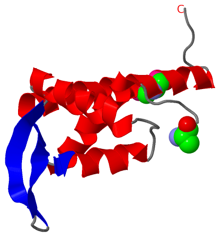
Image Asymmetric Unit
Asymmetric Unit  (Jmol Viewer)
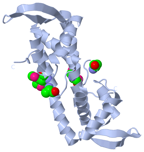
Image Biological Unit 1
Biological Unit 1  (Jmol Viewer)

(-) Description

Title :  THE CRYSTAL STRUCTURE OF SMU.1704 FROM STREPTOCOCCUS MUTANS UA159
 
Authors :  X. -D. Su, X. Liu, T. M. Fu
Date :  29 Dec 09  (Deposition) - 08 Dec 10  (Release) - 08 Dec 10  (Revision)
Method :  X-RAY DIFFRACTION
Resolution :  2.20
Chains :  Asym. Unit :  A
Biol. Unit 1:  A  (2x)
Keywords :  Padr, Transcriptional Factor, Transcription (Keyword Search: [Gene Ontology, PubMed, Web (Google)] )
 
Reference :  X. -D. Su, X. Liu, T. M. Fu
The Crystal Structure Of Smu. 1704 From Streptococcus Mutans Ua159
To Be Published
PubMed: search

(-) Compounds

Molecule 1 - PUTATIVE UNCHARACTERIZED PROTEIN SMU.1704
    Chains: A
    Engineered: YES
    Expression System: ESCHERICHIA COLI
    Expression System Plasmid: PET21B
    Expression System Strain: BL21(DE3)
    Expression System Taxid: 469008
    Expression System Vector Type: PLASMID
    Gene: SMU.1704
    Organism Scientific: STREPTOCOCCUS MUTANS
    Organism Taxid: 210007
    Strain: UA159

 Structural Features

(-) Chains, Units

  1
Asymmetric Unit : A
Biological Unit 1 (2x): A

Summary Information (see also Sequences/Alignments below)

(-) Ligands, Modified Residues, Ions  (1, 2)

Asymmetric Unit (1, 2)
No.NameCountTypeFull Name
1MSE2Mod. Amino AcidSELENOMETHIONINE
Biological Unit 1 (1, 4)
No.NameCountTypeFull Name
1MSE4Mod. Amino AcidSELENOMETHIONINE

(-) Sites  (0, 0)

(no "Site" information available for 3L7W)

(-) SS Bonds  (0, 0)

(no "SS Bond" information available for 3L7W)

(-) Cis Peptide Bonds  (0, 0)

(no "Cis Peptide Bond" information available for 3L7W)

 Sequence-Structure Mapping

(-) SAPs(SNPs)/Variants  (0, 0)

(no "SAP(SNP)/Variant" information available for 3L7W)

(-) PROSITE Motifs  (0, 0)

(no "PROSITE Motif" information available for 3L7W)

(-) Exons   (0, 0)

(no "Exon" information available for 3L7W)

(-) Sequences/Alignments

Asymmetric Unit
   Reformat: Number of residues per line =  ('0' or empty: single-line sequence representation)
  Number of residues per labelling interval =   
  UniProt sequence: complete  aligned part    
   Show mapping: SCOP domains CATH domains Pfam domains Secondary structure (by author)
SAPs(SNPs) PROSITE motifs Exons
(details for a mapped element are shown in a popup box when the mouse pointer rests over it)
Chain A from PDB  Type:PROTEIN  Length:106
 aligned with Q8DSR7_STRMU | Q8DSR7 from UniProtKB/TrEMBL  Length:108

    Alignment length:106
                                    10        20        30        40        50        60        70        80        90       100      
         Q8DSR7_STRMU     1 MYYPVSALLIEYLILAIVSKHDSYGYDISQTIKLIASIKESTLYPILKKLEKAGYLSTYTQEHQGRRRKYYHLTDSGEKHLVYLTKEWSVYKMTIDGIVEGRIRHD 106
               SCOP domains d3l7wa_ A: automated matches                                                                               SCOP domains
               CATH domains ---------------------------------------------------------------------------------------------------------- CATH domains
               Pfam domains -------------PadR-3l7wA01 A:14-82                                                 ------------------------ Pfam domains
         Sec.struct. author .....hhhhhhhhhhhhhhhh.eehhhhhhhhh.....hhhhhhhhhhhhhhh..eeeeeeee..eeeeeeeehhhhhhhhhhhhhhhhhhhhhhhhhhh...... Sec.struct. author
                 SAPs(SNPs) ---------------------------------------------------------------------------------------------------------- SAPs(SNPs)
                    PROSITE ---------------------------------------------------------------------------------------------------------- PROSITE
                 Transcript ---------------------------------------------------------------------------------------------------------- Transcript
                 3l7w A   1 mYYPVSALLIEYLILAIVSKHDSYGYDISQTIKLIASIKESTLYPILKKLEKAGYLSTYTQEHQGRRRKYYHLTDSGEKHLVYLTKEWSVYKmTIDGIVEGRIRHD 106
                            |       10        20        30        40        50        60        70        80        90  |    100      
                            |                                                                                          93-MSE         
                            1-MSE                                                                                                     

   Legend:   → Mismatch (orange background)
  - → Gap (green background, '-', border residues have a numbering label)
    → Modified Residue (blue background, lower-case, 'x' indicates undefined single-letter code, labelled with number + name)
  x → Chemical Group (purple background, 'x', labelled with number + name, e.g. ACE or NH2)
  extra numbering lines below/above indicate numbering irregularities and modified residue names etc., number ends below/above '|'

 Classification and Annotation

(-) SCOP Domains  (1, 1)

Asymmetric Unit

(-) CATH Domains  (0, 0)

(no "CATH Domain" information available for 3L7W)

(-) Pfam Domains  (1, 1)

Asymmetric Unit
(-)
Clan: HTH (544)

(-) Gene Ontology  (0, 0)

Asymmetric Unit(hide GO term definitions)
    (no "Gene Ontology" information available for 3L7W)

 Visualization

(-) Interactive Views

Asymmetric Unit
  Complete Structure
    Jena3D(integrated viewing of ligand, site, SAP, PROSITE, SCOP information)
    WebMol | AstexViewer[tm]@PDBe
(Java Applets, require no local installation except for Java; loading may be slow)
    STRAP
(Java WebStart application, automatic local installation, requires Java; full application with system access!)
    RasMol
(require local installation)
    Molscript (VRML)
(requires installation of a VRML viewer; select preferred view via VRML and generate a mono or stereo PDF format file)
 
  Ligands, Modified Residues, Ions
    MSE  [ RasMol | Jena3D ]  +environment [ RasMol | Jena3D ]
 
  Sites
(no "Sites" information available for 3l7w)
 
  Cis Peptide Bonds
(no "Cis Peptide Bonds" information available for 3l7w)
 
Biological Unit
  Complete Structure
    Biological Unit 1  [ Jena3D ]

(-) Still Images

Jmol
  protein: cartoon or spacefill or dots and stick; nucleic acid: cartoon and stick; ligands: spacefill; active site: stick
Molscript
  protein, nucleic acid: cartoon; ligands: spacefill; active site: ball and stick

 Databases and Analysis Tools

(-) Databases

Access by PDB/NDB ID
  3l7w
    Family and Domain Information: ProDom | SYSTERS
    General Structural Information: GlycoscienceDB | MMDB | NDB | OCA | PDB | PDBe | PDBj | PDBsum | PDBWiki | PQS | PROTEOPEDIA
    Orientation in Membranes: OPM
    Protein Surface: SURFACE
    Secondary Structure: DSSP (structure derived) | HSSP (homology derived)
    Structural Genomics: GeneCensus
    Structural Neighbours: CE | VAST
    Structure Classification: CATH | Dali | SCOP
    Validation and Original Data: BMRB Data View | BMRB Restraints Grid | EDS | PROCHECK | RECOORD | WHAT_CHECK
 
Access by UniProt ID/Accession number
  Q8DSR7_STRMU | Q8DSR7
    Comparative Protein Structure Models: ModBase
    Genomic Information: Ensembl
    Protein-protein Interaction: DIP
    Sequence, Family and Domain Information: InterPro | Pfam | SMART | UniProtKB/TrEMBL
 
Access by Enzyme Classificator   (EC Number)
  (no 'Enzyme Classificator' available)
    General Enzyme Information: BRENDA | EC-PDB | Enzyme | IntEnz
    Pathway: KEGG | MetaCyc
 
Access by Disease Identifier   (MIM ID)
  (no 'MIM ID' available)
    Disease Information: OMIM
 
Access by GenAge ID
  (no 'GenAge ID' available)
    Age Related Information: GenAge

(-) Analysis Tools

Access by PDB/NDB ID
    Domain Information: XDom
    Interatomic Contacts of Structural Units: CSU
    Ligand-protein Contacts: LPC
    Protein Cavities: castP
    Sequence and Secondary Structure: PDBCartoon
    Structure Alignment: STRAP(Java WebStart application, automatic local installation, requires Java; full application with system access!)
    Structure and Sequence Browser: STING
 
Access by UniProt ID/Accession number
  Q8DSR7_STRMU | Q8DSR7
    Protein Disorder Prediction: DisEMBL | FoldIndex | GLOBPLOT (for more information see DisProt)

 Related Entries

(-) Entries Sharing at Least One Protein Chain (UniProt ID)

(no "Entries Sharing at Least One Protein Chain" available for 3L7W)

(-) Related Entries Specified in the PDB File

(no "Related Entries Specified in the PDB File" available for 3L7W)