Show PDB file:   
         Plain Text   HTML   (compressed file size)
QuickSearch:   
by PDB,NDB,UniProt,PROSITE Code or Search Term(s)  
(-)Asymmetric Unit
(-)Asym. Unit - sites
(-)Biological Unit 1
(-)Biol. Unit 1 - sites
collapse expand < >
Image Asymmetric Unit
Asymmetric Unit  (Jmol Viewer)
Image Asym. Unit - sites
Asym. Unit - sites  (Jmol Viewer)
Image Biological Unit 1
Biological Unit 1  (Jmol Viewer)
Image Biol. Unit 1 - sites
Biol. Unit 1 - sites  (Jmol Viewer)

(-) Description

Title :  CRYSTAL STRUCTURE OF VSET UNDER CONDITION B
 
Authors :  H. Wei, M. M. Zhou
Date :  10 Nov 09  (Deposition) - 10 Nov 10  (Release) - 10 Nov 10  (Revision)
Method :  X-RAY DIFFRACTION
Resolution :  1.85
Chains :  Asym. Unit :  A
Biol. Unit 1:  A  (2x)
Keywords :  Set Domain, Viral Protein (Keyword Search: [Gene Ontology, PubMed, Web (Google))
 
Reference :  H. Wei, M. M. Zhou
Dimerization Of A Viral Set Protein Endows Its Function.
Proc. Natl. Acad. Sci. Usa V. 107 18433 2010
PubMed-ID: 20937900  |  Reference-DOI: 10.1073/PNAS.1009911107

(-) Compounds

Molecule 1 - A612L PROTEIN
    ChainsA
    Organism CommonPBCV-1
    Organism ScientificPARAMECIUM BURSARIA CHLORELLA VIRUS 1
    Organism Taxid10506

 Structural Features

(-) Chains, Units

  1
Asymmetric Unit A
Biological Unit 1 (2x)A

Summary Information (see also Sequences/Alignments below)

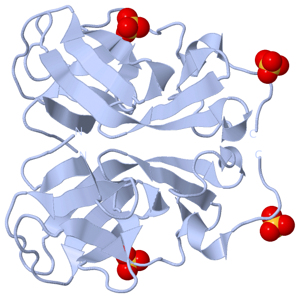
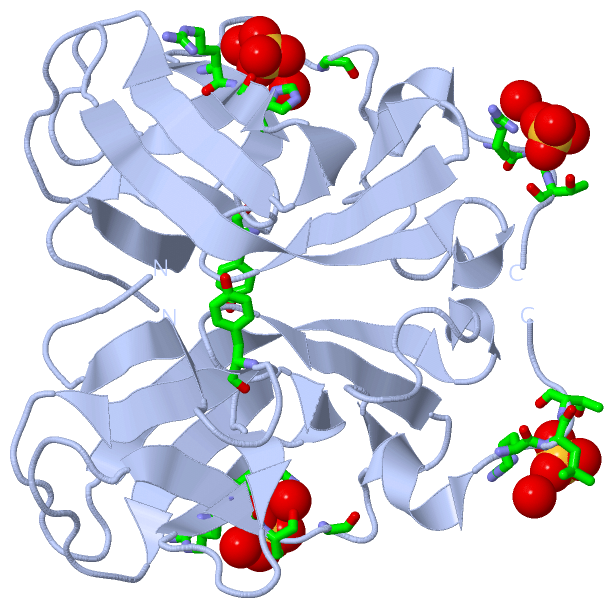
(-) Ligands, Modified Residues, Ions  (1, 2)

Asymmetric Unit (1, 2)
No.NameCountTypeFull Name
1SO42Ligand/IonSULFATE ION
Biological Unit 1 (1, 4)
No.NameCountTypeFull Name
1SO44Ligand/IonSULFATE ION

(-) Sites  (2, 2)

Asymmetric Unit (2, 2)
No.NameEvidenceResiduesDescription
1AC1SOFTWAREARG A:77 , HIS A:78 , ILE A:103 , SER A:104 , GLY A:106 , HOH A:150 , HOH A:183BINDING SITE FOR RESIDUE SO4 A 120
2AC2SOFTWARETYR A:16 , ARG A:115 , LEU A:116 , THR A:117 , HOH A:171BINDING SITE FOR RESIDUE SO4 A 121

(-) SS Bonds  (0, 0)

(no "SS Bond" information available for 3KMJ)

(-) Cis Peptide Bonds  (0, 0)

(no "Cis Peptide Bond" information available for 3KMJ)

 Sequence-Structure Mapping

(-) SAPs(SNPs)/Variants  (0, 0)

(no "SAP(SNP)/Variant" information available for 3KMJ)

(-) PROSITE Motifs  (0, 0)

(no "PROSITE Motif" information available for 3KMJ)

(-) Exons   (0, 0)

(no "Exon" information available for 3KMJ)

(-) Sequences/Alignments

Asymmetric Unit
   Reformat: Number of residues per line =  ('0' or empty: single-line sequence representation)
  Number of residues per labelling interval =   
  UniProt sequence: complete  aligned part    
   Show mapping: SCOP domains CATH domains Pfam domains Secondary structure (by author)
SAPs(SNPs) PROSITE motifs Exons
(details for a mapped element are shown in a popup box when the mouse pointer rests over it)
Chain A from PDB  Type:PROTEIN  Length:119
 aligned with O41094_PBCV1 | O41094 from UniProtKB/TrEMBL  Length:119

    Alignment length:119
                                    10        20        30        40        50        60        70        80        90       100       110         
         O41094_PBCV1     1 MFNDRVIVKKSPLGGYGVFARKSFEKGELVEECLCIVRHNDDWGTALEDYLFSRKNMSAMALGFGAIFNHSKDPNARHELTAGLKRMRIFTIKPIAIGEEITISYGDDYWLSRPRLTQN 119
               SCOP domains d3kmja_ A: Viral histone H3 Lysine 27 Methyltransferase                                                                 SCOP domains
               CATH domains ----------------------------------------------------------------------------------------------------------------------- CATH domains
               Pfam domains ------------------------------------------SET-3kmjA01 A:43-106                                            ------------- Pfam domains
         Sec.struct. author .....eeeee.....eeeee........eeeeeeeeeee.hhh........eee..eeeee.......ee.....eeeee.hhh.eeeeee........eeee.....hhhhh...... Sec.struct. author
                 SAPs(SNPs) ----------------------------------------------------------------------------------------------------------------------- SAPs(SNPs)
                    PROSITE ----------------------------------------------------------------------------------------------------------------------- PROSITE
                 Transcript ----------------------------------------------------------------------------------------------------------------------- Transcript
                 3kmj A   1 MFNDRVIVKKSPLGGYGVFARKSFEKGELVEECLCIVRHNDDWGTALEDYLFSRKNMSAMALGFGAIFNHSKDPNARHELTAGLKRMRIFTIKPIAIGEEITISYGDDYWLSRPRLTQN 119
                                    10        20        30        40        50        60        70        80        90       100       110         

   Legend:   → Mismatch (orange background)
  - → Gap (green background, '-', border residues have a numbering label)
    → Modified Residue (blue background, lower-case, 'x' indicates undefined single-letter code, labelled with number + name)
  x → Chemical Group (purple background, 'x', labelled with number + name, e.g. ACE or NH2)
  extra numbering lines below/above indicate numbering irregularities and modified residue names etc., number ends below/above '|'

 Classification and Annotation

(-) SCOP Domains  (1, 1)

Asymmetric Unit

(-) CATH Domains  (0, 0)

(no "CATH Domain" information available for 3KMJ)

(-) Pfam Domains  (1, 1)

Asymmetric Unit

(-) Gene Ontology  (3, 3)

Asymmetric Unit(hide GO term definitions)
Chain A   (O41094_PBCV1 | O41094)
molecular function
    GO:0008168    methyltransferase activity    Catalysis of the transfer of a methyl group to an acceptor molecule.
    GO:0005515    protein binding    Interacting selectively and non-covalently with any protein or protein complex (a complex of two or more proteins that may include other nonprotein molecules).
biological process
    GO:0032259    methylation    The process in which a methyl group is covalently attached to a molecule.

 Visualization

(-) Interactive Views

Asymmetric Unit
  Complete Structure
    Jena3D(integrated viewing of ligand, site, SAP, PROSITE, SCOP information)
    WebMol | AstexViewer[tm]@PDBe
(Java Applets, require no local installation except for Java; loading may be slow)
    STRAP
(Java WebStart application, automatic local installation, requires Java; full application with system access!)
    RasMol
(require local installation)
    Molscript (VRML)
(requires installation of a VRML viewer; select preferred view via VRML and generate a mono or stereo PDF format file)
 
  Ligands, Modified Residues, Ions
    SO4  [ RasMol | Jena3D ]  +environment [ RasMol | Jena3D ]
 
  Sites
    AC1  [ RasMol ]  +environment [ RasMol ]
    AC2  [ RasMol ]  +environment [ RasMol ]
 
  Cis Peptide Bonds
(no "Cis Peptide Bonds" information available for 3kmj)
 
Biological Unit
  Complete Structure
    Biological Unit 1  [ Jena3D ]

(-) Still Images

Jmol
  protein: cartoon or spacefill or dots and stick; nucleic acid: cartoon and stick; ligands: spacefill; active site: stick
Molscript
  protein, nucleic acid: cartoon; ligands: spacefill; active site: ball and stick

 Databases and Analysis Tools

(-) Databases

Access by PDB/NDB ID
  3kmj
    Family and Domain InformationProDom | SYSTERS
    General Structural InformationGlycoscienceDB | MMDB | NDB | OCA | PDB | PDBe | PDBj | PDBsum | PDBWiki | PQS | PROTEOPEDIA
    Orientation in MembranesOPM
    Protein SurfaceSURFACE
    Secondary StructureDSSP (structure derived) | HSSP (homology derived)
    Structural GenomicsGeneCensus
    Structural NeighboursCE | VAST
    Structure ClassificationCATH | Dali | SCOP
    Validation and Original DataBMRB Data View | BMRB Restraints Grid | EDS | PROCHECK | RECOORD | WHAT_CHECK
 
Access by UniProt ID/Accession number
  O41094_PBCV1 | O41094
    Comparative Protein Structure ModelsModBase
    Genomic InformationEnsembl
    Protein-protein InteractionDIP
    Sequence, Family and Domain InformationInterPro | Pfam | SMART | UniProtKB/TrEMBL
 
Access by Enzyme Classificator   (EC Number)
  (no 'Enzyme Classificator' available)
    General Enzyme InformationBRENDA | EC-PDB | Enzyme | IntEnz
    PathwayKEGG | MetaCyc
 
Access by Disease Identifier   (MIM ID)
  (no 'MIM ID' available)
    Disease InformationOMIM
 
Access by GenAge ID
  (no 'GenAge ID' available)
    Age Related InformationGenAge

(-) Analysis Tools

Access by PDB/NDB ID
    Domain InformationXDom
    Interatomic Contacts of Structural UnitsCSU
    Ligand-protein ContactsLPC
    Protein CavitiescastP
    Sequence and Secondary StructurePDBCartoon
    Structure AlignmentSTRAP(Java WebStart application, automatic local installation, requires Java; full application with system access!)
    Structure and Sequence BrowserSTING
 
Access by UniProt ID/Accession number
  O41094_PBCV1 | O41094
    Protein Disorder PredictionDisEMBL | FoldIndex | GLOBPLOT (for more information see DisProt)

 Related Entries

(-) Entries Sharing at Least One Protein Chain (UniProt ID)

UniProtKB/TrEMBL
        O41094_PBCV1 | O410941n3j 2g46 3kma 3kmt

(-) Related Entries Specified in the PDB File

3kma CRYSTAL STRUCTURE OF VSET UNDER CONDITION A
3kmt CRYSTAL STRUCTURE OF VSET/SAH/H3 TERNARY COMPLEX