Show PDB file:   
         Plain Text   HTML   (compressed file size)
QuickSearch:   
by PDB,NDB,UniProt,PROSITE Code or Search Term(s)  
(-)Asymmetric Unit
(-)Asym. Unit - sites
(-)Biological Unit 1
(-)Biol. Unit 1 - sites
(-)Biological Unit 2
(-)Biological Unit 3
collapse expand < >
Image Asymmetric Unit
Asymmetric Unit  (Jmol Viewer)
Image Asym. Unit - sites
Asym. Unit - sites  (Jmol Viewer)
Image Biological Unit 1
Biological Unit 1  (Jmol Viewer)
Image Biol. Unit 1 - sites
Biol. Unit 1 - sites  (Jmol Viewer)
Image Biological Unit 2
Biological Unit 2  (Jmol Viewer)
Image Biological Unit 3
Biological Unit 3  (Jmol Viewer)

(-) Description

Title :  CRYSTAL STRUCTURE OF PUTATIVE PHOSPHATASE (DUF442) (YP_001181608.1) FROM SHEWANELLA PUTREFACIENS CN-32 AT 1.40 A RESOLUTION
 
Authors :  Joint Center For Structural Genomics (Jcsg)
Date :  02 Apr 09  (Deposition) - 21 Apr 09  (Release) - 13 Jul 11  (Revision)
Method :  X-RAY DIFFRACTION
Resolution :  1.40
Chains :  Asym. Unit :  A,B
Biol. Unit 1:  A  (1x)
Biol. Unit 2:  B  (1x)
Biol. Unit 3:  A,B  (1x)
Keywords :  Yp_001181608. 1, Putative Phosphatase (Duf442), Structural Genomics, Joint Center For Structural Genomics, Jcsg, Protein Structure Initiative, Psi-2, Tyrosine Phosphatase, Hydrolase (Keyword Search: [Gene Ontology, PubMed, Web (Google))
 
Reference :  Joint Center For Structural Genomics (Jcsg)
Crystal Structure Of Putative Phosphatase (Duf442) (Yp_001181608. 1) From Shewanella Putrefaciens Cn-32 At 1. 40 A Resolution
To Be Published
PubMed: search

(-) Compounds

Molecule 1 - PUTATIVE PHOSPHATASE (DUF442)
    ChainsA, B
    EngineeredYES
    Expression SystemESCHERICHIA COLI
    Expression System PlasmidSPEEDET
    Expression System StrainHK100
    Expression System Taxid562
    Expression System Vector TypePLASMID
    GeneSPUTCN32_0073, YP_001181608.1
    Organism ScientificSHEWANELLA PUTREFACIENS CN-32
    Organism Taxid319224

 Structural Features

(-) Chains, Units

  12
Asymmetric Unit AB
Biological Unit 1 (1x)A 
Biological Unit 2 (1x) B
Biological Unit 3 (1x)AB

Summary Information (see also Sequences/Alignments below)

(-) Ligands, Modified Residues, Ions  (4, 20)

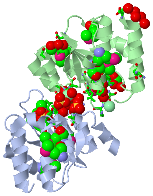
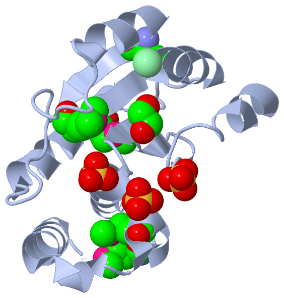
Asymmetric Unit (4, 20)
No.NameCountTypeFull Name
1CL3Ligand/IonCHLORIDE ION
2EDO3Ligand/Ion1,2-ETHANEDIOL
3MSE10Mod. Amino AcidSELENOMETHIONINE
4SO44Ligand/IonSULFATE ION
Biological Unit 1 (3, 9)
No.NameCountTypeFull Name
1CL-1Ligand/IonCHLORIDE ION
2EDO1Ligand/Ion1,2-ETHANEDIOL
3MSE5Mod. Amino AcidSELENOMETHIONINE
4SO43Ligand/IonSULFATE ION
Biological Unit 2 (3, 8)
No.NameCountTypeFull Name
1CL-1Ligand/IonCHLORIDE ION
2EDO2Ligand/Ion1,2-ETHANEDIOL
3MSE5Mod. Amino AcidSELENOMETHIONINE
4SO41Ligand/IonSULFATE ION
Biological Unit 3 (3, 17)
No.NameCountTypeFull Name
1CL-1Ligand/IonCHLORIDE ION
2EDO3Ligand/Ion1,2-ETHANEDIOL
3MSE10Mod. Amino AcidSELENOMETHIONINE
4SO44Ligand/IonSULFATE ION

(-) Sites  (10, 10)

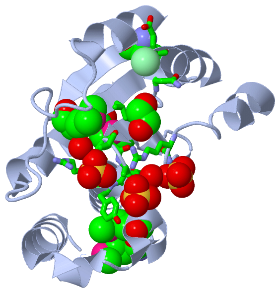
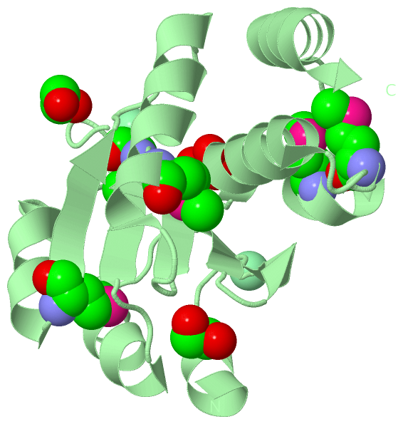
Asymmetric Unit (10, 10)
No.NameEvidenceResiduesDescription
01AC1SOFTWAREASN A:50 , GLU A:51 , ASN B:50 , GLU B:51BINDING SITE FOR RESIDUE CL A 1
02AC2SOFTWARECYS A:125 , LEU A:126 , ALA A:127 , ASN A:128 , TYR A:129 , ARG A:130 , HOH A:248 , HIS B:178 , HOH B:209 , HOH B:302BINDING SITE FOR RESIDUE SO4 A 5
03AC3SOFTWAREARG A:34 , TYR A:129 , HOH A:227 , HOH A:250 , CL B:2 , HOH B:268 , HOH B:388BINDING SITE FOR RESIDUE SO4 A 6
04AC4SOFTWAREARG A:34 , ALA A:35 , HOH A:227 , ARG B:34 , HOH B:336BINDING SITE FOR RESIDUE SO4 A 7
05AC5SOFTWAREARG A:34 , LEU A:48 , ASN A:50 , HOH B:11 , ASN B:50 , HOH B:223BINDING SITE FOR RESIDUE EDO A 8
06AC6SOFTWARESO4 A:6 , ARG A:34 , ALA B:35 , HOH B:238BINDING SITE FOR RESIDUE CL B 2
07AC7SOFTWARESER B:73 , LYS B:74BINDING SITE FOR RESIDUE CL B 3
08AC8SOFTWARECYS B:125 , LEU B:126 , ALA B:127 , ASN B:128 , TYR B:129 , ARG B:130 , HOH B:220BINDING SITE FOR RESIDUE SO4 B 4
09AC9SOFTWAREALA B:60 , VAL B:62 , ASP B:120 , GLN B:166 , HOH B:233 , HOH B:309BINDING SITE FOR RESIDUE EDO B 9
10BC1SOFTWAREPRO B:70 , ASP B:71 , SER B:72 , TYR B:93 , HOH B:366BINDING SITE FOR RESIDUE EDO B 10

(-) SS Bonds  (0, 0)

(no "SS Bond" information available for 3GXH)

(-) Cis Peptide Bonds  (0, 0)

(no "Cis Peptide Bond" information available for 3GXH)

 Sequence-Structure Mapping

(-) SAPs(SNPs)/Variants  (0, 0)

(no "SAP(SNP)/Variant" information available for 3GXH)

(-) PROSITE Motifs  (0, 0)

(no "PROSITE Motif" information available for 3GXH)

(-) Exons   (0, 0)

(no "Exon" information available for 3GXH)

(-) Sequences/Alignments

Asymmetric Unit
   Reformat: Number of residues per line =  ('0' or empty: single-line sequence representation)
  Number of residues per labelling interval =   
  UniProt sequence: complete  aligned part    
   Show mapping: SCOP domains CATH domains Pfam domains Secondary structure (by author)
SAPs(SNPs) PROSITE motifs Exons
(details for a mapped element are shown in a popup box when the mouse pointer rests over it)
Chain A from PDB  Type:PROTEIN  Length:156
 aligned with A4Y1H6_SHEPC | A4Y1H6 from UniProtKB/TrEMBL  Length:178

    Alignment length:156
                                    32        42        52        62        72        82        92       102       112       122       132       142       152       162       172      
         A4Y1H6_SHEPC    23 NIESIENLQGIRALQQQAPQLLSSGLPNEQQFSLLKQAGVDVVINLMPDSSKDAHPDEGKLVTQAGMDYVYIPVDWQNPKVEDVEAFFAAMDQHKGKDVLVHCLANYRASAFAYLYQLKQGQNPNMAQTMTPWNDELAIYPKWQALLTEVSAKYGH 178
               SCOP domains ------------------------------------------------------------------------------------------------------------------------------------------------------------ SCOP domains
               CATH domains ------------------------------------------------------------------------------------------------------------------------------------------------------------ CATH domains
               Pfam domains ------------------------------------------------------------------------------------------------------------------------------------------------------------ Pfam domains
         Sec.struct. author hhhhhhh.......eeee..eeee...hhhhhhhhhhh...eeee...........hhhhhhhhh..eeee........hhhhhhhhhhhhhhh....eeee...hhhhhhhhhhhhhhh....hhhhhhhhhh.hhhhhhhhhhhhhhhhhhh.. Sec.struct. author
                 SAPs(SNPs) ------------------------------------------------------------------------------------------------------------------------------------------------------------ SAPs(SNPs)
                    PROSITE ------------------------------------------------------------------------------------------------------------------------------------------------------------ PROSITE
                 Transcript ------------------------------------------------------------------------------------------------------------------------------------------------------------ Transcript
                 3gxh A  23 NIESIENLQGIRALQQQAPQLLSSGLPNEQQFSLLKQAGVDVVINLmPDSSKDAHPDEGKLVTQAGmDYVYIPVDWQNPKVEDVEAFFAAmDQHKGKDVLVHCLANYRASAFAYLYQLKQGQNPNmAQTmTPWNDELAIYPKWQALLTEVSAKYGH 178
                                    32        42        52        62      | 72        82      | 92       102       112|      122       132       142     | 152       162       172      
                                                                         69-MSE              89-MSE                 113-MSE                            148-MSE                          
                                                                                                                                                           152-MSE                      

Chain B from PDB  Type:PROTEIN  Length:151
 aligned with A4Y1H6_SHEPC | A4Y1H6 from UniProtKB/TrEMBL  Length:178

    Alignment length:153
                                    35        45        55        65        75        85        95       105       115       125       135       145       155       165       175   
         A4Y1H6_SHEPC    26 SIENLQGIRALQQQAPQLLSSGLPNEQQFSLLKQAGVDVVINLMPDSSKDAHPDEGKLVTQAGMDYVYIPVDWQNPKVEDVEAFFAAMDQHKGKDVLVHCLANYRASAFAYLYQLKQGQNPNMAQTMTPWNDELAIYPKWQALLTEVSAKYGH 178
               SCOP domains --------------------------------------------------------------------------------------------------------------------------------------------------------- SCOP domains
               CATH domains --------------------------------------------------------------------------------------------------------------------------------------------------------- CATH domains
               Pfam domains --------------------------------------------------------------------------------------------------------------------------------------------------------- Pfam domains
         Sec.struct. author hhhhhh.....eeee..eeee...hhhhhhhhhhh...eeee...........hhhhhhhhh..eeee........hhhhhhhhhhhhhhh....eeee...hhhhhhhhhhhhhhh....hhhhhhhhhh--...hhhhhhhhhhhhhhhh. Sec.struct. author
                 SAPs(SNPs) --------------------------------------------------------------------------------------------------------------------------------------------------------- SAPs(SNPs)
                    PROSITE --------------------------------------------------------------------------------------------------------------------------------------------------------- PROSITE
                 Transcript --------------------------------------------------------------------------------------------------------------------------------------------------------- Transcript
                 3gxh B  26 SIENLQGIRALQQQAPQLLSSGLPNEQQFSLLKQAGVDVVINLmPDSSKDAHPDEGKLVTQAGmDYVYIPVDWQNPKVEDVEAFFAAmDQHKGKDVLVHCLANYRASAFAYLYQLKQGQNPNmAQTmTPWN--LAIYPKWQALLTEVSAKYGH 178
                                    35        45        55        65   |    75        85   |    95       105       115       125       135       145  |   |155|  |   165       175   
                                                                      69-MSE              89-MSE                 113-MSE                            148-MSE 156  |                   
                                                                                                                                                        152-MSE159                   

   Legend:   → Mismatch (orange background)
  - → Gap (green background, '-', border residues have a numbering label)
    → Modified Residue (blue background, lower-case, 'x' indicates undefined single-letter code, labelled with number + name)
  x → Chemical Group (purple background, 'x', labelled with number + name, e.g. ACE or NH2)
  extra numbering lines below/above indicate numbering irregularities and modified residue names etc., number ends below/above '|'

 Classification and Annotation

(-) SCOP Domains  (0, 0)

(no "SCOP Domain" information available for 3GXH)

(-) CATH Domains  (0, 0)

(no "CATH Domain" information available for 3GXH)

(-) Pfam Domains  (0, 0)

(no "Pfam Domain" information available for 3GXH)

(-) Gene Ontology  (1, 1)

Asymmetric Unit(hide GO term definitions)
Chain A,B   (A4Y1H6_SHEPC | A4Y1H6)
molecular function
    GO:0016787    hydrolase activity    Catalysis of the hydrolysis of various bonds, e.g. C-O, C-N, C-C, phosphoric anhydride bonds, etc. Hydrolase is the systematic name for any enzyme of EC class 3.

 Visualization

(-) Interactive Views

Asymmetric Unit
  Complete Structure
    Jena3D(integrated viewing of ligand, site, SAP, PROSITE, SCOP information)
    WebMol | AstexViewer[tm]@PDBe
(Java Applets, require no local installation except for Java; loading may be slow)
    STRAP
(Java WebStart application, automatic local installation, requires Java; full application with system access!)
    RasMol
(require local installation)
    Molscript (VRML)
(requires installation of a VRML viewer; select preferred view via VRML and generate a mono or stereo PDF format file)
 
  Ligands, Modified Residues, Ions
    CL  [ RasMol | Jena3D ]  +environment [ RasMol | Jena3D ]
    EDO  [ RasMol | Jena3D ]  +environment [ RasMol | Jena3D ]
    MSE  [ RasMol | Jena3D ]  +environment [ RasMol | Jena3D ]
    SO4  [ RasMol | Jena3D ]  +environment [ RasMol | Jena3D ]
 
  Sites
    AC1  [ RasMol ]  +environment [ RasMol ]
    AC2  [ RasMol ]  +environment [ RasMol ]
    AC3  [ RasMol ]  +environment [ RasMol ]
    AC4  [ RasMol ]  +environment [ RasMol ]
    AC5  [ RasMol ]  +environment [ RasMol ]
    AC6  [ RasMol ]  +environment [ RasMol ]
    AC7  [ RasMol ]  +environment [ RasMol ]
    AC8  [ RasMol ]  +environment [ RasMol ]
    AC9  [ RasMol ]  +environment [ RasMol ]
    BC1  [ RasMol ]  +environment [ RasMol ]
 
  Cis Peptide Bonds
(no "Cis Peptide Bonds" information available for 3gxh)
 
Biological Units
  Complete Structure
    Biological Unit 1  [ Jena3D ]
    Biological Unit 2  [ Jena3D ]
    Biological Unit 3  [ Jena3D ]

(-) Still Images

Jmol
  protein: cartoon or spacefill or dots and stick; nucleic acid: cartoon and stick; ligands: spacefill; active site: stick
Molscript
  protein, nucleic acid: cartoon; ligands: spacefill; active site: ball and stick

 Databases and Analysis Tools

(-) Databases

Access by PDB/NDB ID
  3gxh
    Family and Domain InformationProDom | SYSTERS
    General Structural InformationGlycoscienceDB | MMDB | NDB | OCA | PDB | PDBe | PDBj | PDBsum | PDBWiki | PQS | PROTEOPEDIA
    Orientation in MembranesOPM
    Protein SurfaceSURFACE
    Secondary StructureDSSP (structure derived) | HSSP (homology derived)
    Structural GenomicsGeneCensus
    Structural NeighboursCE | VAST
    Structure ClassificationCATH | Dali | SCOP
    Validation and Original DataBMRB Data View | BMRB Restraints Grid | EDS | PROCHECK | RECOORD | WHAT_CHECK
 
Access by UniProt ID/Accession number
  A4Y1H6_SHEPC | A4Y1H6
    Comparative Protein Structure ModelsModBase
    Genomic InformationEnsembl
    Protein-protein InteractionDIP
    Sequence, Family and Domain InformationInterPro | Pfam | SMART | UniProtKB/TrEMBL
 
Access by Enzyme Classificator   (EC Number)
  (no 'Enzyme Classificator' available)
    General Enzyme InformationBRENDA | EC-PDB | Enzyme | IntEnz
    PathwayKEGG | MetaCyc
 
Access by Disease Identifier   (MIM ID)
  (no 'MIM ID' available)
    Disease InformationOMIM
 
Access by GenAge ID
  (no 'GenAge ID' available)
    Age Related InformationGenAge

(-) Analysis Tools

Access by PDB/NDB ID
    Domain InformationXDom
    Interatomic Contacts of Structural UnitsCSU
    Ligand-protein ContactsLPC
    Protein CavitiescastP
    Sequence and Secondary StructurePDBCartoon
    Structure AlignmentSTRAP(Java WebStart application, automatic local installation, requires Java; full application with system access!)
    Structure and Sequence BrowserSTING
 
Access by UniProt ID/Accession number
  A4Y1H6_SHEPC | A4Y1H6
    Protein Disorder PredictionDisEMBL | FoldIndex | GLOBPLOT (for more information see DisProt)

 Related Entries

(-) Entries Sharing at Least One Protein Chain (UniProt ID)

UniProtKB/TrEMBL
        A4Y1H6_SHEPC | A4Y1H63gxg

(-) Related Entries Specified in the PDB File

(no "Related Entries Specified in the PDB File" available for 3GXH)