|
|
|
|
 Description
|
|
Compounds
|
||||||||||||||||||||||||||||||||||||||||||||||||||||||||||||
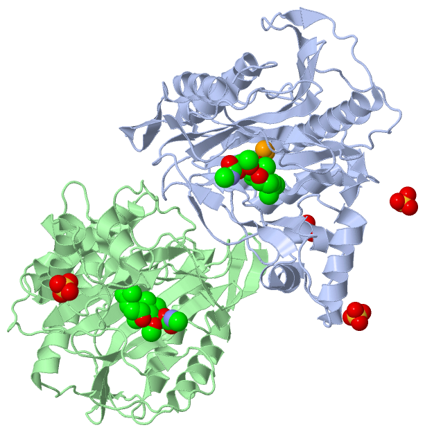
Chains, Units
Summary Information (see also Sequences/Alignments below) |
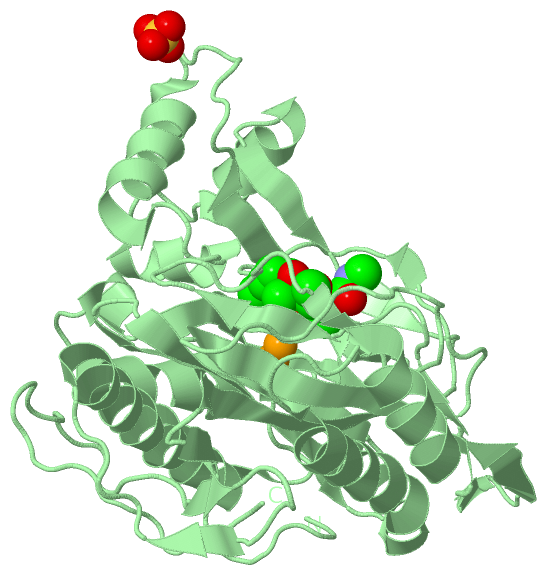
Ligands, Modified Residues, Ions (3, 10)
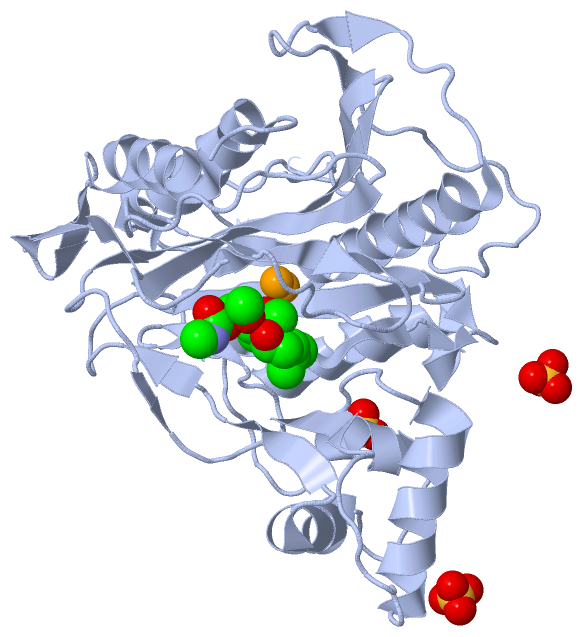
Asymmetric Unit (3, 10)
|
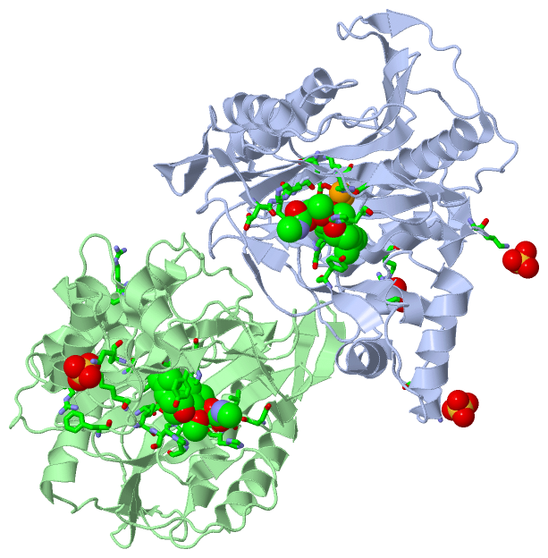
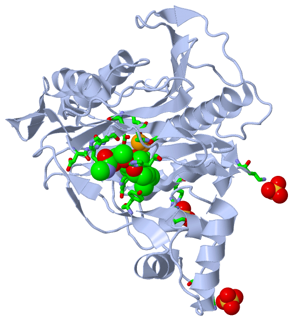
Sites (10, 10)
Asymmetric Unit (10, 10)
|
SS Bonds (0, 0)| (no "SS Bond" information available for 3FMR) |
Cis Peptide Bonds (3, 3)
Asymmetric Unit
|
||||||||||||||||
SAPs(SNPs)/Variants (1, 2)
Asymmetric Unit (1, 2)
|
||||||||||||||||||||||||||||||||||||||||||||||||||||||||||||||||||||||||||||||||||||||||||||||||||||||||||||||||||||||||||||||||||||||||||||||||||||||||||||||||||||||||||||||
PROSITE Motifs (1, 2)
Asymmetric Unit (1, 2)
|
||||||||||||||||||||||||||||||||||||||||||||||||||||||||||||||||||||||||
Exons (0, 0)| (no "Exon" information available for 3FMR) |
Sequences/Alignments
Asymmetric UnitChain A from PDB Type:PROTEIN Length:356 aligned with MAP2_ENCCU | Q8SR45 from UniProtKB/Swiss-Prot Length:358 Alignment length:356 12 22 32 42 52 62 72 82 92 102 112 122 132 142 152 162 172 182 192 202 212 222 232 242 252 262 272 282 292 302 312 322 332 342 352 MAP2_ENCCU 3 CILLNQAEELPIEFLPKDGVYGKGKLFDSRNMEIENFTESDILQDARRAAEAHRRARYRVQSIVRPGITLLEIVRSIEDSTRTLLKGERNNGIGFPAGMSMNSCAAHYTVNPGEQDIVLKEDDVLKIDFGTHSDGRIMDSAFTVAFKENLEPLLVAAREGTETGIKSLGVDVRVCDIGRDINEVISSYEVEIGGRMWPIRPISDLHGHSISQFRIHGGISIPAVNNRDTTRIKGDSFYAVETFATTGKGSIDDRPPCSHFVLNTYKSRKLFNKDLIKVYEFVKDSLGTLPFSPRHLDYYGLVKGGSLKSVNLLTMMGLLTPYPPLNDIDGCKVAQFEHTVYLSEHGKEVLTRGDDY 358 SCOP domains -------------------------------------------------------------------------------------------------------------------------------------------------------------------------------------------------------------------------------------------------------------------------------------------------------------------------------------------------------------------- SCOP domains CATH domains ----------------------------------------3fmrA01 A:43-250,A:331-354 Creatinase/methionine aminopeptidase superfamily 3fmrA02 A:251-330 'winged helix' repressor DNA binding domain 3fmrA01 ---- CATH domains Pfam domains -------------------------------------------------------------------------------------------------------------------------------------------------------------------------------------------------------------------------------------------------------------------------------------------------------------------------------------------------------------------- Pfam domains SAPs(SNPs) ---------------------------------------------------------------------------------------------------------------------------------------------------------------------------------------------------------------------------------------------------------------------------------------------F---------------------------------------------------------------------- SAPs(SNPs) PROSITE --------------------------------------------------------------------------------------------------------------------------MAP_2 ------------------------------------------------------------------------------------------------------------------------------------------------------------------------------------------------------------------------- PROSITE Transcript -------------------------------------------------------------------------------------------------------------------------------------------------------------------------------------------------------------------------------------------------------------------------------------------------------------------------------------------------------------------- Transcript 3fmr A 3 CILLNQAEELPIEFLPKDGVYGKGKLFDSRNMEIENFTESDILQDARRAAEAHRRARYRVQSIVRPGITLLEIVRSIEDSTRTLLKGERNNGIGFPAGMSMNSCAAHYTVNPGEQDIVLKEDDVLKIDFGTHSDGRIMDSAFTVAFKENLEPLLVAAREGTETGIKSLGVDVRVCDIGRDINEVISSYEVEIGGRMWPIRPISDLHGHSISQFRIHGGISIPAVNNRDTTRIKGDSFYAVETFATTGKGSIDDRPPCSHFVLNTYKSRKLFNKDLIKVYEFVKDSLGTLPFSPRHLDYYGLVKGGSLKSVNLLTMMGLLTPYPPLNDIDGCKVAQFEHTVYLSEHGKEVLTRGDDY 358 12 22 32 42 52 62 72 82 92 102 112 122 132 142 152 162 172 182 192 202 212 222 232 242 252 262 272 282 292 302 312 322 332 342 352 Chain B from PDB Type:PROTEIN Length:356 aligned with MAP2_ENCCU | Q8SR45 from UniProtKB/Swiss-Prot Length:358 Alignment length:356 12 22 32 42 52 62 72 82 92 102 112 122 132 142 152 162 172 182 192 202 212 222 232 242 252 262 272 282 292 302 312 322 332 342 352 MAP2_ENCCU 3 CILLNQAEELPIEFLPKDGVYGKGKLFDSRNMEIENFTESDILQDARRAAEAHRRARYRVQSIVRPGITLLEIVRSIEDSTRTLLKGERNNGIGFPAGMSMNSCAAHYTVNPGEQDIVLKEDDVLKIDFGTHSDGRIMDSAFTVAFKENLEPLLVAAREGTETGIKSLGVDVRVCDIGRDINEVISSYEVEIGGRMWPIRPISDLHGHSISQFRIHGGISIPAVNNRDTTRIKGDSFYAVETFATTGKGSIDDRPPCSHFVLNTYKSRKLFNKDLIKVYEFVKDSLGTLPFSPRHLDYYGLVKGGSLKSVNLLTMMGLLTPYPPLNDIDGCKVAQFEHTVYLSEHGKEVLTRGDDY 358 SCOP domains -------------------------------------------------------------------------------------------------------------------------------------------------------------------------------------------------------------------------------------------------------------------------------------------------------------------------------------------------------------------- SCOP domains CATH domains ----------------------------------------3fmrB01 B:43-250,B:331-354 Creatinase/methionine aminopeptidase superfamily 3fmrB02 B:251-330 'winged helix' repressor DNA binding domain 3fmrB01 ---- CATH domains Pfam domains -------------------------------------------------------------------------------------------------------------------------------------------------------------------------------------------------------------------------------------------------------------------------------------------------------------------------------------------------------------------- Pfam domains SAPs(SNPs) ---------------------------------------------------------------------------------------------------------------------------------------------------------------------------------------------------------------------------------------------------------------------------------------------F---------------------------------------------------------------------- SAPs(SNPs) PROSITE --------------------------------------------------------------------------------------------------------------------------MAP_2 ------------------------------------------------------------------------------------------------------------------------------------------------------------------------------------------------------------------------- PROSITE Transcript -------------------------------------------------------------------------------------------------------------------------------------------------------------------------------------------------------------------------------------------------------------------------------------------------------------------------------------------------------------------- Transcript 3fmr B 3 CILLNQAEELPIEFLPKDGVYGKGKLFDSRNMEIENFTESDILQDARRAAEAHRRARYRVQSIVRPGITLLEIVRSIEDSTRTLLKGERNNGIGFPAGMSMNSCAAHYTVNPGEQDIVLKEDDVLKIDFGTHSDGRIMDSAFTVAFKENLEPLLVAAREGTETGIKSLGVDVRVCDIGRDINEVISSYEVEIGGRMWPIRPISDLHGHSISQFRIHGGISIPAVNNRDTTRIKGDSFYAVETFATTGKGSIDDRPPCSHFVLNTYKSRKLFNKDLIKVYEFVKDSLGTLPFSPRHLDYYGLVKGGSLKSVNLLTMMGLLTPYPPLNDIDGCKVAQFEHTVYLSEHGKEVLTRGDDY 358 12 22 32 42 52 62 72 82 92 102 112 122 132 142 152 162 172 182 192 202 212 222 232 242 252 262 272 282 292 302 312 322 332 342 352
|
||||||||||||||||||||
SCOP Domains (0, 0)| (no "SCOP Domain" information available for 3FMR) |
CATH Domains (2, 4)
Asymmetric Unit
 |
Pfam Domains (0, 0)| (no "Pfam Domain" information available for 3FMR) |
Gene Ontology (10, 10)|
Asymmetric Unit(hide GO term definitions) Chain A,B (MAP2_ENCCU | Q8SR45)
|
||||||||||||||||||||||||||||||||||||||||||||||||||||||||||||||||||||||||||||||
Interactive Views
|
|||||||||||||||||||||||||||||||||||||||||||||||||||||||||||||||||||||||||||||||||||||||||||||||||||||||||||||||||||||||||||||||||||||||||||||||||||||||||||||||||||||||||||||||||||||||||||||||||||||||||||||||||||||||||||||||||||||||||
Still Images
|
||||||||||||||||
Databases
|
||||||||||||||||||||||||||||||||||||||||||||||||||||||||||||||||||||||||||||||||||||||||||||||||||||||||||||||||||||||||||||||||||||||||||||||||||||||||||||||||
Analysis Tools
|
|||||||||||||||||||||||||||||||||||||||||||||||||||||||||||||
Entries Sharing at Least One Protein Chain (UniProt ID)
Related Entries Specified in the PDB File
|
|