|
|
|
|
 Description
|
|
Compounds
|
||||||||||||||||||||||||||||||||||||||||||||||||||||||||||||||||||||||||||||||||||
Chains, Units
Summary Information (see also Sequences/Alignments below) |
Ligands, Modified Residues, Ions (3, 3)| Asymmetric/Biological Unit (3, 3) |
Sites (3, 3)
Asymmetric Unit (3, 3)
|
SS Bonds (2, 2)
Asymmetric/Biological Unit
|
||||||||||||
Cis Peptide Bonds (1, 1)
Asymmetric/Biological Unit
|
||||||||
SAPs(SNPs)/Variants (0, 0)| (no "SAP(SNP)/Variant" information available for 3DV4) |
PROSITE Motifs (0, 0)| (no "PROSITE Motif" information available for 3DV4) |
Exons (0, 0)| (no "Exon" information available for 3DV4) |
Sequences/Alignments
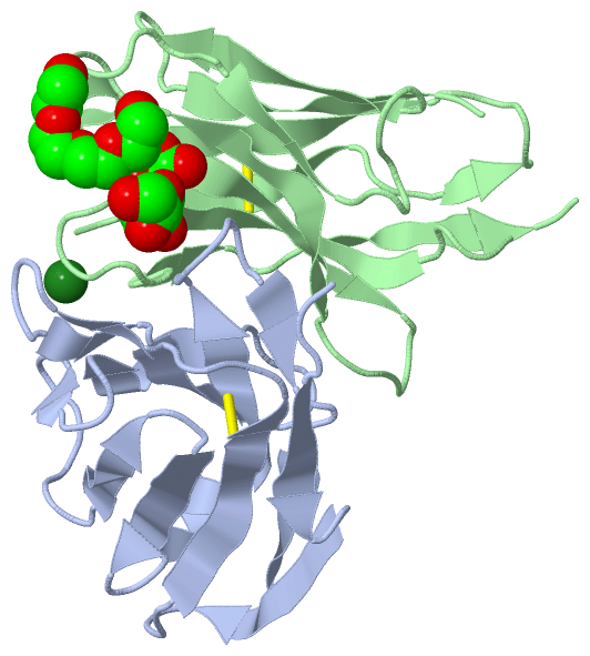
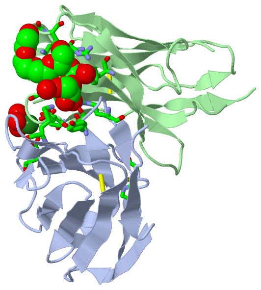
Asymmetric/Biological UnitChain A from PDB Type:PROTEIN Length:112 aligned with A0N262_MOUSE | A0N262 from UniProtKB/TrEMBL Length:115 Alignment length:112 98 97 | 11 21 31 41 51 61 71 81 91 | 100 110 A0N262_MOUSE 2 DIQLTQSPSSLAVSAGEKVTMSCKSSQSVLYSSNQKNYLAWYQQKPGQSPKLLIYWASTRESGVPDRFTGSGSGTDFTLTISSVQAEDLAVYYCHQ-YLSHTFGGGTKLEIK 112 SCOP domains d3dv4a_ A: automated matches SCOP domains CATH domains 3dv4A00 A:1-106 Immunoglobulins CATH domains Pfam domains ---------------------------------------------------------------------------------------------------------------- Pfam domains SAPs(SNPs) ---------------------------------------------------------------------------------------------------------------- SAPs(SNPs) PROSITE ---------------------------------------------------------------------------------------------------------------- PROSITE Transcript ---------------------------------------------------------------------------------------------------------------- Transcript 3dv4 A 1 DIVLTQSPSSLAVSAGERVTMSCKSSQSLFKSRNQKNYLAWYQQKPGQSPKLLIYWASTRESGVPDRFTGSGSGTDFTLTINGVQAEDLAVYYCKQSYNLRTFGGGTKLELK 106 10 20 27C||| 34 44 54 64 74 84 94 104 27A||||| 27B|||| 27C||| 27D|| 27E| 27F Chain B from PDB Type:PROTEIN Length:119 aligned with A2NU21_MOUSE | A2NU21 from UniProtKB/TrEMBL Length:119 Alignment length:121 119 28 38 48 58 68 78 88 98 108 118| - - A2NU21_MOUSE 19 EVKLVESGGGLVQPGGSLSLSCAASGFTFTDYYMSWVRQLPGKALEWLGFIRNKANGYTTEYSASVKGRFTISRDNSQSILYLQMNALRAEDSATYYCAKD-------------------- - SCOP domains d3dv4b_ B: automated matches SCOP domains CATH domains 3dv4B00 B:1-113 Immunoglobulins CATH domains Pfam domains ------------------------------------------------------------------------------------------------------------------------- Pfam domains SAPs(SNPs) ------------------------------------------------------------------------------------------------------------------------- SAPs(SNPs) PROSITE ------------------------------------------------------------------------------------------------------------------------- PROSITE Transcript ------------------------------------------------------------------------------------------------------------------------- Transcript 3dv4 B 1 EVKLVESGGGLVQPGGSLRLSCATSGFTFTDYYMSWVRQPPGKALEWLGFIRNKAKGYTVEYSASVKGRFTISRDNSQSILYLQMNT--AEDSATYYCARDGYYVDAMDYWGQGTSVTVSS 113 10 20 30 40 50 ||| 57 67 77 || 84 94 |102 112 52A|| 82A| 84 100A| 52B| 82B 100B 52C
|
||||||||||||||||||||
SCOP Domains (1, 2)
Asymmetric/Biological Unit
 |
CATH Domains (1, 2)
Asymmetric/Biological Unit
|
Pfam Domains (0, 0)| (no "Pfam Domain" information available for 3DV4) |
Gene Ontology (0, 0)|
Asymmetric/Biological Unit(hide GO term definitions)
(no "Gene Ontology" information available for 3DV4)
|
Interactive Views
|
|||||||||||||||||||||||||||||||||||||||||||||||||||||||||||||||||||||||||||||||||||||||||||||||||||||||||||||||||||||||||||||||||||||||||||||||||||
Still Images
|
||||||||||||||||
Databases
|
||||||||||||||||||||||||||||||||||||||||||||||||||||||||||||||||||||||||||||||||||||||||||||||||||||||||||||||||||||||||||||||||||||||||||||||||||||||||||||||||||||||||||||||||||||||||||
Analysis Tools
|
||||||||||||||||||||||||||||||||||||||||||||||||||||||||||||||||||||||||
Entries Sharing at Least One Protein Chain (UniProt ID)
Related Entries Specified in the PDB File
|
|