Show PDB file:   
         Plain Text   HTML   (compressed file size)
QuickSearch:   
by PDB,NDB,UniProt,PROSITE Code or Search Term(s)  
(-)Asym./Biol. Unit
(-)Asym./Biol. Unit - sites
collapse expand < >
Image Asym./Biol. Unit
Asym./Biol. Unit  (Jmol Viewer)
Image Asym./Biol. Unit - sites
Asym./Biol. Unit - sites  (Jmol Viewer)

(-) Description

Title :  CRYSTAL STRUCTURE OF VITAMIN D HYDROXYLASE CYTOCHROME P450 105A1 (R73A/R84A MUTANT) IN COMPLEX WITH 1ALPHA,25-DIHYDROXYVITAMIN D3
 
Authors :  K. Hayashi, H. Sugimoto, R. Shinkyo, M. Yamada, S. Ikeda, S. Ikushiro, M. Kamakura, Y. Shiro, T. Sakaki
Date :  18 Apr 08  (Deposition) - 04 Nov 08  (Release) - 24 Feb 09  (Revision)
Method :  X-RAY DIFFRACTION
Resolution :  1.70
Chains :  Asym./Biol. Unit :  A
Keywords :  P450, Beta Prism, Heme, Iron, Metal-Binding, Monooxygenase, Oxidoreductase (Keyword Search: [Gene Ontology, PubMed, Web (Google))
 
Reference :  K. Hayashi, H. Sugimoto, R. Shinkyo, M. Yamada, S. Ikeda, S. Ikushiro, M. Kamakura, Y. Shiro, T. Sakaki
Structure-Based Design Of A Highly Active Vitamin D Hydroxylase From Streptomyces Griseolus Cyp105A1
Biochemistry V. 47 11964 2008
PubMed-ID: 18937506  |  Reference-DOI: 10.1021/BI801222D
(for further references see the PDB file header)

(-) Compounds

Molecule 1 - CYTOCHROME P450-SU1
    ChainsA
    EC Number1.14.14.1
    EngineeredYES
    Expression SystemESCHERICHIA COLI
    Expression System PlasmidPKK223-3
    Expression System StrainJM109
    Expression System Taxid562
    Expression System Vector TypePLASMID
    GeneCYP105A1
    MutationYES
    Organism ScientificSTREPTOMYCES GRISEOLUS
    Organism Taxid1909
    StrainATCC 11796
    SynonymP450-CVA1, CYP105A1

 Structural Features

(-) Chains, Units

  1
Asymmetric/Biological Unit A

Summary Information (see also Sequences/Alignments below)
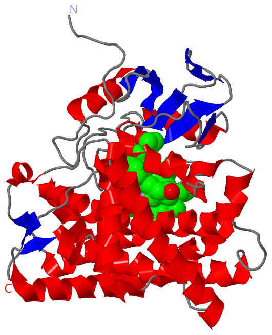
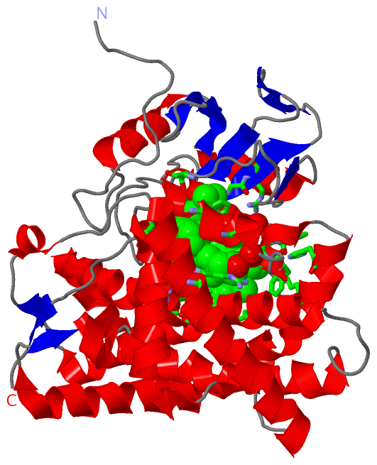
(-) Ligands, Modified Residues, Ions  (2, 2)

Asymmetric/Biological Unit (2, 2)
No.NameCountTypeFull Name
1HEM1Ligand/IonPROTOPORPHYRIN IX CONTAINING FE
2VDX1Ligand/Ion5-{2-[1-(5-HYDROXY-1,5-DIMETHYL-HEXYL)-7A-METHYL-OCTAHYDRO-INDEN-4-YLIDENE]-ETHYLIDENE}-4-METHYLENE-CYCLOHEXANE-1,3-DIOL

(-) Sites  (2, 2)

Asymmetric Unit (2, 2)
No.NameEvidenceResiduesDescription
1AC1SOFTWAREPHE A:95 , ILE A:96 , HIS A:103 , ARG A:107 , LEU A:241 , ALA A:244 , GLY A:245 , THR A:248 , THR A:249 , ALA A:294 , ARG A:297 , ALA A:347 , PHE A:348 , GLY A:349 , VAL A:352 , HIS A:353 , CYS A:355 , GLY A:357 , ALA A:361 , HOH A:506 , HOH A:525 , HOH A:529 , HOH A:579BINDING SITE FOR RESIDUE HEM A 413
2AC2SOFTWARETHR A:81 , PHE A:85 , VAL A:88 , ARG A:193 , ILE A:293 , GLY A:296 , THR A:395 , ILE A:396 , HOH A:560 , HOH A:593BINDING SITE FOR RESIDUE VDX A 501

(-) SS Bonds  (0, 0)

(no "SS Bond" information available for 3CV9)

(-) Cis Peptide Bonds  (2, 2)

Asymmetric/Biological Unit
No.Residues
1Phe A:16 -Pro A:17
2Pro A:100 -Pro A:101

 Sequence-Structure Mapping

(-) SAPs(SNPs)/Variants  (0, 0)

(no "SAP(SNP)/Variant" information available for 3CV9)

(-) PROSITE Motifs  (1, 1)

Asymmetric/Biological Unit (1, 1)
 PROSITEUniProtKBPDB
No.IDACDescriptionIDLocationCountLocation
1CYTOCHROME_P450PS00086 Cytochrome P450 cysteine heme-iron ligand signature.CPXE_STRGO348-357  1A:348-357

(-) Exons   (0, 0)

(no "Exon" information available for 3CV9)

(-) Sequences/Alignments

Asymmetric/Biological Unit
   Reformat: Number of residues per line =  ('0' or empty: single-line sequence representation)
  Number of residues per labelling interval =   
  UniProt sequence: complete  aligned part    
   Show mapping: SCOP domains CATH domains Pfam domains Secondary structure (by author)
SAPs(SNPs) PROSITE motifs Exons
(details for a mapped element are shown in a popup box when the mouse pointer rests over it)
Chain A from PDB  Type:PROTEIN  Length:401
 aligned with CPXE_STRGO | P18326 from UniProtKB/Swiss-Prot  Length:406

    Alignment length:401
                                                                                                                                                                                                                                                                                                                                                                                                                                        406  
                                    17        27        37        47        57        67        77        87        97       107       117       127       137       147       157       167       177       187       197       207       217       227       237       247       257       267       277       287       297       307       317       327       337       347       357       367       377       387       397        |- 
           CPXE_STRGO     8 PQTTDAPAFPSNRSCPYQLPDGYAQLRDTPGPLHRVTLYDGRQAWVVTKHEAARKLLGDPRLSSNRTDDNFPATSPRFEAVRESPQAFIGLDPPEHGTRRRMTISEFTVKRIKGMRPEVEEVVHGFLDEMLAAGPTADLVSQFALPVPSMVICRLLGVPYADHEFFQDASKRLVQSTDAQSALTARNDLAGYLDGLITQFQTEPGAGLVGALVADQLANGEIDREELISTAMLLLIAGHETTASMTSLSVITLLDHPEQYAALRADRSLVPGAVEELLRYLAIADIAGGRVATADIEVEGHLIRAGEGVIVVNSIANRDGTVYEDPDALDIHRSARHHLAFGFGVHQCLGQNLARLELEVILNALMDRVPTLRLAVPVEQLVLRPGTTIQGVNELPVTW--   -
               SCOP domains d3cv9a_ A: automated matches                                                                                                                                                                                                                                                                                                                                                                                      SCOP domains
               CATH domains 3cv9A00 A:8-408 Cytochrome p450                                                                                                                                                                                                                                                                                                                                                                                   CATH domains
               Pfam domains ----------------------------------------------------------------------------------------------------------------------------------------------------------------------------------------------------------------------------------------------------------------------------------------------------------------------------------------------------------------------------------------------------------------- Pfam domains
         Sec.struct. author ...................hhhhhhhhhh...eeeee.....eeeee.hhhhhhhhhhh..ee...........hhhhhhhhh...hhhhh..hhhhhhhhhhhhhhhhhhhhhhhhhhhhhhhhhhhhhhhhh..eehhhhh.hhhhhhhhhhhhh.hhhhhhhhhhhhhhhhhh.hhhhhhhhhhhhhhhhhhhhhhhhhh...hhhhhhhhh.......hhhhhhhhhhhhhhhhhhhhhhhhhhhhhhhhhhhhhhhhhhhh..hhhhhhhhhhhhhh......eeee...eee..eee....eeeehhhhhh................................hhhhhhhhhhhhhhhhhhhh...ee..hhhhh..............eee... Sec.struct. author
                 SAPs(SNPs) ----------------------------------------------------------------------------------------------------------------------------------------------------------------------------------------------------------------------------------------------------------------------------------------------------------------------------------------------------------------------------------------------------------------- SAPs(SNPs)
                    PROSITE ----------------------------------------------------------------------------------------------------------------------------------------------------------------------------------------------------------------------------------------------------------------------------------------------------------------------------------------------------CYTOCHROME--------------------------------------------------- PROSITE
                 Transcript ----------------------------------------------------------------------------------------------------------------------------------------------------------------------------------------------------------------------------------------------------------------------------------------------------------------------------------------------------------------------------------------------------------------- Transcript
                 3cv9 A   8 PQTTDAPAFPSNRSCPYQLPDGYAQLRDTPGPLHRVTLYDGRQAWVVTKHEAARKLLGDPRLSSNATDDNFPATSPAFEAVRESPQAFIGLDPPEHGTRRRMTISEFTVKRIKGMRPEVEEVVHGFLDEMLAAGPTADLVSQFALPVPSMVICRLLGVPYADHEFFQDASKRLVQSTDAQSALTARNDLAGYLDGLITQFQTEPGAGLVGALVADQLANGEIDREELISTAMLLLIAGHETTASMTSLSVITLLDHPEQYAALRADRSLVPGAVEELLRYLAIADIAGGRVATADIEVEGQLIRAGEGVIVVNSIANRDGTVYEDPDALDIHRSARHHLAFGFGVHQCLGQNLARLELEVILNALMDRVPTLRLAVPVEQLVLRPGTTIQGVNELPVTWHH 408
                                    17        27        37        47        57        67        77        87        97       107       117       127       137       147       157       167       177       187       197       207       217       227       237       247       257       267       277       287       297       307       317       327       337       347       357       367       377       387       397       407 

   Legend:   → Mismatch (orange background)
  - → Gap (green background, '-', border residues have a numbering label)
    → Modified Residue (blue background, lower-case, 'x' indicates undefined single-letter code, labelled with number + name)
  x → Chemical Group (purple background, 'x', labelled with number + name, e.g. ACE or NH2)
  extra numbering lines below/above indicate numbering irregularities and modified residue names etc., number ends below/above '|'

 Classification and Annotation

(-) SCOP Domains  (1, 1)

Asymmetric/Biological Unit

(-) CATH Domains  (1, 1)

Asymmetric/Biological Unit

(-) Pfam Domains  (0, 0)

(no "Pfam Domain" information available for 3CV9)

(-) Gene Ontology  (12, 12)

Asymmetric/Biological Unit(hide GO term definitions)
Chain A   (CPXE_STRGO | P18326)
molecular function
    GO:0004498    calcidiol 1-monooxygenase activity    Catalysis of the reaction: calcidiol + H(+) + NADPH + O(2) = calcitriol + H(2)O + NADP(+).
    GO:0020037    heme binding    Interacting selectively and non-covalently with heme, any compound of iron complexed in a porphyrin (tetrapyrrole) ring.
    GO:0005506    iron ion binding    Interacting selectively and non-covalently with iron (Fe) ions.
    GO:0046872    metal ion binding    Interacting selectively and non-covalently with any metal ion.
    GO:0004497    monooxygenase activity    Catalysis of the incorporation of one atom from molecular oxygen into a compound and the reduction of the other atom of oxygen to water.
    GO:0016491    oxidoreductase activity    Catalysis of an oxidation-reduction (redox) reaction, a reversible chemical reaction in which the oxidation state of an atom or atoms within a molecule is altered. One substrate acts as a hydrogen or electron donor and becomes oxidized, while the other acts as hydrogen or electron acceptor and becomes reduced.
    GO:0016705    oxidoreductase activity, acting on paired donors, with incorporation or reduction of molecular oxygen    Catalysis of an oxidation-reduction (redox) reaction in which hydrogen or electrons are transferred from each of two donors, and molecular oxygen is reduced or incorporated into a donor.
    GO:0016709    oxidoreductase activity, acting on paired donors, with incorporation or reduction of molecular oxygen, NAD(P)H as one donor, and incorporation of one atom of oxygen    Catalysis of an oxidation-reduction (redox) reaction in which hydrogen or electrons are transferred from NADH or NADPH and one other donor, and one atom of oxygen is incorporated into one donor.
biological process
    GO:0036378    calcitriol biosynthetic process from calciol    Conversion of vitamin D3 from its largely inactive form (calciol, also called cholecalciferol) into a hormonally active form (calcitriol). Conversion requires 25-hydroxylation of calciol in the liver to form calcidiol, and subsequent 1,alpha-hydroxylation of calcidiol in the kidney to form calcitriol.
    GO:0055114    oxidation-reduction process    A metabolic process that results in the removal or addition of one or more electrons to or from a substance, with or without the concomitant removal or addition of a proton or protons.
    GO:0070640    vitamin D3 metabolic process    The chemical reactions and pathways involving vitamin D3, (3S,5Z,7E)-9,10-secocholesta-5,7,10(19)-trien-3-ol.
cellular component
    GO:0005737    cytoplasm    All of the contents of a cell excluding the plasma membrane and nucleus, but including other subcellular structures.

 Visualization

(-) Interactive Views

Asymmetric/Biological Unit
  Complete Structure
    Jena3D(integrated viewing of ligand, site, SAP, PROSITE, SCOP information)
    WebMol | AstexViewer[tm]@PDBe
(Java Applets, require no local installation except for Java; loading may be slow)
    STRAP
(Java WebStart application, automatic local installation, requires Java; full application with system access!)
    RasMol
(require local installation)
    Molscript (VRML)
(requires installation of a VRML viewer; select preferred view via VRML and generate a mono or stereo PDF format file)
 
  Ligands, Modified Residues, Ions
    HEM  [ RasMol | Jena3D ]  +environment [ RasMol | Jena3D ]
    VDX  [ RasMol | Jena3D ]  +environment [ RasMol | Jena3D ]
 
  Sites
    AC1  [ RasMol ]  +environment [ RasMol ]
    AC2  [ RasMol ]  +environment [ RasMol ]
 
  Cis Peptide Bonds
    Phe A:16 - Pro A:17   [ RasMol ]  
    Pro A:100 - Pro A:101   [ RasMol ]  
 

(-) Still Images

Jmol
  protein: cartoon or spacefill or dots and stick; nucleic acid: cartoon and stick; ligands: spacefill; active site: stick
Molscript
  protein, nucleic acid: cartoon; ligands: spacefill; active site: ball and stick

 Databases and Analysis Tools

(-) Databases

Access by PDB/NDB ID
  3cv9
    Family and Domain InformationProDom | SYSTERS
    General Structural InformationGlycoscienceDB | MMDB | NDB | OCA | PDB | PDBe | PDBj | PDBsum | PDBWiki | PQS | PROTEOPEDIA
    Orientation in MembranesOPM
    Protein SurfaceSURFACE
    Secondary StructureDSSP (structure derived) | HSSP (homology derived)
    Structural GenomicsGeneCensus
    Structural NeighboursCE | VAST
    Structure ClassificationCATH | Dali | SCOP
    Validation and Original DataBMRB Data View | BMRB Restraints Grid | EDS | PROCHECK | RECOORD | WHAT_CHECK
 
Access by UniProt ID/Accession number
  CPXE_STRGO | P18326
    Comparative Protein Structure ModelsModBase
    Genomic InformationEnsembl
    Protein-protein InteractionDIP
    Sequence, Family and Domain InformationInterPro | Pfam | SMART | UniProtKB/SwissProt
 
Access by Enzyme Classificator   (EC Number)
  1.14.14.1
    General Enzyme InformationBRENDA | EC-PDB | Enzyme | IntEnz
    PathwayKEGG | MetaCyc
 
Access by Disease Identifier   (MIM ID)
  (no 'MIM ID' available)
    Disease InformationOMIM
 
Access by GenAge ID
  (no 'GenAge ID' available)
    Age Related InformationGenAge

(-) Analysis Tools

Access by PDB/NDB ID
    Domain InformationXDom
    Interatomic Contacts of Structural UnitsCSU
    Ligand-protein ContactsLPC
    Protein CavitiescastP
    Sequence and Secondary StructurePDBCartoon
    Structure AlignmentSTRAP(Java WebStart application, automatic local installation, requires Java; full application with system access!)
    Structure and Sequence BrowserSTING
 
Access by UniProt ID/Accession number
  CPXE_STRGO | P18326
    Protein Disorder PredictionDisEMBL | FoldIndex | GLOBPLOT (for more information see DisProt)

 Related Entries

(-) Entries Sharing at Least One Protein Chain (UniProt ID)

UniProtKB/Swiss-Prot
        CPXE_STRGO | P183262zbx 2zby 2zbz 3cv8 5x7e

(-) Related Entries Specified in the PDB File

2zbx WILD TYPE, IMIDAZOLE BOUND
2zby R84A MUTANT, SUBSTRATE FREE
2zbz R84A MUTANT IN COMPLEX WITH PRODUCT
3cv8 R84F MUTANT, SUBSTRATE FREE