|
|
|
|
 Description
|
|
Compounds
|
||||||||||||||||||||||||||||||||||||
Chains, Units
Summary Information (see also Sequences/Alignments below) |
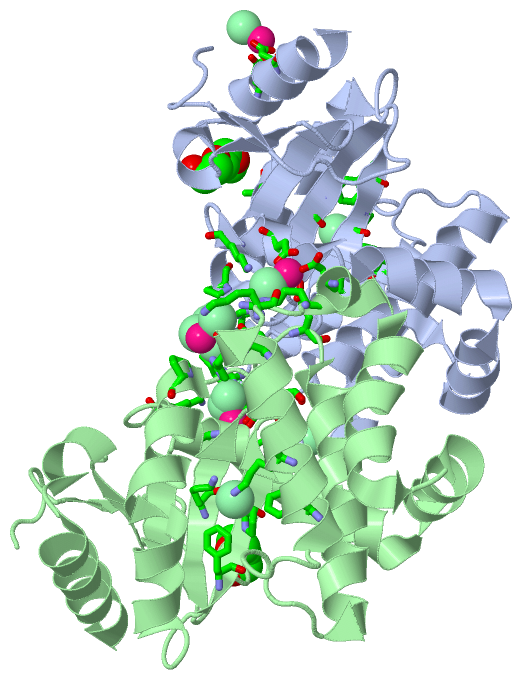
Ligands, Modified Residues, Ions (4, 18)
Asymmetric Unit (4, 18)
|
Sites (17, 17)
Asymmetric Unit (17, 17)
|
SS Bonds (0, 0)| (no "SS Bond" information available for 3C7M) |
Cis Peptide Bonds (2, 2)
Asymmetric Unit
|
||||||||||||
SAPs(SNPs)/Variants (0, 0)| (no "SAP(SNP)/Variant" information available for 3C7M) |
PROSITE Motifs (0, 0)| (no "PROSITE Motif" information available for 3C7M) |
Exons (0, 0)| (no "Exon" information available for 3C7M) |
Sequences/Alignments
Asymmetric UnitChain A from PDB Type:PROTEIN Length:195 aligned with DSBL_ECOL6 | P0A4L7 from UniProtKB/Swiss-Prot Length:222 Alignment length:195 37 47 57 67 77 87 97 107 117 127 137 147 157 167 177 187 197 207 217 DSBL_ECOL6 28 FTEGTDYMVLEKPIPNADKTLIKVFSYACPFCYKYDKAVTGPVSEKVKDIVAFTPFHLETKGEYGKQASEVFAVLINKDKAAGISLFDANSQFKKAKFAYYAAYHDKKERWSDGKDPAAFIKTGLDAAGMSQADFEAALKEPAVQETLEKWKASYDVAKIQGVPAYVVNGKYLIYTKSIKSIDAMADLIRELASK 222 SCOP domains d3c7ma_ A: automated matches SCOP domains CATH domains --------------------------------------------------------------------------------------------------------------------------------------------------------------------------------------------------- CATH domains Pfam domains --------------------------------------------------------------------------------------------------------------------------------------------------------------------------------------------------- Pfam domains SAPs(SNPs) --------------------------------------------------------------------------------------------------------------------------------------------------------------------------------------------------- SAPs(SNPs) PROSITE --------------------------------------------------------------------------------------------------------------------------------------------------------------------------------------------------- PROSITE Transcript --------------------------------------------------------------------------------------------------------------------------------------------------------------------------------------------------- Transcript 3c7m A 1 FTEGTDYMVLEKPIPNADKTLIKVFSYACPFCYKYDKAVTGPVSEKVKDIVAFTPFHLETKGEYGKQASEVFAVLINKDKAAGISLFDANSQFKKAKFAYYAAYHDKKERWSDGKDPAAFIKTGLDAAGMSQADFEAALKEPAVQETLEKWKASYDVAKIQGVPAYVVNGKYLIYTKSIKSIDAMADLIRELASK 195 10 20 30 40 50 60 70 80 90 100 110 120 130 140 150 160 170 180 190 Chain B from PDB Type:PROTEIN Length:195 aligned with DSBL_ECOL6 | P0A4L7 from UniProtKB/Swiss-Prot Length:222 Alignment length:195 37 47 57 67 77 87 97 107 117 127 137 147 157 167 177 187 197 207 217 DSBL_ECOL6 28 FTEGTDYMVLEKPIPNADKTLIKVFSYACPFCYKYDKAVTGPVSEKVKDIVAFTPFHLETKGEYGKQASEVFAVLINKDKAAGISLFDANSQFKKAKFAYYAAYHDKKERWSDGKDPAAFIKTGLDAAGMSQADFEAALKEPAVQETLEKWKASYDVAKIQGVPAYVVNGKYLIYTKSIKSIDAMADLIRELASK 222 SCOP domains d3c7mb_ B: automated matches SCOP domains CATH domains --------------------------------------------------------------------------------------------------------------------------------------------------------------------------------------------------- CATH domains Pfam domains --------------------------------------------------------------------------------------------------------------------------------------------------------------------------------------------------- Pfam domains SAPs(SNPs) --------------------------------------------------------------------------------------------------------------------------------------------------------------------------------------------------- SAPs(SNPs) PROSITE --------------------------------------------------------------------------------------------------------------------------------------------------------------------------------------------------- PROSITE Transcript --------------------------------------------------------------------------------------------------------------------------------------------------------------------------------------------------- Transcript 3c7m B 1 FTEGTDYMVLEKPIPNADKTLIKVFSYACPFCYKYDKAVTGPVSEKVKDIVAFTPFHLETKGEYGKQASEVFAVLINKDKAAGISLFDANSQFKKAKFAYYAAYHDKKERWSDGKDPAAFIKTGLDAAGMSQADFEAALKEPAVQETLEKWKASYDVAKIQGVPAYVVNGKYLIYTKSIKSIDAMADLIRELASK 195 10 20 30 40 50 60 70 80 90 100 110 120 130 140 150 160 170 180 190
|
||||||||||||||||||||
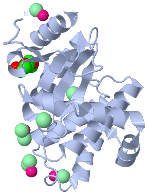
SCOP Domains (1, 2)
Asymmetric Unit
 |
CATH Domains (0, 0)| (no "CATH Domain" information available for 3C7M) |
Pfam Domains (0, 0)| (no "Pfam Domain" information available for 3C7M) |
Gene Ontology (3, 3)|
Asymmetric Unit(hide GO term definitions) Chain A,B (DSBL_ECOL6 | P0A4L7)
|
||||||||||||||||||||||||||||||||||||
Interactive Views
|
||||||||||||||||||||||||||||||||||||||||||||||||||||||||||||||||||||||||||||||||||||||||||||||||||||||||||||||||||||||||||||||||||||||||||||||||||||||||||||||||||||||||||||||||||||||||||||||||||||||||||||||||||||||||||||||||||||||||||||||||||||||||||||||||||||||||||||||||||||||||||
Still Images
|
||||||||||||||||
Databases
|
||||||||||||||||||||||||||||||||||||||||||||||||||||||||||||||||||||||||||||||||||||||||||||||||||||||||||||||||||||||||||||||||||||||||||||||||||||||||||||||||
Analysis Tools
|
|||||||||||||||||||||||||||||||||||||||||||||||||||||||||||||
Entries Sharing at Least One Protein Chain (UniProt ID)
Related Entries Specified in the PDB File
|
|