|
|
|
|
 Description
|
|
Compounds
|
||||||||||||||||||||||||||||||||||||||||||||||||
Chains, Units
Summary Information (see also Sequences/Alignments below) |
Ligands, Modified Residues, Ions (3, 3)| Asymmetric/Biological Unit (3, 3) |
Sites (2, 2)
Asymmetric Unit (2, 2)
|
SS Bonds (0, 0)| (no "SS Bond" information available for 3AB9) |
Cis Peptide Bonds (0, 0)| (no "Cis Peptide Bond" information available for 3AB9) |
SAPs(SNPs)/Variants (0, 0)| (no "SAP(SNP)/Variant" information available for 3AB9) |
PROSITE Motifs (2, 2)
Asymmetric/Biological Unit (2, 2)
|
||||||||||||||||||||||||||||||||
Exons (0, 0)| (no "Exon" information available for 3AB9) |
Sequences/Alignments
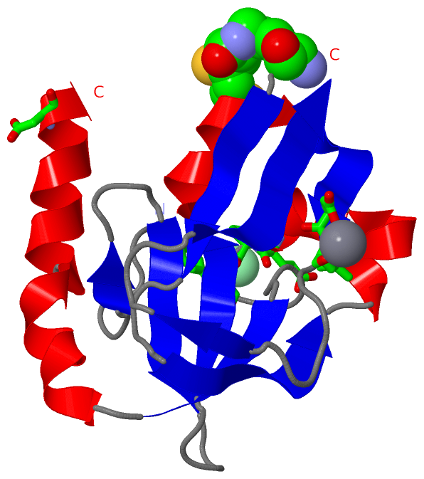
Asymmetric/Biological UnitChain A from PDB Type:PROTEIN Length:127 aligned with GCSH_ECOLI | P0A6T9 from UniProtKB/Swiss-Prot Length:129 Alignment length:127 12 22 32 42 52 62 72 82 92 102 112 122 GCSH_ECOLI 3 NVPAELKYSKEHEWLRKEADGTYTVGITEHAQELLGDMVFVDLPEVGATVSAGDDCAVAESVKAASDIYAPVSGEIVAVNDALSDSPELVNSEPYAGGWIFKIKASDESELESLLDATAYEALLEDE 129 SCOP domains d3ab9a_ A: Protein H of glycine cleavage system SCOP domains CATH domains 3ab9A00 A:2-128 [code=2.40.50.100, no name defined] CATH domains Pfam domains ------------------------------------------------------------------------------------------------------------------------------- Pfam domains SAPs(SNPs) ------------------------------------------------------------------------------------------------------------------------------- SAPs(SNPs) PROSITE (1) ---------------------BIOTINYL_LIPOYL PDB: A:23-105 UniProt: 24-106 ----------------------- PROSITE (1) PROSITE (2) ----------------------------------------------LIPOYL PDB: A:48-77 --------------------------------------------------- PROSITE (2) Transcript ------------------------------------------------------------------------------------------------------------------------------- Transcript 3ab9 A 2 NVPAELKYSKEHEWLRKEADGTYTVGITEHAQELLGDMVFVDLPEVGATVSAGDDCAVAESVkAASDIYAPVSGEIVAVNDALSDSPELVNSEPYAGGWIFKIKASDESELESLLDATAYEALLEDE 128 11 21 31 41 51 61 | 71 81 91 101 111 121 64-LA2
|
||||||||||||||||||||
SCOP Domains (1, 1)
Asymmetric/Biological Unit
 |
CATH Domains (1, 1)
Asymmetric/Biological Unit
|
Pfam Domains (0, 0)| (no "Pfam Domain" information available for 3AB9) |
Gene Ontology (5, 5)|
Asymmetric/Biological Unit(hide GO term definitions) Chain A (GCSH_ECOLI | P0A6T9)
|
||||||||||||||||||||||||||||||||||||||||||||||||
Interactive Views
|
|||||||||||||||||||||||||||||||||||||||||||||||||||||||||||||||||||||||||||||||||||||||||||||||||||||||||||||||||||||||||||||||||||||||||||
Still Images
|
||||||||||||||||
Databases
|
||||||||||||||||||||||||||||||||||||||||||||||||||||||||||||||||||||||||||||||||||||||||||||||||||||||||||||||||||||||||||||||||||||||||||||||||||||||||||||||||
Analysis Tools
|
|||||||||||||||||||||||||||||||||||||||||||||||||||||||||||||
Entries Sharing at Least One Protein Chain (UniProt ID)
Related Entries Specified in the PDB File
|
|