Show PDB file:   
         Plain Text   HTML   (compressed file size)
QuickSearch:   
by PDB,NDB,UniProt,PROSITE Code or Search Term(s)  
(-)Asym./Biol. Unit - manually
(-)Asym./Biol. Unit
(-)Asym./Biol. Unit - sites
collapse expand < >
Image Asym./Biol. Unit - manually
Asym./Biol. Unit - manually  (Jmol Viewer)
Image Asym./Biol. Unit
Asym./Biol. Unit  (Jmol Viewer)
Image Asym./Biol. Unit - sites
Asym./Biol. Unit - sites  (Jmol Viewer)

(-) Description

Title :  REFINED STRUCTURES AT 2 ANGSTROMS AND 2.2 ANGSTROMS OF THE TWO FORMS OF THE H-PROTEIN, A LIPOAMIDE-CONTAINING PROTEIN OF THE GLYCINE DECARBOXYLASE COMPLEX
 
Authors :  S. Pares, C. Cohen-Addad
Date :  11 Jan 95  (Deposition) - 31 Mar 95  (Release) - 24 Feb 09  (Revision)
Method :  X-RAY DIFFRACTION
Resolution :  2.20
Chains :  Asym./Biol. Unit :  A
Keywords :  Oxidoreductases(Acting On Ch-Nh2 Donor) (Keyword Search: [Gene Ontology, PubMed, Web (Google))
 
Reference :  C. Cohen-Addad, S. Pares, L. Sieker, M. Neuburger, R. Douce
The Lipoamide Arm In The Glycine Decarboxylase Complex Is Not Freely Swinging.
Nat. Struct. Biol. V. 2 63 1995
PubMed-ID: 7719855  |  Reference-DOI: 10.1038/NSB0195-63
(for further references see the PDB file header)

(-) Compounds

Molecule 1 - H-PROTEIN
    ChainsA
    EC Number1.4.4.2
    EngineeredYES
    Organism CommonPEA
    Organism ScientificPISUM SATIVUM
    Organism Taxid3888

 Structural Features

(-) Chains, Units

  1
Asymmetric/Biological Unit A

Summary Information (see also Sequences/Alignments below)

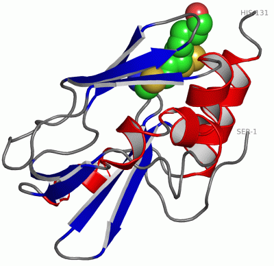
(-) Ligands, Modified Residues, Ions  (1, 1)

Asymmetric/Biological Unit (1, 1)
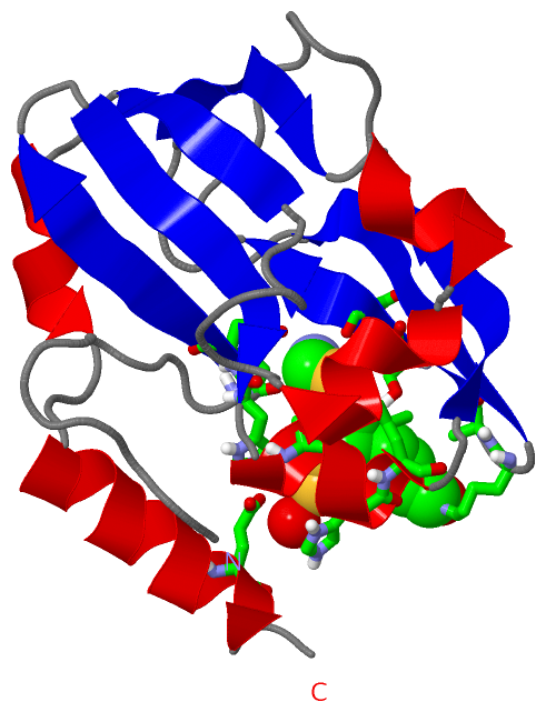
No.NameCountTypeFull Name
1OSS1Ligand/Ion6-(HYDROXYETHYLDITHIO)-8-(AMINOMETHYLTHIO)OCTANOICACID

(-) Sites  (1, 1)

Asymmetric Unit (1, 1)
No.NameEvidenceResiduesDescription
1AC1SOFTWARESER A:12 , HIS A:13 , GLU A:14 , ALA A:31 , HIS A:34 , LEU A:35 , LYS A:63 , ALA A:64 , SER A:66 , ASP A:67 , GLU A:127 , HOH A:219BINDING SITE FOR RESIDUE OSS A 132

(-) SS Bonds  (0, 0)

(no "SS Bond" information available for 1HTP)

(-) Cis Peptide Bonds  (0, 0)

(no "Cis Peptide Bond" information available for 1HTP)

 Sequence-Structure Mapping

(-) SAPs(SNPs)/Variants  (0, 0)

(no "SAP(SNP)/Variant" information available for 1HTP)

(-) PROSITE Motifs  (2, 2)

Asymmetric/Biological Unit (2, 2)
 PROSITEUniProtKBPDB
No.IDACDescriptionIDLocationCountLocation
1BIOTINYL_LIPOYLPS50968 Biotinyl/lipoyl domain profile.GCSH_PEA56-138  1A:22-104
2LIPOYLPS00189 2-oxo acid dehydrogenases acyltransferase component lipoyl binding site.GCSH_PEA81-110  1A:47-76

(-) Exons   (0, 0)

(no "Exon" information available for 1HTP)

(-) Sequences/Alignments

Asymmetric/Biological Unit
   Reformat: Number of residues per line =  ('0' or empty: single-line sequence representation)
  Number of residues per labelling interval =   
  UniProt sequence: complete  aligned part    
   Show mapping: SCOP domains CATH domains Pfam domains Secondary structure (by author)
SAPs(SNPs) PROSITE motifs Exons
(details for a mapped element are shown in a popup box when the mouse pointer rests over it)
Chain A from PDB  Type:PROTEIN  Length:131
 aligned with GCSH_PEA | P16048 from UniProtKB/Swiss-Prot  Length:165

    Alignment length:131
                                    44        54        64        74        84        94       104       114       124       134       144       154       164 
             GCSH_PEA    35 SNVLDGLKYAPSHEWVKHEGSVATIGITDHAQDHLGEVVFVELPEPGVSVTKGKGFGAVESVKATSDVNSPISGEVIEVNTGLTGKPGLINSSPYEDGWMIKIKPTSPDELESLLGAKEYTKFCEEEDAAH 165
               SCOP domains d1htpa_ A: Protein H of glycine cleavage system                                                                                     SCOP domains
               CATH domains 1htpA00 A:1-131  [code=2.40.50.100, no name defined]                                                                                CATH domains
               Pfam domains ----------------------------------------------------------------------------------------------------------------------------------- Pfam domains
         Sec.struct. author .............eeeeee..eeeeee.hhhhhhh..eeeee............eeeeeee...eeeee....eeeeee.hhhhh.hhhhhh........eeeee..hhhhhh...hhhhhhhhhhhh... Sec.struct. author
                 SAPs(SNPs) ----------------------------------------------------------------------------------------------------------------------------------- SAPs(SNPs)
                PROSITE (1) ---------------------BIOTINYL_LIPOYL  PDB: A:22-104 UniProt: 56-138                                     --------------------------- PROSITE (1)
                PROSITE (2) ----------------------------------------------LIPOYL  PDB: A:47-76          ------------------------------------------------------- PROSITE (2)
                 Transcript ----------------------------------------------------------------------------------------------------------------------------------- Transcript
                 1htp A   1 SNVLDGLKYAPSHEWVKHEGSVATIGITDHAQDHLGEVVFVELPEPGVSVTKGKGFGAVESVKATSDVNSPISGEVIEVNTGLTGKPGLINSSPYEDGWMIKIKPTSPDELESLLGAKEYTKFCEEEDAAH 131
                                    10        20        30        40        50        60        70        80        90       100       110       120       130 
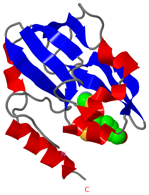
   Legend:   → Mismatch (orange background)
  - → Gap (green background, '-', border residues have a numbering label)
    → Modified Residue (blue background, lower-case, 'x' indicates undefined single-letter code, labelled with number + name)
  x → Chemical Group (purple background, 'x', labelled with number + name, e.g. ACE or NH2)
  extra numbering lines below/above indicate numbering irregularities and modified residue names etc., number ends below/above '|'

 Classification and Annotation

(-) SCOP Domains  (1, 1)

Asymmetric/Biological Unit

(-) CATH Domains  (1, 1)

Asymmetric/Biological Unit
(-)
Class: Mainly Beta (13760)

(-) Pfam Domains  (0, 0)

(no "Pfam Domain" information available for 1HTP)

(-) Gene Ontology  (3, 3)

Asymmetric/Biological Unit(hide GO term definitions)
Chain A   (GCSH_PEA | P16048)
biological process
    GO:0019464    glycine decarboxylation via glycine cleavage system    The chemical reactions and pathways resulting in the breakdown of glycine by oxidative cleavage to carbon dioxide, ammonia, and a methylene group, mediated by enzymes of the glycine cleavage complex.
cellular component
    GO:0005960    glycine cleavage complex    A protein complex that catalyzes the reversible oxidation of glycine. In E. coli, it has four components: dihydrolipoamide dehydrogenase, glycine dehydrogenase (decarboxylating), lipoyl-GcvH-protein and aminomethyltransferase, also known as L, P, H, and T.
    GO:0005739    mitochondrion    A semiautonomous, self replicating organelle that occurs in varying numbers, shapes, and sizes in the cytoplasm of virtually all eukaryotic cells. It is notably the site of tissue respiration.

 Visualization

(-) Interactive Views

Asymmetric/Biological Unit
  Complete Structure
    Jena3D(integrated viewing of ligand, site, SAP, PROSITE, SCOP information)
    WebMol | AstexViewer[tm]@PDBe
(Java Applets, require no local installation except for Java; loading may be slow)
    STRAP
(Java WebStart application, automatic local installation, requires Java; full application with system access!)
    RasMol
(require local installation)
    Molscript (VRML)
(requires installation of a VRML viewer; select preferred view via VRML and generate a mono or stereo PDF format file)
 
  Ligands, Modified Residues, Ions
    OSS  [ RasMol | Jena3D ]  +environment [ RasMol | Jena3D ]
 
  Sites
    AC1  [ RasMol ]  +environment [ RasMol ]
 
  Cis Peptide Bonds
(no "Cis Peptide Bonds" information available for 1htp)
 

(-) Still Images

Jmol
  protein: cartoon or spacefill or dots and stick; nucleic acid: cartoon and stick; ligands: spacefill; active site: stick
Molscript
  protein, nucleic acid: cartoon; ligands: spacefill; active site: ball and stick

 Databases and Analysis Tools

(-) Databases

Access by PDB/NDB ID
  1htp
    Family and Domain InformationProDom | SYSTERS
    General Structural InformationGlycoscienceDB | MMDB | NDB | OCA | PDB | PDBe | PDBj | PDBsum | PDBWiki | PQS | PROTEOPEDIA
    Orientation in MembranesOPM
    Protein SurfaceSURFACE
    Secondary StructureDSSP (structure derived) | HSSP (homology derived)
    Structural GenomicsGeneCensus
    Structural NeighboursCE | VAST
    Structure ClassificationCATH | Dali | SCOP
    Validation and Original DataBMRB Data View | BMRB Restraints Grid | EDS | PROCHECK | RECOORD | WHAT_CHECK
 
Access by UniProt ID/Accession number
  GCSH_PEA | P16048
    Comparative Protein Structure ModelsModBase
    Genomic InformationEnsembl
    Protein-protein InteractionDIP
    Sequence, Family and Domain InformationInterPro | Pfam | SMART | UniProtKB/SwissProt
 
Access by Enzyme Classificator   (EC Number)
  1.4.4.2
    General Enzyme InformationBRENDA | EC-PDB | Enzyme | IntEnz
    PathwayKEGG | MetaCyc
 
Access by Disease Identifier   (MIM ID)
  (no 'MIM ID' available)
    Disease InformationOMIM
 
Access by GenAge ID
  (no 'GenAge ID' available)
    Age Related InformationGenAge

(-) Analysis Tools

Access by PDB/NDB ID
    Domain InformationXDom
    Interatomic Contacts of Structural UnitsCSU
    Ligand-protein ContactsLPC
    Protein CavitiescastP
    Sequence and Secondary StructurePDBCartoon
    Structure AlignmentSTRAP(Java WebStart application, automatic local installation, requires Java; full application with system access!)
    Structure and Sequence BrowserSTING
 
Access by UniProt ID/Accession number
  GCSH_PEA | P16048
    Protein Disorder PredictionDisEMBL | FoldIndex | GLOBPLOT (for more information see DisProt)

 Related Entries

(-) Entries Sharing at Least One Protein Chain (UniProt ID)

UniProtKB/Swiss-Prot
        GCSH_PEA | P160481dxm 1hpc

(-) Related Entries Specified in the PDB File

(no "Related Entries Specified in the PDB File" available for 1HTP)