Show PDB file:   
         Plain Text   HTML   (compressed file size)
QuickSearch:   
by PDB,NDB,UniProt,PROSITE Code or Search Term(s)  
(-)Asym./Biol. Unit
(-)Asym./Biol. Unit - sites
collapse expand < >
Image Asym./Biol. Unit
Asym./Biol. Unit  (Jmol Viewer)
Image Asym./Biol. Unit - sites
Asym./Biol. Unit - sites  (Jmol Viewer)

(-) Description

Title :  CRYSTAL STRUCTURE OF HELICOBACTER PYLORI T4SS PROTEIN CAGL IN A CUBIC CRYSTAL FORM WITH A DISTORTED HELICAL CONFORMATION OF THE RGD-MOTIF
 
Authors :  S. Barden, H. H. Niemann
Date :  20 Nov 12  (Deposition) - 09 Oct 13  (Release) - 27 Nov 13  (Revision)
Method :  X-RAY DIFFRACTION
Resolution :  2.20
Chains :  Asym./Biol. Unit :  A
Keywords :  Protein Binding, Kkqek, Ser Mutant, Adhesion, Rgd Motif, Integrin Binding, Type Iv Secretion, T4S, Virulence, Cag18, Hp0539 (Keyword Search: [Gene Ontology, PubMed, Web (Google))
 
Reference :  S. Barden, S. Lange, N. Tegtmeyer, J. Conradi, N. Sewald, S. Backert, H. H. Niemann
A Helical Rgd Motif Promoting Cell Adhesion: Crystal Structures Of The Helicobacter Pylori Type Iv Secretion System Pilus Protein Cagl
Structure V. 21 1931 2013
PubMed-ID: 24076404  |  Reference-DOI: 10.1016/J.STR.2013.08.018

(-) Compounds

Molecule 1 - CAG PATHOGENICITY ISLAND PROTEIN (CAG18)
    ChainsA
    EngineeredYES
    Expression SystemESCHERICHIA COLI
    Expression System StrainBL21(DE3)
    Expression System Taxid469008
    Expression System VariantCODONPLUS RIL
    Expression System VectorPETM11
    Expression System Vector TypePLASMID
    FragmentRESIDUES 21-237
    MutationYES
    Organism ScientificHELICOBACTER PYLORI
    Organism Taxid85962
    Strain26695
    SynonymCAGL

 Structural Features

(-) Chains, Units

  1
Asymmetric/Biological Unit A

Summary Information (see also Sequences/Alignments below)

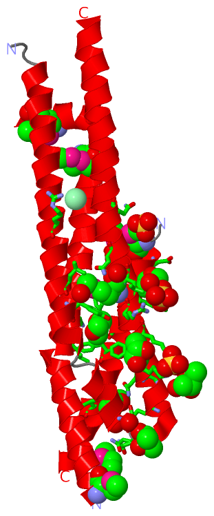
(-) Ligands, Modified Residues, Ions  (5, 15)

Asymmetric/Biological Unit (5, 15)
No.NameCountTypeFull Name
1BU93Ligand/IonMESO-2,3-BUTANEDIOL
2CL1Ligand/IonCHLORIDE ION
3DIO3Ligand/Ion1,4-DIETHYLENE DIOXIDE
4MSE5Mod. Amino AcidSELENOMETHIONINE
5SO43Ligand/IonSULFATE ION

(-) Sites  (10, 10)

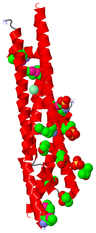
Asymmetric Unit (10, 10)
No.NameEvidenceResiduesDescription
01AC1SOFTWAREARG A:194 , LYS A:195 , HOH A:2111 , HOH A:2112BINDING SITE FOR RESIDUE SO4 A1230
02AC2SOFTWARELYS A:187 , LYS A:191 , ARG A:194 , BU9 A:1235BINDING SITE FOR RESIDUE SO4 A1231
03AC3SOFTWAREMSE A:20 , ASN A:206BINDING SITE FOR RESIDUE SO4 A1232
04AC4SOFTWAREASP A:31 , TYR A:34 , LEU A:79 , LYS A:83 , PHE A:86 , PHE A:189BINDING SITE FOR RESIDUE DIO A1233
05AC5SOFTWAREVAL A:41 , GLU A:182 , ASN A:186 , HOH A:2086BINDING SITE FOR RESIDUE DIO A1234
06AC6SOFTWARELEU A:160 , TYR A:163 , LYS A:187 , LEU A:198 , ASN A:201 , SO4 A:1231 , HOH A:2071 , HOH A:2094 , HOH A:2103BINDING SITE FOR RESIDUE BU9 A1235
07AC7SOFTWAREASP A:22BINDING SITE FOR RESIDUE DIO A1236
08AC8SOFTWAREARG A:153 , LYS A:185BINDING SITE FOR RESIDUE CL A1237
09AC9SOFTWARESER A:177 , GLN A:178 , HOH A:2083BINDING SITE FOR RESIDUE BU9 A1238
10BC1SOFTWARELYS A:187BINDING SITE FOR RESIDUE BU9 A1239

(-) SS Bonds  (0, 0)

(no "SS Bond" information available for 3ZCI)

(-) Cis Peptide Bonds  (0, 0)

(no "Cis Peptide Bond" information available for 3ZCI)

 Sequence-Structure Mapping

(-) SAPs(SNPs)/Variants  (0, 0)

(no "SAP(SNP)/Variant" information available for 3ZCI)

(-) PROSITE Motifs  (0, 0)

(no "PROSITE Motif" information available for 3ZCI)

(-) Exons   (0, 0)

(no "Exon" information available for 3ZCI)

(-) Sequences/Alignments

Asymmetric/Biological Unit
   Reformat: Number of residues per line =  ('0' or empty: single-line sequence representation)
  Number of residues per labelling interval =   
  UniProt sequence: complete  aligned part    
   Show mapping: SCOP domains CATH domains Pfam domains Secondary structure (by author)
SAPs(SNPs) PROSITE motifs Exons
(details for a mapped element are shown in a popup box when the mouse pointer rests over it)
Chain A from PDB  Type:PROTEIN  Length:191
 aligned with O25272_HELPY | O25272 from UniProtKB/TrEMBL  Length:237

    Alignment length:212
                                    27        37        47        57        67        77        87        97       107       117       127       137       147       157       167       177       187       197       207       217       227  
         O25272_HELPY    18 LMAEDITSGLKQLDSTYQETNQQVLKNLDEIFSTTSPSANNEMGEEDALNIKKAAIALRGDLALLKANFEANELFFISEDVIFKTYMSSPELLLTYMKINPLDQNTAEQQCGISDKVLVLYCEGKLKIEQEKQNIRERLETSLKAYQSNIGGTASLITASQTLVESLKNKNFIKGIRKLMLAHNKVFLNYLEELDALERSLEQSKRQYLQER 229
               SCOP domains -------------------------------------------------------------------------------------------------------------------------------------------------------------------------------------------------------------------- SCOP domains
               CATH domains -------------------------------------------------------------------------------------------------------------------------------------------------------------------------------------------------------------------- CATH domains
               Pfam domains -------------------------------------------------------------------------------------------------------------------------------------------------------------------------------------------------------------------- Pfam domains
         Sec.struct. author ..hhhhhhhhhhhhhhhhhhhhhhhhhhhhhh.--------hhhhhhhhhhhhhhhhhhhhhhhhhhhhhhhhhhhhhhhhhhh....hhhhhhhhhhhh-------------...hhhhhhhhhhhhhhhhhhhhhhhhhhhhhhhhhh..hhhhhhh..hhhhhhhhhhhhhhhhhhhhhhhhhhhhhhhhhhhhhhhhhhhhhhhhhh. Sec.struct. author
                 SAPs(SNPs) -------------------------------------------------------------------------------------------------------------------------------------------------------------------------------------------------------------------- SAPs(SNPs)
                    PROSITE -------------------------------------------------------------------------------------------------------------------------------------------------------------------------------------------------------------------- PROSITE
                 Transcript -------------------------------------------------------------------------------------------------------------------------------------------------------------------------------------------------------------------- Transcript
                 3zci A  18 GAmEDITSGLKQLDSTYQETNQQVLKNLDEIFS--------EmGEEDALNIATAAIALRGDLALLKANFEANELFFISEDVIFKTYmSSPELLLTYmKIN-------------SDKVLVLYCEGKLKIEATAQNIRERLETSLKAYQSNIGGTASLITASQTLVESLKNKNFIKGIRKLmLAHNKVFLNYLEELDALERSLEQSKRQYLQER 229
                              |     27        37        47  |      - ||     67        77        87        97      |107      |117         -   |   137       147       157       167       177       187       197       207       217       227  
                              |                            50       59|                                         104-MSE   114-MSE          131                                                               197-MSE                            
                             20-MSE                                  60-MSE                                                  117                                                                                                                

   Legend:   → Mismatch (orange background)
  - → Gap (green background, '-', border residues have a numbering label)
    → Modified Residue (blue background, lower-case, 'x' indicates undefined single-letter code, labelled with number + name)
  x → Chemical Group (purple background, 'x', labelled with number + name, e.g. ACE or NH2)
  extra numbering lines below/above indicate numbering irregularities and modified residue names etc., number ends below/above '|'

 Classification and Annotation

(-) SCOP Domains  (0, 0)

(no "SCOP Domain" information available for 3ZCI)

(-) CATH Domains  (0, 0)

(no "CATH Domain" information available for 3ZCI)

(-) Pfam Domains  (0, 0)

(no "Pfam Domain" information available for 3ZCI)

(-) Gene Ontology  (0, 0)

Asymmetric/Biological Unit(hide GO term definitions)
    (no "Gene Ontology" information available for 3ZCI)

 Visualization

(-) Interactive Views

Asymmetric/Biological Unit
  Complete Structure
    Jena3D(integrated viewing of ligand, site, SAP, PROSITE, SCOP information)
    WebMol | AstexViewer[tm]@PDBe
(Java Applets, require no local installation except for Java; loading may be slow)
    STRAP
(Java WebStart application, automatic local installation, requires Java; full application with system access!)
    RasMol
(require local installation)
    Molscript (VRML)
(requires installation of a VRML viewer; select preferred view via VRML and generate a mono or stereo PDF format file)
 
  Ligands, Modified Residues, Ions
    BU9  [ RasMol | Jena3D ]  +environment [ RasMol | Jena3D ]
    CL  [ RasMol | Jena3D ]  +environment [ RasMol | Jena3D ]
    DIO  [ RasMol | Jena3D ]  +environment [ RasMol | Jena3D ]
    MSE  [ RasMol | Jena3D ]  +environment [ RasMol | Jena3D ]
    SO4  [ RasMol | Jena3D ]  +environment [ RasMol | Jena3D ]
 
  Sites
    AC1  [ RasMol ]  +environment [ RasMol ]
    AC2  [ RasMol ]  +environment [ RasMol ]
    AC3  [ RasMol ]  +environment [ RasMol ]
    AC4  [ RasMol ]  +environment [ RasMol ]
    AC5  [ RasMol ]  +environment [ RasMol ]
    AC6  [ RasMol ]  +environment [ RasMol ]
    AC7  [ RasMol ]  +environment [ RasMol ]
    AC8  [ RasMol ]  +environment [ RasMol ]
    AC9  [ RasMol ]  +environment [ RasMol ]
    BC1  [ RasMol ]  +environment [ RasMol ]
 
  Cis Peptide Bonds
(no "Cis Peptide Bonds" information available for 3zci)
 

(-) Still Images

Jmol
  protein: cartoon or spacefill or dots and stick; nucleic acid: cartoon and stick; ligands: spacefill; active site: stick
Molscript
  protein, nucleic acid: cartoon; ligands: spacefill; active site: ball and stick

 Databases and Analysis Tools

(-) Databases

Access by PDB/NDB ID
  3zci
    Family and Domain InformationProDom | SYSTERS
    General Structural InformationGlycoscienceDB | MMDB | NDB | OCA | PDB | PDBe | PDBj | PDBsum | PDBWiki | PQS | PROTEOPEDIA
    Orientation in MembranesOPM
    Protein SurfaceSURFACE
    Secondary StructureDSSP (structure derived) | HSSP (homology derived)
    Structural GenomicsGeneCensus
    Structural NeighboursCE | VAST
    Structure ClassificationCATH | Dali | SCOP
    Validation and Original DataBMRB Data View | BMRB Restraints Grid | EDS | PROCHECK | RECOORD | WHAT_CHECK
 
Access by UniProt ID/Accession number
  O25272_HELPY | O25272
    Comparative Protein Structure ModelsModBase
    Genomic InformationEnsembl
    Protein-protein InteractionDIP
    Sequence, Family and Domain InformationInterPro | Pfam | SMART | UniProtKB/TrEMBL
 
Access by Enzyme Classificator   (EC Number)
  (no 'Enzyme Classificator' available)
    General Enzyme InformationBRENDA | EC-PDB | Enzyme | IntEnz
    PathwayKEGG | MetaCyc
 
Access by Disease Identifier   (MIM ID)
  (no 'MIM ID' available)
    Disease InformationOMIM
 
Access by GenAge ID
  (no 'GenAge ID' available)
    Age Related InformationGenAge

(-) Analysis Tools

Access by PDB/NDB ID
    Domain InformationXDom
    Interatomic Contacts of Structural UnitsCSU
    Ligand-protein ContactsLPC
    Protein CavitiescastP
    Sequence and Secondary StructurePDBCartoon
    Structure AlignmentSTRAP(Java WebStart application, automatic local installation, requires Java; full application with system access!)
    Structure and Sequence BrowserSTING
 
Access by UniProt ID/Accession number
  O25272_HELPY | O25272
    Protein Disorder PredictionDisEMBL | FoldIndex | GLOBPLOT (for more information see DisProt)

 Related Entries

(-) Entries Sharing at Least One Protein Chain (UniProt ID)

UniProtKB/TrEMBL
        O25272_HELPY | O252723zcj 4cii 4x5u

(-) Related Entries Specified in the PDB File

3zcj CRYSTAL STRUCTURE OF HELICOBACTER PYLORI T4SS PROTEIN CAGL IN A TETRAGONAL CRYSTAL FORM WITH A HELICAL RGD -MOTIF (6 MOL PER ASU)