|
|
|
|
 Description
|
|
Compounds
|
||||||||||||||||||||||||||||
Chains, Units
Summary Information (see also Sequences/Alignments below) |
Ligands, Modified Residues, Ions (4, 10)| Asymmetric/Biological Unit (4, 10) |
Sites (10, 10)
Asymmetric Unit (10, 10)
|
SS Bonds (14, 14)
Asymmetric/Biological Unit
|
||||||||||||||||||||||||||||||||||||||||||||||||||||||||||||
Cis Peptide Bonds (0, 0)| (no "Cis Peptide Bond" information available for 3V9M) |
SAPs(SNPs)/Variants (0, 0)| (no "SAP(SNP)/Variant" information available for 3V9M) |
PROSITE Motifs (2, 4)
Asymmetric/Biological Unit (2, 4)
|
||||||||||||||||||||||||||||||||
Exons (0, 0)| (no "Exon" information available for 3V9M) |
Sequences/Alignments
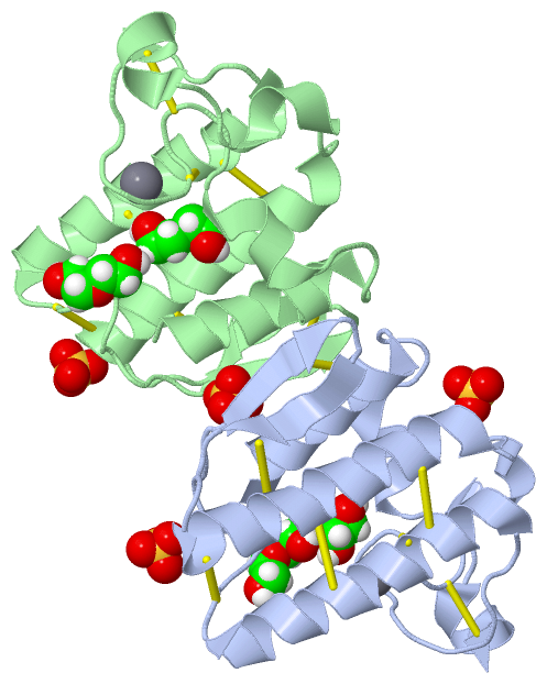
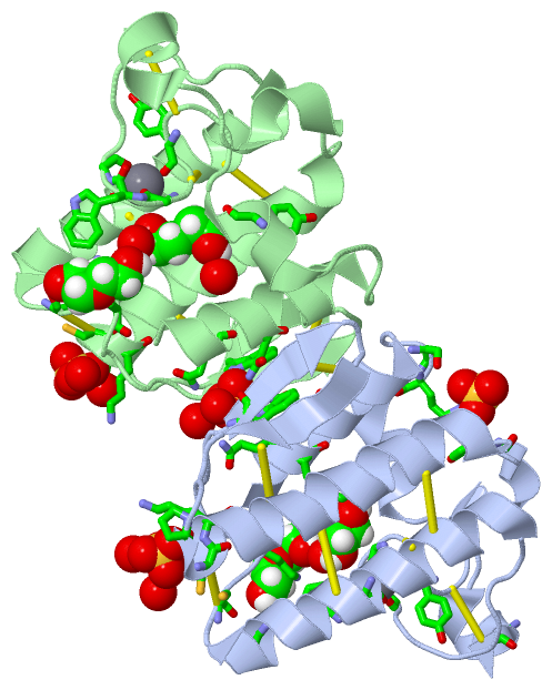
Asymmetric/Biological UnitChain A from PDB Type:PROTEIN Length:118 aligned with PA2BB_PSEAU | P04056 from UniProtKB/Swiss-Prot Length:118 Alignment length:118 10 20 30 40 50 60 70 80 90 100 110 PA2BB_PSEAU 1 NLIQFGNMIQCANKGSRPSLDYADYGCYCGWGGSGTPVDELDRCCQVHDNCYEQAGKKGCFPKLTLYSWKCTGNVPTCNSKPGCKSFVCACDAAAAKCFAKAPYKKENYNIDTKKRCK 118 SCOP domains d3v9ma_ A: automated matches SCOP domains CATH domains ---------------------------------------------------------------------------------------------------------------------- CATH domains Pfam domains ---------------------------------------------------------------------------------------------------------------------- Pfam domains SAPs(SNPs) ---------------------------------------------------------------------------------------------------------------------- SAPs(SNPs) PROSITE -------------------------------------------PA2_HIS ------------------------------------PA2_ASP -------------------- PROSITE Transcript ---------------------------------------------------------------------------------------------------------------------- Transcript 3v9m A 1 NLIQFGNMIQCANKGSRPSLDYADYGCYCGWGGSGTPVDELDRCCQVHDNCYEQAGKKGCFPKLTLYSWKCTGNVPTCNSKPGCKSFVCACDAAAAKCFAKAPYKKENYNIDTKKRCK 118 10 20 30 40 50 60 70 80 90 100 110 Chain B from PDB Type:PROTEIN Length:118 aligned with PA2BB_PSEAU | P04056 from UniProtKB/Swiss-Prot Length:118 Alignment length:118 10 20 30 40 50 60 70 80 90 100 110 PA2BB_PSEAU 1 NLIQFGNMIQCANKGSRPSLDYADYGCYCGWGGSGTPVDELDRCCQVHDNCYEQAGKKGCFPKLTLYSWKCTGNVPTCNSKPGCKSFVCACDAAAAKCFAKAPYKKENYNIDTKKRCK 118 SCOP domains d3v9mb_ B: automated matches SCOP domains CATH domains ---------------------------------------------------------------------------------------------------------------------- CATH domains Pfam domains ---------------------------------------------------------------------------------------------------------------------- Pfam domains SAPs(SNPs) ---------------------------------------------------------------------------------------------------------------------- SAPs(SNPs) PROSITE -------------------------------------------PA2_HIS ------------------------------------PA2_ASP -------------------- PROSITE Transcript ---------------------------------------------------------------------------------------------------------------------- Transcript 3v9m B 1 NLIQFGNMIQCANKGSRPSLDYADYGCYCGWGGSGTPVDELDRCCQVHDNCYEQAGKKGCFPKLTLYSWKCTGNVPTCNSKPGCKSFVCACDAAAAKCFAKAPYKKENYNIDTKKRCK 118 10 20 30 40 50 60 70 80 90 100 110
|
||||||||||||||||||||
SCOP Domains (1, 2)
Asymmetric/Biological Unit
 |
CATH Domains (0, 0)| (no "CATH Domain" information available for 3V9M) |
Pfam Domains (0, 0)| (no "Pfam Domain" information available for 3V9M) |
Gene Ontology (7, 7)|
Asymmetric/Biological Unit(hide GO term definitions) Chain A,B (PA2BB_PSEAU | P04056)
|
||||||||||||||||||||||||||||||||||||||||||||||||||||||||||||
Interactive Views
|
||||||||||||||||||||||||||||||||||||||||||||||||||||||||||||||||||||||||||||||||||||||||||||||||||||||||||||||||||||||||||||||||||||||||||||||||||||||||||||||||||||||||||||||||||||||||||||||||||||||||||
Still Images
|
||||||||||||||||
Databases
|
||||||||||||||||||||||||||||||||||||||||||||||||||||||||||||||||||||||||||||||||||||||||||||||||||||||||||||||||||||||||||||||||||||||||||||||||||||||||||||||||
Analysis Tools
|
|||||||||||||||||||||||||||||||||||||||||||||||||||||||||||||
Entries Sharing at Least One Protein Chain (UniProt ID)
Related Entries Specified in the PDB File
|
|