|
|
|
|
 Description
|
|
Compounds
|
||||||||||||||||||||||||||||||||||||||||||||||||||||||||||||
Chains, Units
Summary Information (see also Sequences/Alignments below) |
Ligands, Modified Residues, Ions (1, 2)
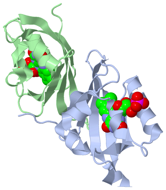
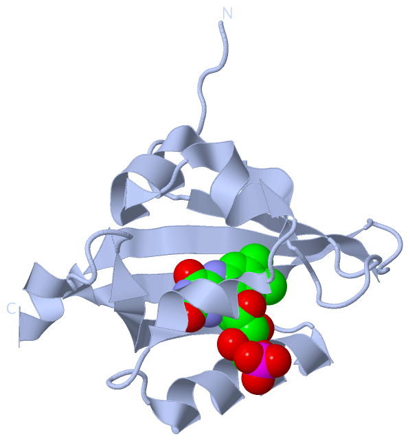
Asymmetric Unit (1, 2)
|
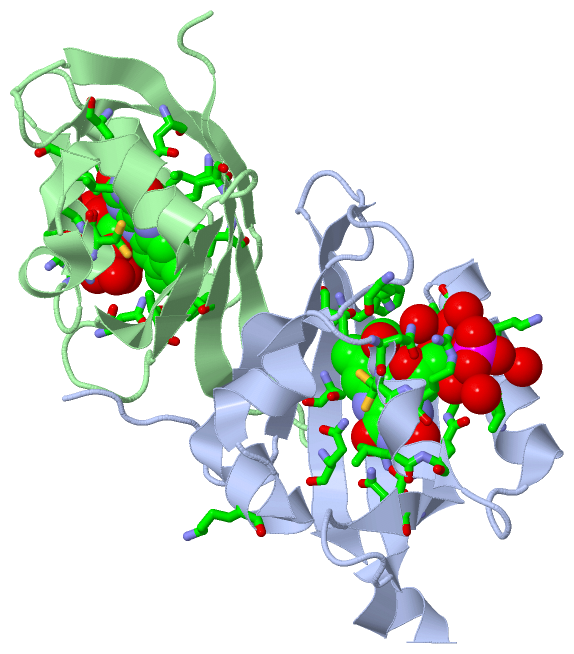
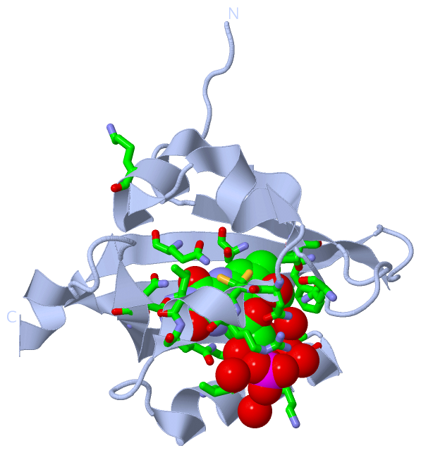
Sites (2, 2)
Asymmetric Unit (2, 2)
|
SS Bonds (0, 0)| (no "SS Bond" information available for 3T50) |
Cis Peptide Bonds (0, 0)| (no "Cis Peptide Bond" information available for 3T50) |
SAPs(SNPs)/Variants (0, 0)| (no "SAP(SNP)/Variant" information available for 3T50) |
PROSITE Motifs (1, 2)
Asymmetric Unit (1, 2)
|
||||||||||||||||||||||||||||||||||||||||||||||||||||||||||||||||||||||||||||||||||||||||||||||||
Exons (0, 0)| (no "Exon" information available for 3T50) |
Sequences/Alignments
Asymmetric UnitChain A from PDB Type:PROTEIN Length:115 aligned with LOVHK_BRUME | Q8YC53 from UniProtKB/Swiss-Prot Length:489 Alignment length:115 35 45 55 65 75 85 95 105 115 125 135 LOVHK_BRUME 26 AVEFTLMPMLITNPHLPDNPIVFANPAFLKLTGYEADEVMGRNCRFLQGHGTDPAHVRAIKSAIAAEKPIDIDIINYKKSGEAFWNRLHISPVHNANGRLQHFVSSQLDVTLELS 140 SCOP domains d3t50a_ A: automated matches SCOP domains CATH domains ------------------------------------------------------------------------------------------------------------------- CATH domains Pfam domains ------------------------------------------------------------------------------------------------------------------- Pfam domains SAPs(SNPs) ------------------------------------------------------------------------------------------------------------------- SAPs(SNPs) PROSITE -------------------------------------------------------------------PAC PDB: A:93-139 UniProt: 93-147 PROSITE Transcript ------------------------------------------------------------------------------------------------------------------- Transcript 3t50 A 26 ASEFTLMPMLITNPHLPDNPIVFANPAFLKLTGYEADEVMGRNCRFLQGHGTDPAHVRAIKSAIAAEKPIDIDIINYKKSGEAFWNRLHISPVHNANGRLQHFVSSQLDVTLELV 140 35 45 55 65 75 85 95 105 115 125 135 Chain B from PDB Type:PROTEIN Length:108 aligned with LOVHK_BRUME | Q8YC53 from UniProtKB/Swiss-Prot Length:489 Alignment length:108 39 49 59 69 79 89 99 109 119 129 LOVHK_BRUME 30 TLMPMLITNPHLPDNPIVFANPAFLKLTGYEADEVMGRNCRFLQGHGTDPAHVRAIKSAIAAEKPIDIDIINYKKSGEAFWNRLHISPVHNANGRLQHFVSSQLDVTL 137 SCOP domains d3t50b_ B: automated matches SCOP domains CATH domains ------------------------------------------------------------------------------------------------------------ CATH domains Pfam domains ------------------------------------------------------------------------------------------------------------ Pfam domains SAPs(SNPs) ------------------------------------------------------------------------------------------------------------ SAPs(SNPs) PROSITE ---------------------------------------------------------------PAC PDB: B:93-137 UniProt: 93-147 PROSITE Transcript ------------------------------------------------------------------------------------------------------------ Transcript 3t50 B 30 TLMPMLITNPHLPDNPIVFANPAFLKLTGYEADEVMGRNCRFLQGHGTDPAHVRAIKSAIAAEKPIDIDIINYKKSGEAFWNRLHISPVHNANGRLQHFVSSQLDVTL 137 39 49 59 69 79 89 99 109 119 129
|
||||||||||||||||||||
SCOP Domains (1, 2)
Asymmetric Unit
 |
CATH Domains (0, 0)| (no "CATH Domain" information available for 3T50) |
Pfam Domains (0, 0)| (no "Pfam Domain" information available for 3T50) |
Gene Ontology (15, 15)|
Asymmetric Unit(hide GO term definitions) Chain A,B (LOVHK_BRUME | Q8YC53)
|
||||||||||||||||||||||||||||||||||||||||||||||||||||||||||||||||||||||||||||||||||||||||||||||||||||||||||||
Interactive Views
|
|||||||||||||||||||||||||||||||||||||||||||||||||||||||||||||||||||||||||||||||||||||||||||||||||||||||||||||||||||||||||||||||||||||||||||||||||||||||||
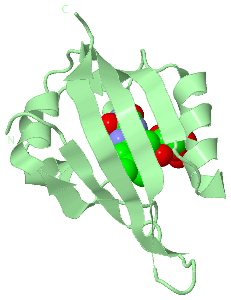
Still Images
|
||||||||||||||||
Databases
|
||||||||||||||||||||||||||||||||||||||||||||||||||||||||||||||||||||||||||||||||||||||||||||||||||||||||||||||||||||||||||||||||||||||||||||||||||||||||||||||||
Analysis Tools
|
|||||||||||||||||||||||||||||||||||||||||||||||||||||||||||||
Entries Sharing at Least One Protein Chain (UniProt ID)
Related Entries Specified in the PDB File
|
|