|
|
|
|
 Description
|
|
Compounds
|
||||||||||||||||||||||||||||||||||||||||||||||||||||||||
Chains, Units
Summary Information (see also Sequences/Alignments below) |
Ligands, Modified Residues, Ions (5, 11)| Asymmetric/Biological Unit (5, 11) |
Sites (11, 11)
Asymmetric Unit (11, 11)
|
SS Bonds (0, 0)| (no "SS Bond" information available for 3S42) |
Cis Peptide Bonds (0, 0)| (no "Cis Peptide Bond" information available for 3S42) |
SAPs(SNPs)/Variants (0, 0)| (no "SAP(SNP)/Variant" information available for 3S42) |
PROSITE Motifs (1, 2)
Asymmetric/Biological Unit (1, 2)
|
||||||||||||||||||||||||
Exons (0, 0)| (no "Exon" information available for 3S42) |
Sequences/Alignments
Asymmetric/Biological UnitChain A from PDB Type:PROTEIN Length:260 aligned with AROD_SALTY | P58687 from UniProtKB/Swiss-Prot Length:252 Alignment length:260 1 |2 12 22 32 42 52 62 72 82 92 102 112 122 132 142 152 162 172 182 192 202 212 222 232 242 252 AROD_SALTY - --------MKTVTVRDLVVGEGAPKIIVSLMGKTITDVKSEALAYREADFDILEWRVDHFANVTTAESVLEAAGAIREIITDKPLLFTFRSAKEGGEQALTTGQYIDLNRAAVDSGLVDMIDLELFTGDDEVKATVGYAHQHNVAVIMSNHDFHKTPAAEEIVQRLRKMQELGADIPKIAVMPQTKADVLTLLTATVEMQERYADRPIITMSMSKTGVISRLAGEVFGSAATFGAVKKASAPGQISVADLRTVLTILHQA 252 SCOP domains d3s42a_ A: automated matches SCOP domains CATH domains -------------------------------------------------------------------------------------------------------------------------------------------------------------------------------------------------------------------------------------------------------------------- CATH domains Pfam domains -------------------------------------------------------------------------------------------------------------------------------------------------------------------------------------------------------------------------------------------------------------------- Pfam domains SAPs(SNPs) -------------------------------------------------------------------------------------------------------------------------------------------------------------------------------------------------------------------------------------------------------------------- SAPs(SNPs) PROSITE -------------------------------------------------------------------------------------------------------------------------DEHYDROQUINASE_I PDB: A:114-14------------------------------------------------------------------------------------------------------------ PROSITE Transcript -------------------------------------------------------------------------------------------------------------------------------------------------------------------------------------------------------------------------------------------------------------------- Transcript 3s42 A -7 NLYFQSNAMKTVTVRDLVVGEGAPKIIVSLMGKTITDVKSEALAYREADFDILEWRVDHFANVTTAESVLEAAGAIREIITDKPLLFTFRSAKEGGEQALTTGQYIDLNRAAVDSGLVDMIDLELFTGDDEVKATVGYAHQHNVAVIMSNHDFHKTPAAEEIVQRLRKMQELGADIPKIAVMPQTKADVLTLLTATVEMQERYADRPIITMSMSKTGVISRLAGEVFGSAATFGAVKKASAPGQISVADLRTVLTILHQA 252 2 12 22 32 42 52 62 72 82 92 102 112 122 132 142 152 162 172 182 192 202 212 222 232 242 252 Chain B from PDB Type:PROTEIN Length:266 aligned with AROD_SALTY | P58687 from UniProtKB/Swiss-Prot Length:252 Alignment length:266 1 - | 6 16 26 36 46 56 66 76 86 96 106 116 126 136 146 156 166 176 186 196 206 216 226 236 246 AROD_SALTY - --------------MKTVTVRDLVVGEGAPKIIVSLMGKTITDVKSEALAYREADFDILEWRVDHFANVTTAESVLEAAGAIREIITDKPLLFTFRSAKEGGEQALTTGQYIDLNRAAVDSGLVDMIDLELFTGDDEVKATVGYAHQHNVAVIMSNHDFHKTPAAEEIVQRLRKMQELGADIPKIAVMPQTKADVLTLLTATVEMQERYADRPIITMSMSKTGVISRLAGEVFGSAATFGAVKKASAPGQISVADLRTVLTILHQA 252 SCOP domains -------------d3s42b1 B:0-252 automated matches SCOP domains CATH domains -------------------------------------------------------------------------------------------------------------------------------------------------------------------------------------------------------------------------------------------------------------------------- CATH domains Pfam domains (1) --------------------------------DHquinase_I-3s42B01 B:19-247 ----- Pfam domains (1) Pfam domains (2) --------------------------------DHquinase_I-3s42B02 B:19-247 ----- Pfam domains (2) SAPs(SNPs) -------------------------------------------------------------------------------------------------------------------------------------------------------------------------------------------------------------------------------------------------------------------------- SAPs(SNPs) PROSITE -------------------------------------------------------------------------------------------------------------------------------DEHYDROQUINASE_I PDB: B:114-14------------------------------------------------------------------------------------------------------------ PROSITE Transcript -------------------------------------------------------------------------------------------------------------------------------------------------------------------------------------------------------------------------------------------------------------------------- Transcript 3s42 B -23 MHHHHHNLYFQSNAMKTVTVRDLVVGEGAPKIIVSLMGKTITDVKSEALAYREADFDILEWRVDHFANVTTAESVLEAAGAIREIITDKPLLFTFRSAKEGGEQALTTGQYIDLNRAAVDSGLVDMIDLELFTGDDEVKATVGYAHQHNVAVIMSNHDFHKTPAAEEIVQRLRKMQELGADIPKIAVMPQTKADVLTLLTATVEMQERYADRPIITMSMSKTGVISRLAGEVFGSAATFGAVKKASAPGQISVADLRTVLTILHQA 252 || -4 6 16 26 36 46 56 66 76 86 96 106 116 126 136 146 156 166 176 186 196 206 216 226 236 246 -18| -7
|
||||||||||||||||||||
SCOP Domains (1, 2)
Asymmetric/Biological Unit
 |
CATH Domains (0, 0)| (no "CATH Domain" information available for 3S42) |
Pfam Domains (1, 2)
Asymmetric/Biological Unit
|
Gene Ontology (7, 7)|
Asymmetric/Biological Unit(hide GO term definitions) Chain A,B (AROD_SALTY | P58687)
|
||||||||||||||||||||||||||||||||||||||||||||||||||||||
Interactive Views
|
||||||||||||||||||||||||||||||||||||||||||||||||||||||||||||||||||||||||||||||||||||||||||||||||||||||||||||||||||||||||||||||||||||||||||||||||||||||||||||||||||||||||||||||||||||||||||||||||||||||||||||||||||||||||
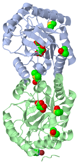
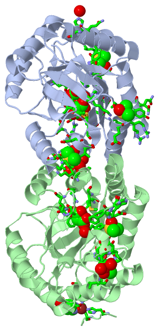
Still Images
|
||||||||||||||||
Databases
|
||||||||||||||||||||||||||||||||||||||||||||||||||||||||||||||||||||||||||||||||||||||||||||||||||||||||||||||||||||||||||||||||||||||||||||||||||||||||||||||||
Analysis Tools
|
|||||||||||||||||||||||||||||||||||||||||||||||||||||||||||||
Entries Sharing at Least One Protein Chain (UniProt ID)
Related Entries Specified in the PDB File
|
|