Show PDB file:   
         Plain Text   HTML   (compressed file size)
QuickSearch:   
by PDB,NDB,UniProt,PROSITE Code or Search Term(s)  
(-)Asym./Biol. Unit
collapse expand < >
Image Asym./Biol. Unit
Asym./Biol. Unit  (Jmol Viewer)

(-) Description

Title :  STRUCTURE OF PSEUDOMONAS FLUORESCENCE LAPD PERIPLASMIC DOMAIN
 
Authors :  H. Sondermann, M. V. A. S. Navarro, D. Chatterjee
Date :  10 Nov 10  (Deposition) - 09 Feb 11  (Release) - 23 Feb 11  (Revision)
Method :  X-RAY DIFFRACTION
Resolution :  1.77
Chains :  Asym./Biol. Unit :  D,F
Keywords :  Binding Protein, Lapg, Lyase (Keyword Search: [Gene Ontology, PubMed, Web (Google))
 
Reference :  M. V. Navarro, P. D. Newell, P. V. Krasteva, D. Chatterjee, D. R. Madden G. A. O'Toole, H. Sondermann
Structural Basis For C-Di-Gmp-Mediated Inside-Out Signaling Controlling Periplasmic Proteolysis.
Plos Biol. V. 9 E1000 2011
PubMed-ID: 21304926  |  Reference-DOI: 10.1371/JOURNAL.PBIO.1000588

(-) Compounds

Molecule 1 - CYCLIC DIMERIC GMP BINDING PROTEIN
    ChainsD, F
    EngineeredYES
    Expression SystemESCHERICHIA COLI
    Expression System Taxid562
    FragmentUNP RESIDUES 22-151
    GeneLAPD, PFL01_0131
    Organism ScientificPSEUDOMONAS FLUORESCENS
    Organism Taxid205922
    StrainPF0-1

 Structural Features

(-) Chains, Units

  12
Asymmetric/Biological Unit DF

Summary Information (see also Sequences/Alignments below)

(-) Ligands, Modified Residues, Ions  (1, 6)

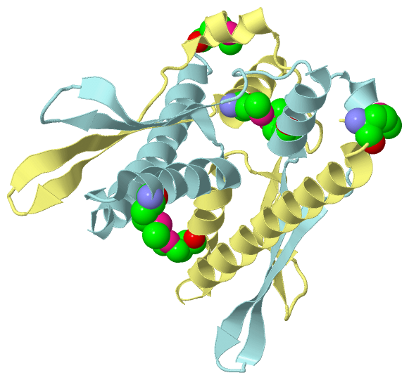
Asymmetric/Biological Unit (1, 6)
No.NameCountTypeFull Name
1MSE6Mod. Amino AcidSELENOMETHIONINE

(-) Sites  (0, 0)

(no "Site" information available for 3PJV)

(-) SS Bonds  (0, 0)

(no "SS Bond" information available for 3PJV)

(-) Cis Peptide Bonds  (0, 0)

(no "Cis Peptide Bond" information available for 3PJV)

 Sequence-Structure Mapping

(-) SAPs(SNPs)/Variants  (0, 0)

(no "SAP(SNP)/Variant" information available for 3PJV)

(-) PROSITE Motifs  (0, 0)

(no "PROSITE Motif" information available for 3PJV)

(-) Exons   (0, 0)

(no "Exon" information available for 3PJV)

(-) Sequences/Alignments

Asymmetric/Biological Unit
   Reformat: Number of residues per line =  ('0' or empty: single-line sequence representation)
  Number of residues per labelling interval =   
  UniProt sequence: complete  aligned part    
   Show mapping: SCOP domains CATH domains Pfam domains Secondary structure (by author)
SAPs(SNPs) PROSITE motifs Exons
(details for a mapped element are shown in a popup box when the mouse pointer rests over it)
Chain D from PDB  Type:PROTEIN  Length:128
 aligned with Q3KK31_PSEPF | Q3KK31 from UniProtKB/TrEMBL  Length:648

    Alignment length:128
                                    32        42        52        62        72        82        92       102       112       122       132       142        
         Q3KK31_PSEPF    23 FMVSLESSRTQYVNQLRSHAQDAATALALSLTPNIDDPAMVELLVSSIFDSGYYSSIRVVDLKTDQTIVERNGIPAVTNVPDWFVKLIGLEPAGGDALVSRGWEQAARVEVVSHPMFALAKLWQSALG 150
               SCOP domains -------------------------------------------------------------------------------------------------------------------------------- SCOP domains
               CATH domains -------------------------------------------------------------------------------------------------------------------------------- CATH domains
               Pfam domains -------------------------------------------------------------------------------------------------------------------------------- Pfam domains
         Sec.struct. author ...hhhhhhhhhhhhhhhhhhhhhhhhhhhhhhhh.hhhhhhhhhhhhhh...eeeeeeee.....eeeeee........hhhhhhhhh.....eeeeeee..eeeeeeeeee.hhhhhhhhhhhhhh Sec.struct. author
                 SAPs(SNPs) -------------------------------------------------------------------------------------------------------------------------------- SAPs(SNPs)
                    PROSITE -------------------------------------------------------------------------------------------------------------------------------- PROSITE
                 Transcript -------------------------------------------------------------------------------------------------------------------------------- Transcript
                 3pjv D  23 FmVSLESSRTQYVNQLRSHAQDAATALALSLTPNIDDPAmVELLVSSIFDSGYYSSIRVVDLKTDQTIVERNGIPAVTNVPDWFVKLIGLEPAGGDALVSRGWEQAARVEVVSHPmFALAKLWQSALG 150
                             |      32        42        52        62        72        82        92       102       112       122       132     | 142        
                             |                                    62-MSE                                                                     138-MSE        
                            24-MSE                                                                                                                          

Chain F from PDB  Type:PROTEIN  Length:127
 aligned with Q3KK31_PSEPF | Q3KK31 from UniProtKB/TrEMBL  Length:648

    Alignment length:127
                                    33        43        53        63        73        83        93       103       113       123       133       143       
         Q3KK31_PSEPF    24 MVSLESSRTQYVNQLRSHAQDAATALALSLTPNIDDPAMVELLVSSIFDSGYYSSIRVVDLKTDQTIVERNGIPAVTNVPDWFVKLIGLEPAGGDALVSRGWEQAARVEVVSHPMFALAKLWQSALG 150
               SCOP domains ------------------------------------------------------------------------------------------------------------------------------- SCOP domains
               CATH domains ------------------------------------------------------------------------------------------------------------------------------- CATH domains
               Pfam domains ------------------------------------------------------------------------------------------------------------------------------- Pfam domains
         Sec.struct. author ...hhhhhhhhhhhhhhhhhhhhhhhhhhhhhhh.hhhhhhhhhhhhhhhh.eeeeeeee....eeeeeee........hhhhhhhhh...eeeeeeeee..eeeeeeeeee.hhhhhhhhhhhhh. Sec.struct. author
                 SAPs(SNPs) ------------------------------------------------------------------------------------------------------------------------------- SAPs(SNPs)
                    PROSITE ------------------------------------------------------------------------------------------------------------------------------- PROSITE
                 Transcript ------------------------------------------------------------------------------------------------------------------------------- Transcript
                 3pjv F  24 mVSLESSRTQYVNQLRSHAQDAATALALSLTPNIDDPAmVELLVSSIFDSGYYSSIRVVDLKTDQTIVERNGIPAVTNVPDWFVKLIGLEPAGGDALVSRGWEQAARVEVVSHPmFALAKLWQSALG 150
                            |       33        43        53        63        73        83        93       103       113       123       133    |  143       
                           24-MSE                                62-MSE                                                                     138-MSE        

   Legend:   → Mismatch (orange background)
  - → Gap (green background, '-', border residues have a numbering label)
    → Modified Residue (blue background, lower-case, 'x' indicates undefined single-letter code, labelled with number + name)
  x → Chemical Group (purple background, 'x', labelled with number + name, e.g. ACE or NH2)
  extra numbering lines below/above indicate numbering irregularities and modified residue names etc., number ends below/above '|'

 Classification and Annotation

(-) SCOP Domains  (0, 0)

(no "SCOP Domain" information available for 3PJV)

(-) CATH Domains  (0, 0)

(no "CATH Domain" information available for 3PJV)

(-) Pfam Domains  (0, 0)

(no "Pfam Domain" information available for 3PJV)

(-) Gene Ontology  (6, 6)

Asymmetric/Biological Unit(hide GO term definitions)
Chain D,F   (Q3KK31_PSEPF | Q3KK31)
molecular function
    GO:0000166    nucleotide binding    Interacting selectively and non-covalently with a nucleotide, any compound consisting of a nucleoside that is esterified with (ortho)phosphate or an oligophosphate at any hydroxyl group on the ribose or deoxyribose.
    GO:0005515    protein binding    Interacting selectively and non-covalently with any protein or protein complex (a complex of two or more proteins that may include other nonprotein molecules).
    GO:0004871    signal transducer activity    Conveys a signal across a cell to trigger a change in cell function or state. A signal is a physical entity or change in state that is used to transfer information in order to trigger a response.
biological process
    GO:0007165    signal transduction    The cellular process in which a signal is conveyed to trigger a change in the activity or state of a cell. Signal transduction begins with reception of a signal (e.g. a ligand binding to a receptor or receptor activation by a stimulus such as light), or for signal transduction in the absence of ligand, signal-withdrawal or the activity of a constitutively active receptor. Signal transduction ends with regulation of a downstream cellular process, e.g. regulation of transcription or regulation of a metabolic process. Signal transduction covers signaling from receptors located on the surface of the cell and signaling via molecules located within the cell. For signaling between cells, signal transduction is restricted to events at and within the receiving cell.
cellular component
    GO:0016021    integral component of membrane    The component of a membrane consisting of the gene products and protein complexes having at least some part of their peptide sequence embedded in the hydrophobic region of the membrane.
    GO:0016020    membrane    A lipid bilayer along with all the proteins and protein complexes embedded in it an attached to it.

 Visualization

(-) Interactive Views

Asymmetric/Biological Unit
  Complete Structure
    Jena3D(integrated viewing of ligand, site, SAP, PROSITE, SCOP information)
    WebMol | AstexViewer[tm]@PDBe
(Java Applets, require no local installation except for Java; loading may be slow)
    STRAP
(Java WebStart application, automatic local installation, requires Java; full application with system access!)
    RasMol
(require local installation)
    Molscript (VRML)
(requires installation of a VRML viewer; select preferred view via VRML and generate a mono or stereo PDF format file)
 
  Ligands, Modified Residues, Ions
    MSE  [ RasMol | Jena3D ]  +environment [ RasMol | Jena3D ]
 
  Sites
(no "Sites" information available for 3pjv)
 
  Cis Peptide Bonds
(no "Cis Peptide Bonds" information available for 3pjv)
 

(-) Still Images

Jmol
  protein: cartoon or spacefill or dots and stick; nucleic acid: cartoon and stick; ligands: spacefill; active site: stick
Molscript
  protein, nucleic acid: cartoon; ligands: spacefill; active site: ball and stick

 Databases and Analysis Tools

(-) Databases

Access by PDB/NDB ID
  3pjv
    Family and Domain InformationProDom | SYSTERS
    General Structural InformationGlycoscienceDB | MMDB | NDB | OCA | PDB | PDBe | PDBj | PDBsum | PDBWiki | PQS | PROTEOPEDIA
    Orientation in MembranesOPM
    Protein SurfaceSURFACE
    Secondary StructureDSSP (structure derived) | HSSP (homology derived)
    Structural GenomicsGeneCensus
    Structural NeighboursCE | VAST
    Structure ClassificationCATH | Dali | SCOP
    Validation and Original DataBMRB Data View | BMRB Restraints Grid | EDS | PROCHECK | RECOORD | WHAT_CHECK
 
Access by UniProt ID/Accession number
  Q3KK31_PSEPF | Q3KK31
    Comparative Protein Structure ModelsModBase
    Genomic InformationEnsembl
    Protein-protein InteractionDIP
    Sequence, Family and Domain InformationInterPro | Pfam | SMART | UniProtKB/TrEMBL
 
Access by Enzyme Classificator   (EC Number)
  (no 'Enzyme Classificator' available)
    General Enzyme InformationBRENDA | EC-PDB | Enzyme | IntEnz
    PathwayKEGG | MetaCyc
 
Access by Disease Identifier   (MIM ID)
  (no 'MIM ID' available)
    Disease InformationOMIM
 
Access by GenAge ID
  (no 'GenAge ID' available)
    Age Related InformationGenAge

(-) Analysis Tools

Access by PDB/NDB ID
    Domain InformationXDom
    Interatomic Contacts of Structural UnitsCSU
    Ligand-protein ContactsLPC
    Protein CavitiescastP
    Sequence and Secondary StructurePDBCartoon
    Structure AlignmentSTRAP(Java WebStart application, automatic local installation, requires Java; full application with system access!)
    Structure and Sequence BrowserSTING
 
Access by UniProt ID/Accession number
  Q3KK31_PSEPF | Q3KK31
    Protein Disorder PredictionDisEMBL | FoldIndex | GLOBPLOT (for more information see DisProt)

 Related Entries

(-) Entries Sharing at Least One Protein Chain (UniProt ID)

UniProtKB/TrEMBL
        Q3KK31_PSEPF | Q3KK313pjt 3pju 3pjw 3pjx

(-) Related Entries Specified in the PDB File

3pjt 3pju 3pjw 3pjx