|
|
|
|
 Description
|
|
Compounds
|
||||||||||||||||||||||||||||||||||||||||
Chains, Units
Summary Information (see also Sequences/Alignments below) |
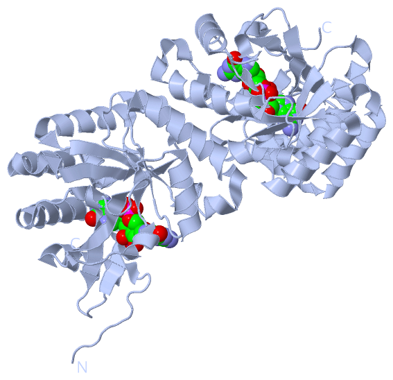
Ligands, Modified Residues, Ions (1, 1)
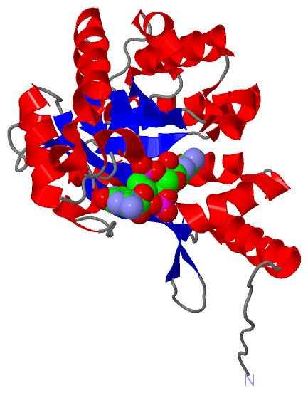
Asymmetric Unit (1, 1)
|
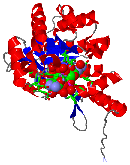
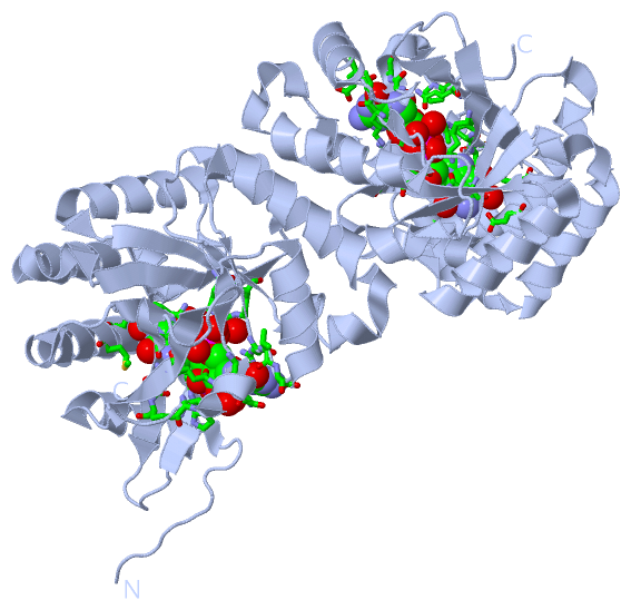
Sites (1, 1)
Asymmetric Unit (1, 1)
|
SS Bonds (0, 0)| (no "SS Bond" information available for 3PJU) |
Cis Peptide Bonds (0, 0)| (no "Cis Peptide Bond" information available for 3PJU) |
SAPs(SNPs)/Variants (0, 0)| (no "SAP(SNP)/Variant" information available for 3PJU) |
PROSITE Motifs (0, 0)| (no "PROSITE Motif" information available for 3PJU) |
Exons (0, 0)| (no "Exon" information available for 3PJU) |
Sequences/Alignments
Asymmetric UnitChain A from PDB Type:PROTEIN Length:249 aligned with Q3KK31_PSEPF | Q3KK31 from UniProtKB/TrEMBL Length:648 Alignment length:249 409 419 429 439 449 459 469 479 489 499 509 519 529 539 549 559 569 579 589 599 609 619 629 639 Q3KK31_PSEPF 400 QSLVADVGDDHHAWHRLLDQALNQRRFELFFQPVVAAQDTQLVLHYKVLSRLLDEQGQTIPAGRFLPWLERFGWTARLDRLMLERVLEQMAGHEESLALNLSSATLADPQALNKVFEILRAHSNLGARLTLEIGEEQLPEQAVLEQLTRRLRELGFSLSLQRFGGRFSMIGNLARLGLAYLKIDGSYIRAIDQESDKRLFIEAIQRAAHSIDLPLIAERVETEGELSVIREMGLYGVQGQLFGEPKPWG 648 SCOP domains --------------------------------------------------------------------------------------------------------------------------------------------------------------------------------------------------------------------------------------------------------- SCOP domains CATH domains --------------------------------------------------------------------------------------------------------------------------------------------------------------------------------------------------------------------------------------------------------- CATH domains Pfam domains --------------EAL-3pjuA01 A:414-644 ---- Pfam domains SAPs(SNPs) --------------------------------------------------------------------------------------------------------------------------------------------------------------------------------------------------------------------------------------------------------- SAPs(SNPs) PROSITE --------------------------------------------------------------------------------------------------------------------------------------------------------------------------------------------------------------------------------------------------------- PROSITE Transcript --------------------------------------------------------------------------------------------------------------------------------------------------------------------------------------------------------------------------------------------------------- Transcript 3pju A 400 QSLVADVGDDHHAWHRLLDQALNQRRFELFFQPVVAAQDTQLVLHYKVLSRLLDEQGQTIPAGRFLPWLERFGWTARLDRLMLERVLEQMAGHEESLALNLSSATLADPQALNKVFEILRAHSNLGARLTLEIGEEQLPEQAVLEQLTRRLRELGFSLSLQRFGGRFSMIGNLARLGLAYLKIDGSYIRAIDQESDKRLFIEAIQRAAHSIDLPLIAERVETEGELSVIREMGLYGVQGQLFGEPKPWG 648 409 419 429 439 449 459 469 479 489 499 509 519 529 539 549 559 569 579 589 599 609 619 629 639
|
||||||||||||||||||||
SCOP Domains (0, 0)| (no "SCOP Domain" information available for 3PJU) |
CATH Domains (0, 0)| (no "CATH Domain" information available for 3PJU) |
Pfam Domains (1, 1)
Asymmetric Unit
|
Gene Ontology (6, 6)|
Asymmetric Unit(hide GO term definitions) Chain A (Q3KK31_PSEPF | Q3KK31)
|
||||||||||||||||||||||||||||||||||||||||||||||||||||||
Interactive Views
|
||||||||||||||||||||||||||||||||||||||||||||||||||||||||||||||||||||||||||||||||||||||||||||||||||||||||||||||||||||||||||||||||||||||||
Still Images
|
||||||||||||||||
Databases
|
||||||||||||||||||||||||||||||||||||||||||||||||||||||||||||||||||||||||||||||||||||||||||||||||||||||||||||||||||||||||||||||||||||||||||||||||||||||||||||||||
Analysis Tools
|
|||||||||||||||||||||||||||||||||||||||||||||||||||||||||||||
Entries Sharing at Least One Protein Chain (UniProt ID)
Related Entries Specified in the PDB File
|
|