Show PDB file:   
         Plain Text   HTML   (compressed file size)
QuickSearch:   
by PDB,NDB,UniProt,PROSITE Code or Search Term(s)  
(-)Asymmetric Unit
(-)Asym. Unit - sites
(-)Biological Unit 1
(-)Biol. Unit 1 - sites
(-)Biological Unit 2
(-)Biological Unit 3
collapse expand < >
Image Asymmetric Unit
Asymmetric Unit  (Jmol Viewer)
Image Asym. Unit - sites
Asym. Unit - sites  (Jmol Viewer)
Image Biological Unit 1
Biological Unit 1  (Jmol Viewer)
Image Biol. Unit 1 - sites
Biol. Unit 1 - sites  (Jmol Viewer)
Image Biological Unit 2
Biological Unit 2  (Jmol Viewer)
Image Biological Unit 3
Biological Unit 3  (Jmol Viewer)

(-) Description

Title :  CRYSTAL STRUCTURE OF THE EXTRACELLULAR DOMAIN OF THE PUTATIVE HISTIDINE KINASE VPHK1S-Z8
 
Authors :  Z. Zhang, W. A. Hendrickson
Date :  24 Jan 10  (Deposition) - 05 May 10  (Release) - 11 Aug 10  (Revision)
Method :  X-RAY DIFFRACTION
Resolution :  1.76
Chains :  Asym. Unit :  A,B
Biol. Unit 1:  A  (1x)
Biol. Unit 2:  B  (1x)
Biol. Unit 3:  A,B  (1x)
Keywords :  Pdc Fold, Signaling Protein (Keyword Search: [Gene Ontology, PubMed, Web (Google))
 
Reference :  Z. Zhang, W. A. Hendrickson
Structural Characterization Of The Predominant Family Of Histidine Kinase Sensor Domains.
J. Mol. Biol. V. 400 335 2010
PubMed-ID: 20435045  |  Reference-DOI: 10.1016/J.JMB.2010.04.049

(-) Compounds

Molecule 1 - PUTATIVE SENSORY BOX/GGDEF FAMILY PROTEIN
    ChainsA, B
    EngineeredYES
    Expression SystemESCHERICHIA COLI
    Expression System PlasmidPET22B+
    Expression System StrainBL21(DE3)
    Expression System Taxid562
    Expression System Vector TypePLASMID
    FragmentEXTRACELLULAR DOMAIN (UNP RESIDUES 28-313)
    GeneVP0354
    Organism ScientificVIBRIO PARAHAEMOLYTICUS
    Organism Taxid670

 Structural Features

(-) Chains, Units

  12
Asymmetric Unit AB
Biological Unit 1 (1x)A 
Biological Unit 2 (1x) B
Biological Unit 3 (1x)AB

Summary Information (see also Sequences/Alignments below)

(-) Ligands, Modified Residues, Ions  (3, 13)

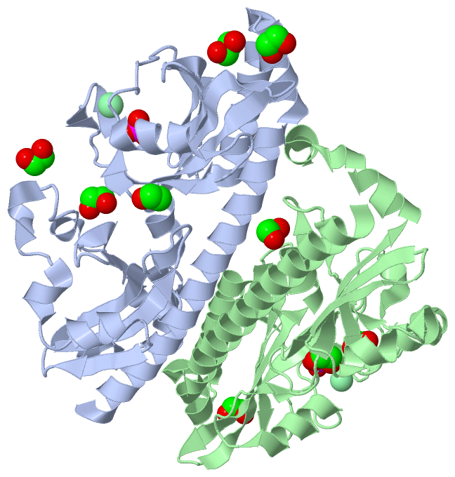
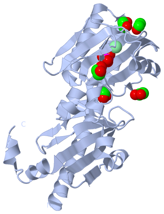
Asymmetric Unit (3, 13)
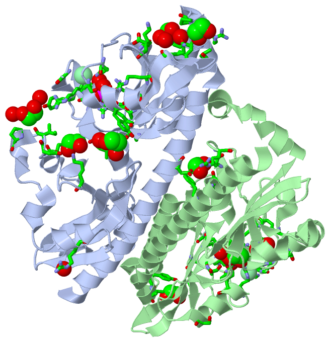
No.NameCountTypeFull Name
1CL2Ligand/IonCHLORIDE ION
2EDO9Ligand/Ion1,2-ETHANEDIOL
3PO42Ligand/IonPHOSPHATE ION
Biological Unit 1 (2, 6)
No.NameCountTypeFull Name
1CL-1Ligand/IonCHLORIDE ION
2EDO5Ligand/Ion1,2-ETHANEDIOL
3PO41Ligand/IonPHOSPHATE ION
Biological Unit 2 (2, 5)
No.NameCountTypeFull Name
1CL-1Ligand/IonCHLORIDE ION
2EDO4Ligand/Ion1,2-ETHANEDIOL
3PO41Ligand/IonPHOSPHATE ION
Biological Unit 3 (2, 11)
No.NameCountTypeFull Name
1CL-1Ligand/IonCHLORIDE ION
2EDO9Ligand/Ion1,2-ETHANEDIOL
3PO42Ligand/IonPHOSPHATE ION

(-) Sites  (13, 13)

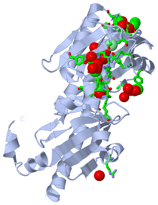
Asymmetric Unit (13, 13)
No.NameEvidenceResiduesDescription
01AC1SOFTWAREGLN A:106 , ARG A:108 , LYS A:138 , ARG A:141 , TYR A:143 , GLU A:163 , ARG A:164 , ARG A:178 , ASN A:196 , HOH A:334BINDING SITE FOR RESIDUE PO4 A 321
02AC2SOFTWARELYS A:138 , ARG A:141 , GLY A:167BINDING SITE FOR RESIDUE CL A 350
03AC3SOFTWARETYR A:78 , PRO A:82 , ASP A:111 , LYS A:117 , HOH A:382 , HOH A:515BINDING SITE FOR RESIDUE EDO A 2
04AC4SOFTWAREHOH A:25 , TYR A:171 , ILE A:238 , PRO A:240 , HOH A:570BINDING SITE FOR RESIDUE EDO A 3
05AC5SOFTWARETYR A:146 , ALA A:157 , HOH A:535 , HOH A:571BINDING SITE FOR RESIDUE EDO A 4
06AC6SOFTWAREILE A:160 , LYS A:220 , TYR A:235 , ASP A:237BINDING SITE FOR RESIDUE EDO A 5
07AC7SOFTWARELEU A:79 , ARG A:80 , VAL A:185 , GLN A:190 , SER B:149BINDING SITE FOR RESIDUE EDO A 9
08AC8SOFTWAREGLN B:106 , ARG B:108 , LYS B:138 , ARG B:141 , TYR B:143 , GLU B:163 , ARG B:164 , ARG B:178 , ASN B:196 , HOH B:366BINDING SITE FOR RESIDUE PO4 B 321
09AC9SOFTWARELYS B:138 , ARG B:141 , LYS B:166 , GLY B:167BINDING SITE FOR RESIDUE CL B 351
10BC1SOFTWAREILE B:160 , LYS B:220 , ILE B:226 , TYR B:235 , ASP B:237BINDING SITE FOR RESIDUE EDO B 1
11BC2SOFTWAREASP A:60 , GLN B:47 , TYR B:50 , SER B:209 , ARG B:212BINDING SITE FOR RESIDUE EDO B 6
12BC3SOFTWAREGLY B:159 , ILE B:160 , LYS B:220 , SER B:282 , HOH B:431BINDING SITE FOR RESIDUE EDO B 7
13BC4SOFTWAREARG A:257 , HOH A:345 , TYR B:78 , LEU B:79 , LYS B:117BINDING SITE FOR RESIDUE EDO B 8

(-) SS Bonds  (0, 0)

(no "SS Bond" information available for 3LID)

(-) Cis Peptide Bonds  (3, 3)

Asymmetric Unit
No.Residues
1Tyr A:171 -Pro A:172
2Ile A:309 -Gln A:310
3Tyr B:171 -Pro B:172

 Sequence-Structure Mapping

(-) SAPs(SNPs)/Variants  (0, 0)

(no "SAP(SNP)/Variant" information available for 3LID)

(-) PROSITE Motifs  (0, 0)

(no "PROSITE Motif" information available for 3LID)

(-) Exons   (0, 0)

(no "Exon" information available for 3LID)

(-) Sequences/Alignments

Asymmetric Unit
   Reformat: Number of residues per line =  ('0' or empty: single-line sequence representation)
  Number of residues per labelling interval =   
  UniProt sequence: complete  aligned part    
   Show mapping: SCOP domains CATH domains Pfam domains Secondary structure (by author)
SAPs(SNPs) PROSITE motifs Exons
(details for a mapped element are shown in a popup box when the mouse pointer rests over it)
Chain A from PDB  Type:PROTEIN  Length:279
 aligned with Q87SR8_VIBPA | Q87SR8 from UniProtKB/TrEMBL  Length:626

    Alignment length:279
                                    42        52        62        72        82        92       102       112       122       132       142       152       162       172       182       192       202       212       222       232       242       252       262       272       282       292       302         
         Q87SR8_VIBPA    33 LANNVENTAKEALHQLAYTGREYNNIQDQIETISDLLGHSQSLYDYLREPSKANLTILENMWSSVARNQKLYKQIRFLDTSGTEKVRIKYDFKTSIAGPSLILRDKSAREYFKYAQSLDNEQISAWGIELERDKGELVYPLSPSLRILMPISVNDVRQGYLVLNVDIEYLSSLLNYSPVRDFHIELVKHKGFYIASPDESRLYGDIIPERSQFNFSNMYPDIWPRVVSEQAGYSYSGEHLIAFSSIKFVSNEPLHLIIDLSNEQLSKRATRDINDLIQE 311
               SCOP domains d3lida1 A:33-206 GGDEF family protein VP0354                                                                                                                                  d3lida2 A:207-311 GGDEF family protein VP0354                                                             SCOP domains
               CATH domains --------------------------------------------------------------------------------------------------------------------------------------------------------------------------------------------------------------------------------------------------------------------------------------- CATH domains
               Pfam domains --------------------------------------------------------------------------------------------------------------------------------------------------------------------------------------------------------------------------------------------------------------------------------------- Pfam domains
         Sec.struct. author hhhhhhhhhhhhhhhhhhhhhhhhhhhhhhhhhhhhhhhhhhhhhhhhh.hhhhhhhhhhhhhhhhhhhh.eeeeeeee....eeeeeeee....eeee.........hhhhhhhhhh.....ee...ee.ee..ee.....eeeeeeeeeee..eeeeeeeeeeehhhhhhhhhee....ee.eeee....eee..hhhhh....hhhhh..hhhhhh..hhhhhhh...eeee...eeeeeeee.......eeeeeeeehhhhhhh...hhhhhh.. Sec.struct. author
                 SAPs(SNPs) --------------------------------------------------------------------------------------------------------------------------------------------------------------------------------------------------------------------------------------------------------------------------------------- SAPs(SNPs)
                    PROSITE --------------------------------------------------------------------------------------------------------------------------------------------------------------------------------------------------------------------------------------------------------------------------------------- PROSITE
                 Transcript --------------------------------------------------------------------------------------------------------------------------------------------------------------------------------------------------------------------------------------------------------------------------------------- Transcript
                 3lid A  33 LANNVENTAKEALHQLAYTGREYNNIQDQIETISDLLGHSQSLYDYLREPSKANLTILENMWSSVARNQKLYKQIRFLDTSGTEKVRIKYDFKTSIAGPSLILRDKSAREYFKYAQSLDNEQISAWGIELERDKGELVYPLSPSLRILMPISVNDVRQGYLVLNVDIEYLSSLLNYSPVRDFHIELVKHKGFYIASPDESRLYGDIIPERSQFNFSNMYPDIWPRVVSEQAGYSYSGEHLIAFSSIKFVSNEPLHLIIDLSNEQLSKRATRDINDLIQE 311
                                    42        52        62        72        82        92       102       112       122       132       142       152       162       172       182       192       202       212       222       232       242       252       262       272       282       292       302         

Chain B from PDB  Type:PROTEIN  Length:276
 aligned with Q87SR8_VIBPA | Q87SR8 from UniProtKB/TrEMBL  Length:626

    Alignment length:276
                                    45        55        65        75        85        95       105       115       125       135       145       155       165       175       185       195       205       215       225       235       245       255       265       275       285       295       305      
         Q87SR8_VIBPA    36 NVENTAKEALHQLAYTGREYNNIQDQIETISDLLGHSQSLYDYLREPSKANLTILENMWSSVARNQKLYKQIRFLDTSGTEKVRIKYDFKTSIAGPSLILRDKSAREYFKYAQSLDNEQISAWGIELERDKGELVYPLSPSLRILMPISVNDVRQGYLVLNVDIEYLSSLLNYSPVRDFHIELVKHKGFYIASPDESRLYGDIIPERSQFNFSNMYPDIWPRVVSEQAGYSYSGEHLIAFSSIKFVSNEPLHLIIDLSNEQLSKRATRDINDLIQE 311
               SCOP domains d3lidb1 B:36-206 GGDEF family protein VP0354                                                                                                                               d3lidb2 B:207-311 GGDEF family protein VP0354                                                             SCOP domains
               CATH domains ------------------------------------------------------------------------------------------------------------------------------------------------------------------------------------------------------------------------------------------------------------------------------------ CATH domains
               Pfam domains ------------------------------------------------------------------------------------------------------------------------------------------------------------------------------------------------------------------------------------------------------------------------------------ Pfam domains
         Sec.struct. author hhhhhhhhhhhhhhhhhhhhhhhhhhhhhhhhhhhhhhhhhhhhhh.hhhhhhhhhhhhhhhhhhhh.eeeeeeee....eeeeeeee....eeee.........hhhhhhhhhh.....ee...ee.ee..ee.....eeeeeeeeeee..eeeeeeeeeeehhhhhhhhhee....ee.eeee....eee..hhhhh....hhhhh..hhhhhh..hhhhhh...eeeee...eeeeeeee.......eeeeeeeehhhhhhhhhhhhhhhhhh Sec.struct. author
                 SAPs(SNPs) ------------------------------------------------------------------------------------------------------------------------------------------------------------------------------------------------------------------------------------------------------------------------------------ SAPs(SNPs)
                    PROSITE ------------------------------------------------------------------------------------------------------------------------------------------------------------------------------------------------------------------------------------------------------------------------------------ PROSITE
                 Transcript ------------------------------------------------------------------------------------------------------------------------------------------------------------------------------------------------------------------------------------------------------------------------------------ Transcript
                 3lid B  36 NVENTAKEALHQLAYTGREYNNIQDQIETISDLLGHSQSLYDYLREPSKANLTILENMWSSVARNQKLYKQIRFLDTSGTEKVRIKYDFKTSIAGPSLILRDKSAREYFKYAQSLDNEQISAWGIELERDKGELVYPLSPSLRILMPISVNDVRQGYLVLNVDIEYLSSLLNYSPVRDFHIELVKHKGFYIASPDESRLYGDIIPERSQFNFSNMYPDIWPRVVSEQAGYSYSGEHLIAFSSIKFVSNEPLHLIIDLSNEQLSKRATRDINDLIQE 311
                                    45        55        65        75        85        95       105       115       125       135       145       155       165       175       185       195       205       215       225       235       245       255       265       275       285       295       305      

   Legend:   → Mismatch (orange background)
  - → Gap (green background, '-', border residues have a numbering label)
    → Modified Residue (blue background, lower-case, 'x' indicates undefined single-letter code, labelled with number + name)
  x → Chemical Group (purple background, 'x', labelled with number + name, e.g. ACE or NH2)
  extra numbering lines below/above indicate numbering irregularities and modified residue names etc., number ends below/above '|'

 Classification and Annotation

(-) SCOP Domains  (1, 4)

Asymmetric Unit

(-) CATH Domains  (0, 0)

(no "CATH Domain" information available for 3LID)

(-) Pfam Domains  (0, 0)

(no "Pfam Domain" information available for 3LID)

(-) Gene Ontology  (4, 4)

Asymmetric Unit(hide GO term definitions)
Chain A,B   (Q87SR8_VIBPA | Q87SR8)
molecular function
    GO:0000155    phosphorelay sensor kinase activity    Catalysis of the phosphorylation of a histidine residue in response to detection of an extracellular signal such as a chemical ligand or change in environment, to initiate a change in cell state or activity. The two-component sensor is a histidine kinase that autophosphorylates a histidine residue in its active site. The phosphate is then transferred to an aspartate residue in a downstream response regulator, to trigger a response.
biological process
    GO:0000160    phosphorelay signal transduction system    A conserved series of molecular signals found in prokaryotes and eukaryotes; involves autophosphorylation of a histidine kinase and the transfer of the phosphate group to an aspartate that then acts as a phospho-donor to response regulator proteins.
    GO:0023014    signal transduction by protein phosphorylation    A process in which the transfer of one or more phosphate groups to a substrate transmits a signal to the phosphorylated substrate.
cellular component
    GO:0005622    intracellular    The living contents of a cell; the matter contained within (but not including) the plasma membrane, usually taken to exclude large vacuoles and masses of secretory or ingested material. In eukaryotes it includes the nucleus and cytoplasm.

 Visualization

(-) Interactive Views

Asymmetric Unit
  Complete Structure
    Jena3D(integrated viewing of ligand, site, SAP, PROSITE, SCOP information)
    WebMol | AstexViewer[tm]@PDBe
(Java Applets, require no local installation except for Java; loading may be slow)
    STRAP
(Java WebStart application, automatic local installation, requires Java; full application with system access!)
    RasMol
(require local installation)
    Molscript (VRML)
(requires installation of a VRML viewer; select preferred view via VRML and generate a mono or stereo PDF format file)
 
  Ligands, Modified Residues, Ions
    CL  [ RasMol | Jena3D ]  +environment [ RasMol | Jena3D ]
    EDO  [ RasMol | Jena3D ]  +environment [ RasMol | Jena3D ]
    PO4  [ RasMol | Jena3D ]  +environment [ RasMol | Jena3D ]
 
  Sites
    AC1  [ RasMol ]  +environment [ RasMol ]
    AC2  [ RasMol ]  +environment [ RasMol ]
    AC3  [ RasMol ]  +environment [ RasMol ]
    AC4  [ RasMol ]  +environment [ RasMol ]
    AC5  [ RasMol ]  +environment [ RasMol ]
    AC6  [ RasMol ]  +environment [ RasMol ]
    AC7  [ RasMol ]  +environment [ RasMol ]
    AC8  [ RasMol ]  +environment [ RasMol ]
    AC9  [ RasMol ]  +environment [ RasMol ]
    BC1  [ RasMol ]  +environment [ RasMol ]
    BC2  [ RasMol ]  +environment [ RasMol ]
    BC3  [ RasMol ]  +environment [ RasMol ]
    BC4  [ RasMol ]  +environment [ RasMol ]
 
  Cis Peptide Bonds
    Ile A:309 - Gln A:310   [ RasMol ]  
    Tyr A:171 - Pro A:172   [ RasMol ]  
    Tyr B:171 - Pro B:172   [ RasMol ]  
 
Biological Units
  Complete Structure
    Biological Unit 1  [ Jena3D ]
    Biological Unit 2  [ Jena3D ]
    Biological Unit 3  [ Jena3D ]

(-) Still Images

Jmol
  protein: cartoon or spacefill or dots and stick; nucleic acid: cartoon and stick; ligands: spacefill; active site: stick
Molscript
  protein, nucleic acid: cartoon; ligands: spacefill; active site: ball and stick

 Databases and Analysis Tools

(-) Databases

Access by PDB/NDB ID
  3lid
    Family and Domain InformationProDom | SYSTERS
    General Structural InformationGlycoscienceDB | MMDB | NDB | OCA | PDB | PDBe | PDBj | PDBsum | PDBWiki | PQS | PROTEOPEDIA
    Orientation in MembranesOPM
    Protein SurfaceSURFACE
    Secondary StructureDSSP (structure derived) | HSSP (homology derived)
    Structural GenomicsGeneCensus
    Structural NeighboursCE | VAST
    Structure ClassificationCATH | Dali | SCOP
    Validation and Original DataBMRB Data View | BMRB Restraints Grid | EDS | PROCHECK | RECOORD | WHAT_CHECK
 
Access by UniProt ID/Accession number
  Q87SR8_VIBPA | Q87SR8
    Comparative Protein Structure ModelsModBase
    Genomic InformationEnsembl
    Protein-protein InteractionDIP
    Sequence, Family and Domain InformationInterPro | Pfam | SMART | UniProtKB/TrEMBL
 
Access by Enzyme Classificator   (EC Number)
  (no 'Enzyme Classificator' available)
    General Enzyme InformationBRENDA | EC-PDB | Enzyme | IntEnz
    PathwayKEGG | MetaCyc
 
Access by Disease Identifier   (MIM ID)
  (no 'MIM ID' available)
    Disease InformationOMIM
 
Access by GenAge ID
  (no 'GenAge ID' available)
    Age Related InformationGenAge

(-) Analysis Tools

Access by PDB/NDB ID
    Domain InformationXDom
    Interatomic Contacts of Structural UnitsCSU
    Ligand-protein ContactsLPC
    Protein CavitiescastP
    Sequence and Secondary StructurePDBCartoon
    Structure AlignmentSTRAP(Java WebStart application, automatic local installation, requires Java; full application with system access!)
    Structure and Sequence BrowserSTING
 
Access by UniProt ID/Accession number
  Q87SR8_VIBPA | Q87SR8
    Protein Disorder PredictionDisEMBL | FoldIndex | GLOBPLOT (for more information see DisProt)

 Related Entries

(-) Entries Sharing at Least One Protein Chain (UniProt ID)

UniProtKB/TrEMBL
        Q87SR8_VIBPA | Q87SR82p7j 3lie

(-) Related Entries Specified in the PDB File

3li8 3li9 3lia 3lib 3lic 3lie 3lif