Show PDB file:   
         Plain Text   HTML   (compressed file size)
QuickSearch:   
by PDB,NDB,UniProt,PROSITE Code or Search Term(s)  
(-)Asymmetric Unit
(-)Biological Unit 1
(-)Biological Unit 2
collapse expand < >
Image Asymmetric Unit
Asymmetric Unit  (Jmol Viewer)
Image Biological Unit 1
Biological Unit 1  (Jmol Viewer)
Image Biological Unit 2
Biological Unit 2  (Jmol Viewer)

(-) Description

Title :  CRYSTAL STRUCTURE OF HYDRATED BIOTIN PROTEIN LIGASE FROM M. TUBERCULOSIS
 
Authors :  V. Gupta, R. K. Gupta, G. Khare, D. M. Salunke, A. K. Tyagi
Date :  16 Dec 09  (Deposition) - 09 Mar 10  (Release) - 09 Mar 10  (Revision)
Method :  X-RAY DIFFRACTION
Resolution :  2.80
Chains :  Asym. Unit :  A,B
Biol. Unit 1:  A  (1x)
Biol. Unit 2:  B  (1x)
Keywords :  Bira, Hydrated Structure, Disordered Ligand Binding Loops, Ligase (Keyword Search: [Gene Ontology, PubMed, Web (Google))
 
Reference :  V. Gupta, R. K. Gupta, G. Khare, D. M. Salunke, A. Surolia, A. K. Tyagi
Structural Ordering Of Disordered Ligand-Binding Loops Of Biotin Protein Ligase Into Active Conformations As A Consequence Of Dehydration.
Plos One V. 5 E9222 2010
PubMed-ID: 20169168  |  Reference-DOI: 10.1371/JOURNAL.PONE.0009222

(-) Compounds

Molecule 1 - BIRA BIFUNCTIONAL PROTEIN
    ChainsA, B
    EC Number6.3.4.15
    EngineeredYES
    Expression SystemESCHERICHIA COLI
    Expression System PlasmidPASK-IBA43PLUS
    Expression System StrainBL21 (DE3)
    Expression System Taxid562
    Expression System Vector TypePLASMID
    GeneBIRA, MT3379, RV3279C
    Organism ScientificMYCOBACTERIUM TUBERCULOSIS
    Organism Taxid83332
    StrainH37RV
    SynonymBIOTIN PROTEIN LIGASE

 Structural Features

(-) Chains, Units

  12
Asymmetric Unit AB
Biological Unit 1 (1x)A 
Biological Unit 2 (1x) B

Summary Information (see also Sequences/Alignments below)

(-) Ligands, Modified Residues, Ions  (0, 0)

(no "Ligand,Modified Residues,Ions" information available for 3L2Z)

(-) Sites  (0, 0)

(no "Site" information available for 3L2Z)

(-) SS Bonds  (0, 0)

(no "SS Bond" information available for 3L2Z)

(-) Cis Peptide Bonds  (6, 6)
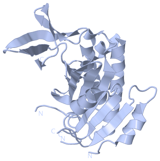
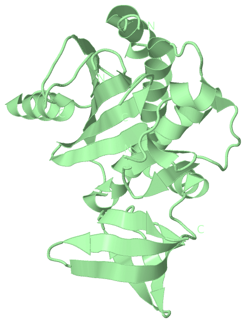
Asymmetric Unit
No.Residues
1Ile A:21 -Gly A:22
2Ala A:23 -Gly A:24
3Trp A:128 -Pro A:129
4Gln A:148 -Pro A:149
5Trp B:128 -Pro B:129
6Gln B:148 -Pro B:149

 Sequence-Structure Mapping

(-) SAPs(SNPs)/Variants  (0, 0)

(no "SAP(SNP)/Variant" information available for 3L2Z)

(-) PROSITE Motifs  (0, 0)

(no "PROSITE Motif" information available for 3L2Z)

(-) Exons   (0, 0)

(no "Exon" information available for 3L2Z)

(-) Sequences/Alignments

Asymmetric Unit
   Reformat: Number of residues per line =  ('0' or empty: single-line sequence representation)
  Number of residues per labelling interval =   
  UniProt sequence: complete  aligned part    
   Show mapping: SCOP domains CATH domains Pfam domains Secondary structure (by author)
SAPs(SNPs) PROSITE motifs Exons
(details for a mapped element are shown in a popup box when the mouse pointer rests over it)
Chain A from PDB  Type:PROTEIN  Length:240
 aligned with P96884_MYCTO | P96884 from UniProtKB/TrEMBL  Length:266

    Alignment length:258
                                    17        27        37        47        57        67        77        87        97       107       117       127       137       147       157       167       177       187       197       207       217       227       237       247       257        
         P96884_MYCTO     8 RPPLDERSLRDQLIGAGSGWRQLDVVAQTGSTNADLLARAASGADIDGVVLIAEHQTAGRGRHGRGWAATARAQIILSVGVRVVDVPVQAWGWLSLAAGLAVLDSVAPLIAVPPAETGLKWPNDVLARGGKLAGILAEVAQPFVVLGVGLNVTQAPEEVDPDATSLLDLGVAAPDRNRIASRLLRELEARIIQWRNANPQLAADYRARSLTIGSRVRVELPGGQDVVGIARDIDDQGRLCLDVGGRTVVVSAGDVVHL 265
               SCOP domains ------------------------------------------------------------------------------------------------------------------------------------------------------------------------------------------------------------------------------------------------------------------ SCOP domains
               CATH domains ------------------------------------------------------------------------------------------------------------------------------------------------------------------------------------------------------------------------------------------------------------------ CATH domains
               Pfam domains ------------------------------------------------------------------------------------------------------------------------------------------------------------------------------------------------------------------------------------------------------------------ Pfam domains
         Sec.struct. author ....hhhhhhhhhh.......eeeee....hhhhhhhhhhhh......eeeee....------------....eeeeeeeee....hhhhhhhhhhhhhhhhhhhhh.....hhh.eeee...eeee..eeeeeeeeeee..eeeeeeeee...------..............hhhhhhhhhhhhhhhhhhhhhh.hhhhhhhhhhh......eeeeee...eeeeeeeeee.....eeeee..eeeee....eee. Sec.struct. author
                 SAPs(SNPs) ------------------------------------------------------------------------------------------------------------------------------------------------------------------------------------------------------------------------------------------------------------------ SAPs(SNPs)
                    PROSITE ------------------------------------------------------------------------------------------------------------------------------------------------------------------------------------------------------------------------------------------------------------------ PROSITE
                 Transcript ------------------------------------------------------------------------------------------------------------------------------------------------------------------------------------------------------------------------------------------------------------------ Transcript
                 3l2z A   8 RPPLDERSLRDQLIGAGSGWRQLDVVAQTGSTNADLLARAASGADIDGVVLIAEHQT------------TARAQIILSVGVRVVDVPVQAWGWLSLAAGLAVLDSVAPLIAVPPAETGLKWPNDVLARGGKLAGILAEVAQPFVVLGVGLNVTQ------PDATSLLDLGVAAPDRNRIASRLLRELEARIIQWRNANPQLAADYRARSLTIGSRVRVELPGGQDVVGIARDIDDQGRLCLDVGGRTVVVSAGDVVHL 265
                                    17        27        37        47        57      |  -        77        87        97       107       117       127       137       147       157   |     -|      177       187       197       207       217       227       237       247       257        
                                                                                   64           77                                                                                 161    168                                                                                                 

Chain B from PDB  Type:PROTEIN  Length:243
 aligned with P96884_MYCTO | P96884 from UniProtKB/TrEMBL  Length:266

    Alignment length:259
                                    17        27        37        47        57        67        77        87        97       107       117       127       137       147       157       167       177       187       197       207       217       227       237       247       257         
         P96884_MYCTO     8 RPPLDERSLRDQLIGAGSGWRQLDVVAQTGSTNADLLARAASGADIDGVVLIAEHQTAGRGRHGRGWAATARAQIILSVGVRVVDVPVQAWGWLSLAAGLAVLDSVAPLIAVPPAETGLKWPNDVLARGGKLAGILAEVAQPFVVLGVGLNVTQAPEEVDPDATSLLDLGVAAPDRNRIASRLLRELEARIIQWRNANPQLAADYRARSLTIGSRVRVELPGGQDVVGIARDIDDQGRLCLDVGGRTVVVSAGDVVHLR 266
               SCOP domains ------------------------------------------------------------------------------------------------------------------------------------------------------------------------------------------------------------------------------------------------------------------- SCOP domains
               CATH domains ------------------------------------------------------------------------------------------------------------------------------------------------------------------------------------------------------------------------------------------------------------------- CATH domains
               Pfam domains ------------------------------------------------------------------------------------------------------------------------------------------------------------------------------------------------------------------------------------------------------------------- Pfam domains
         Sec.struct. author ....hhhhhhhhhh.......eeeee....hhhhhhhhhhhh......eeeee.....-----------....eeeeeeeee....hhhhhhhhhhhhhhhhhhhhhhhh.......eee...eeee..eeeeeeeeeee..eeeeeeeee....-----.....hhhhh....hhhhhhhhhhhhhhhhhhhhhh.hhhhhhhhhhh......eeeeee...eeeeeeeee.......eeee..eeeee...eeee.. Sec.struct. author
                 SAPs(SNPs) ------------------------------------------------------------------------------------------------------------------------------------------------------------------------------------------------------------------------------------------------------------------- SAPs(SNPs)
                    PROSITE ------------------------------------------------------------------------------------------------------------------------------------------------------------------------------------------------------------------------------------------------------------------- PROSITE
                 Transcript ------------------------------------------------------------------------------------------------------------------------------------------------------------------------------------------------------------------------------------------------------------------- Transcript
                 3l2z B   8 RPPLDERSLRDQLIGAGSGWRQLDVVAQTGSTNADLLARAASGADIDGVVLIAEHQTA-----------TARAQIILSVGVRVVDVPVQAWGWLSLAAGLAVLDSVAPLIAVPPAETGLKWPNDVLARGGKLAGILAEVAQPFVVLGVGLNVTQA-----PDATSLLDLGVAAPDRNRIASRLLRELEARIIQWRNANPQLAADYRARSLTIGSRVRVELPGGQDVVGIARDIDDQGRLCLDVGGRTVVVSAGDVVHLR 266
                                    17        27        37        47        57       | -        77        87        97       107       117       127       137       147       157    |    -|      177       187       197       207       217       227       237       247       257         
                                                                                    65          77                                                                                  162   168                                                                                                  

   Legend:   → Mismatch (orange background)
  - → Gap (green background, '-', border residues have a numbering label)
    → Modified Residue (blue background, lower-case, 'x' indicates undefined single-letter code, labelled with number + name)
  x → Chemical Group (purple background, 'x', labelled with number + name, e.g. ACE or NH2)
  extra numbering lines below/above indicate numbering irregularities and modified residue names etc., number ends below/above '|'

 Classification and Annotation

(-) SCOP Domains  (0, 0)

(no "SCOP Domain" information available for 3L2Z)

(-) CATH Domains  (0, 0)

(no "CATH Domain" information available for 3L2Z)

(-) Pfam Domains  (0, 0)

(no "Pfam Domain" information available for 3L2Z)

(-) Gene Ontology  (6, 6)

Asymmetric Unit(hide GO term definitions)
Chain A,B   (P96884_MYCTO | P96884)
molecular function
    GO:0004077    biotin-[acetyl-CoA-carboxylase] ligase activity    Catalysis of the reaction: ATP + biotin + apo-(acetyl-CoA:carbon-dioxide ligase (ADP forming)) = AMP + diphosphate + (acetyl-CoA:carbon-dioxide ligase (ADP forming)).
    GO:0018271    biotin-protein ligase activity    Catalysis of the reaction: ATP + biotin + protein = AMP + diphosphate + biotin-protein.
    GO:0016874    ligase activity    Catalysis of the joining of two substances, or two groups within a single molecule, with the concomitant hydrolysis of the diphosphate bond in ATP or a similar triphosphate.
biological process
    GO:0006768    biotin metabolic process    The chemical reactions and pathways involving biotin, cis-tetrahydro-2-oxothieno(3,4-d)imidazoline-4-valeric acid; the (+) enantiomer is very widely distributed in cells and serves as a carrier in a number of enzymatic beta-carboxylation reactions.
    GO:0006464    cellular protein modification process    The covalent alteration of one or more amino acids occurring in proteins, peptides and nascent polypeptides (co-translational, post-translational modifications) occurring at the level of an individual cell. Includes the modification of charged tRNAs that are destined to occur in a protein (pre-translation modification).
    GO:0009305    protein biotinylation    The addition of biotin (vitamin B7 / vitamin H) to a protein amino acid.

 Visualization

(-) Interactive Views

Asymmetric Unit
  Complete Structure
    Jena3D(integrated viewing of ligand, site, SAP, PROSITE, SCOP information)
    WebMol | AstexViewer[tm]@PDBe
(Java Applets, require no local installation except for Java; loading may be slow)
    STRAP
(Java WebStart application, automatic local installation, requires Java; full application with system access!)
    RasMol
(require local installation)
    Molscript (VRML)
(requires installation of a VRML viewer; select preferred view via VRML and generate a mono or stereo PDF format file)
 
  Ligands, Modified Residues, Ions
(no "Ligands, Modified Residues, Ions" information available for 3l2z)
 
  Sites
(no "Sites" information available for 3l2z)
 
  Cis Peptide Bonds
    Ala A:23 - Gly A:24   [ RasMol ]  
    Gln A:148 - Pro A:149   [ RasMol ]  
    Gln B:148 - Pro B:149   [ RasMol ]  
    Ile A:21 - Gly A:22   [ RasMol ]  
    Trp A:128 - Pro A:129   [ RasMol ]  
    Trp B:128 - Pro B:129   [ RasMol ]  
 
Biological Units
  Complete Structure
    Biological Unit 1  [ Jena3D ]
    Biological Unit 2  [ Jena3D ]

(-) Still Images

Jmol
  protein: cartoon or spacefill or dots and stick; nucleic acid: cartoon and stick; ligands: spacefill; active site: stick
Molscript
  protein, nucleic acid: cartoon; ligands: spacefill; active site: ball and stick

 Databases and Analysis Tools

(-) Databases

Access by PDB/NDB ID
  3l2z
    Family and Domain InformationProDom | SYSTERS
    General Structural InformationGlycoscienceDB | MMDB | NDB | OCA | PDB | PDBe | PDBj | PDBsum | PDBWiki | PQS | PROTEOPEDIA
    Orientation in MembranesOPM
    Protein SurfaceSURFACE
    Secondary StructureDSSP (structure derived) | HSSP (homology derived)
    Structural GenomicsGeneCensus
    Structural NeighboursCE | VAST
    Structure ClassificationCATH | Dali | SCOP
    Validation and Original DataBMRB Data View | BMRB Restraints Grid | EDS | PROCHECK | RECOORD | WHAT_CHECK
 
Access by UniProt ID/Accession number
  P96884_MYCTO | P96884
    Comparative Protein Structure ModelsModBase
    Genomic InformationEnsembl
    Protein-protein InteractionDIP
    Sequence, Family and Domain InformationInterPro | Pfam | SMART | UniProtKB/TrEMBL
 
Access by Enzyme Classificator   (EC Number)
  6.3.4.15
    General Enzyme InformationBRENDA | EC-PDB | Enzyme | IntEnz
    PathwayKEGG | MetaCyc
 
Access by Disease Identifier   (MIM ID)
  (no 'MIM ID' available)
    Disease InformationOMIM
 
Access by GenAge ID
  (no 'GenAge ID' available)
    Age Related InformationGenAge

(-) Analysis Tools

Access by PDB/NDB ID
    Domain InformationXDom
    Interatomic Contacts of Structural UnitsCSU
    Ligand-protein ContactsLPC
    Protein CavitiescastP
    Sequence and Secondary StructurePDBCartoon
    Structure AlignmentSTRAP(Java WebStart application, automatic local installation, requires Java; full application with system access!)
    Structure and Sequence BrowserSTING
 
Access by UniProt ID/Accession number
  P96884_MYCTO | P96884
    Protein Disorder PredictionDisEMBL | FoldIndex | GLOBPLOT (for more information see DisProt)

 Related Entries

(-) Entries Sharing at Least One Protein Chain (UniProt ID)

UniProtKB/Swiss-Prot
        P96884_MYCTO | P968844op0
UniProtKB/TrEMBL
        P96884_MYCTO | P968842cgh 3l1a 3rux

(-) Related Entries Specified in the PDB File

3l1a DEHYDRATED STRUCTURE