|
|
|
|
 Description
|
|
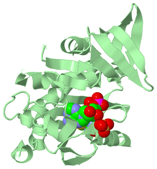
Compounds
|
||||||||||||||||||||||||||||||||||||||||||||||||||||||||
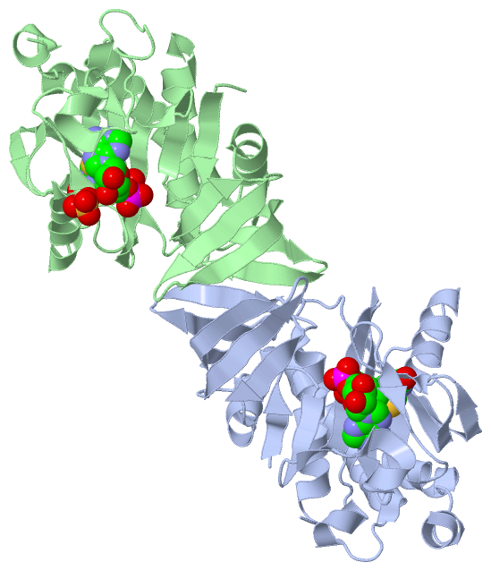
Chains, Units
Summary Information (see also Sequences/Alignments below) |
Ligands, Modified Residues, Ions (2, 3)| Asymmetric Unit (2, 3) Biological Unit 1 (1, 1) Biological Unit 2 (2, 2) |
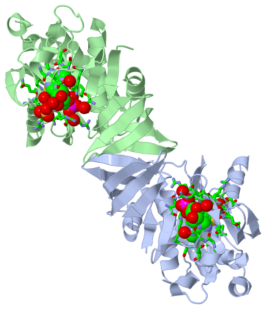
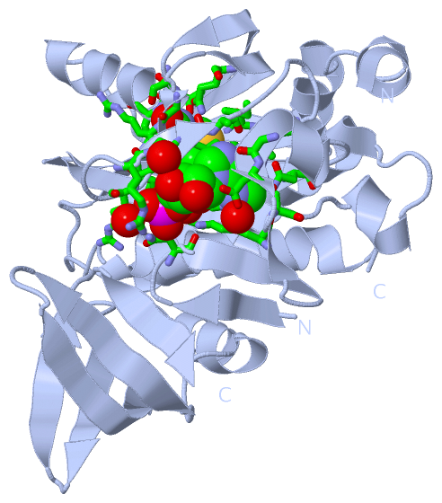
Sites (3, 3)
Asymmetric Unit (3, 3)
|
SS Bonds (0, 0)| (no "SS Bond" information available for 4OP0) |
Cis Peptide Bonds (7, 7)
Asymmetric Unit
|
||||||||||||||||||||||||||||||||
SAPs(SNPs)/Variants (0, 0)| (no "SAP(SNP)/Variant" information available for 4OP0) |
PROSITE Motifs (0, 0)| (no "PROSITE Motif" information available for 4OP0) |
Exons (0, 0)| (no "Exon" information available for 4OP0) |
Sequences/Alignments
Asymmetric Unit
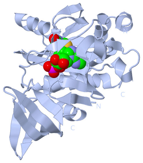
Chain A from PDB Type:PROTEIN Length:259
SCOP domains ------------------------------------------------------------------------------------------------------------------------------------------------------------------------------------------------------------------------------------------------------------------- SCOP domains
CATH domains ------------------------------------------------------------------------------------------------------------------------------------------------------------------------------------------------------------------------------------------------------------------- CATH domains
Pfam domains ------------------------------------------------------------------------------------------------------------------------------------------------------------------------------------------------------------------------------------------------------------------- Pfam domains
SAPs(SNPs) ------------------------------------------------------------------------------------------------------------------------------------------------------------------------------------------------------------------------------------------------------------------- SAPs(SNPs)
PROSITE ------------------------------------------------------------------------------------------------------------------------------------------------------------------------------------------------------------------------------------------------------------------- PROSITE
Transcript ------------------------------------------------------------------------------------------------------------------------------------------------------------------------------------------------------------------------------------------------------------------- Transcript
4op0 A 1 MADRDRLRPPLDERSLRDQLIGAGSGWRQLDVVAQTGSTNADLLARAASGADIDGVVLIAEHQTAGRGRHGRGWAATARAQIILSVGVRVVDVPVQAWGWLSLAAGLAVLDSVAPLITGLKWPNDVLARGGKLAGILAEVAQPFVVLGVGLNVTQAPEEVDPDATSLLDLGVAAPDRNRIASRLLRELEARIIQWRNANPQLAADYRARSLTIGSRVRVELPGGQDVVGIARDIDDQGRLCLDVGGRTVVVSAGDVVHL 265
10 20 30 40 50 60 70 80 90 100 110 |126 136 146 156 166 176 186 196 206 216 226 236 246 256
117|
124
Chain B from PDB Type:PROTEIN Length:257
SCOP domains ----------------------------------------------------------------------------------------------------------------------------------------------------------------------------------------------------------------------------------------------------------------- SCOP domains
CATH domains ----------------------------------------------------------------------------------------------------------------------------------------------------------------------------------------------------------------------------------------------------------------- CATH domains
Pfam domains ----------------------------------------------------------------------------------------------------------------------------------------------------------------------------------------------------------------------------------------------------------------- Pfam domains
SAPs(SNPs) ----------------------------------------------------------------------------------------------------------------------------------------------------------------------------------------------------------------------------------------------------------------- SAPs(SNPs)
PROSITE ----------------------------------------------------------------------------------------------------------------------------------------------------------------------------------------------------------------------------------------------------------------- PROSITE
Transcript ----------------------------------------------------------------------------------------------------------------------------------------------------------------------------------------------------------------------------------------------------------------- Transcript
4op0 B 3 DRDRLRPPLDERSLRDQLIGSGWRQLDVVAQTGSTNADLLARAASGADIDGVVLIAEHQTAGRGRHGRGWAATARAQIILSVGVRVVDVPVQAWGWLSLAAGLAVLDSVAPLIAETGLKWPNDVLARGGKLAGILAEVAQPFVVLGVGLNVTQAPEEVDPDATSLLDLGVAAPDRNRIASRLLRELEARIIQWRNANPQLAADYRARSLTIGSRVRVELPGGQDVVGIARDIDDQGRLCLDVGGRTVVVSAGDVVHL 265
12 24 34 44 54 64 74 84 94 104 114 || 128 138 148 158 168 178 188 198 208 218 228 238 248 258
21| 117|
24 122
|
||||||||||||||||||||
SCOP Domains (0, 0)| (no "SCOP Domain" information available for 4OP0) |
CATH Domains (0, 0)| (no "CATH Domain" information available for 4OP0) |
Pfam Domains (0, 0)| (no "Pfam Domain" information available for 4OP0) |
Gene Ontology (6, 9)|
Asymmetric Unit(hide GO term definitions) |
Interactive Views
|
|||||||||||||||||||||||||||||||||||||||||||||||||||||||||||||||||||||||||||||||||||||||||||||||||||||||||||||||||||||||||||||||||||||||||||||||||||||||||||||||||||||||||||||||||||||||||||||||||||||||||||||
Still Images
|
||||||||||||||||
Databases
|
||||||||||||||||||||||||||||||||||||||||||||||||||||||||||||||||||||||||||||||||||||||||||||||||||||||||||||||||||||||||||||||||||||||||||||||||||||||||||||||||||||||||||||||||||||||||||
Analysis Tools
|
||||||||||||||||||||||||||||||||||||||||||||||||||||||||||||||||||||||||
Entries Sharing at Least One Protein Chain (UniProt ID)
Related Entries Specified in the PDB File
|
|