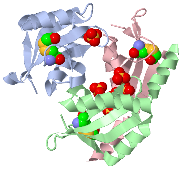
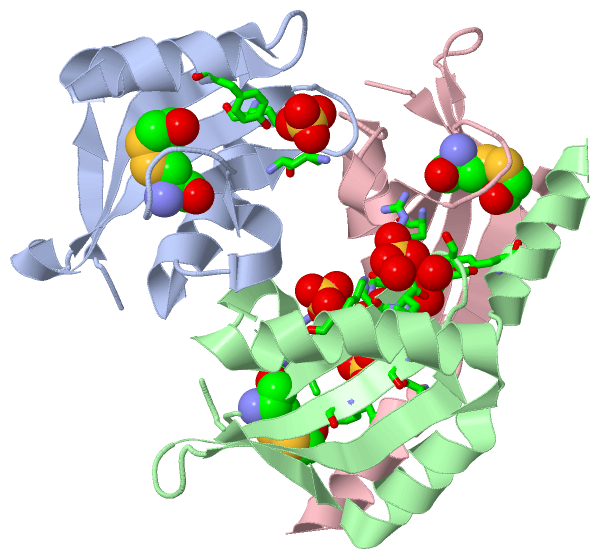
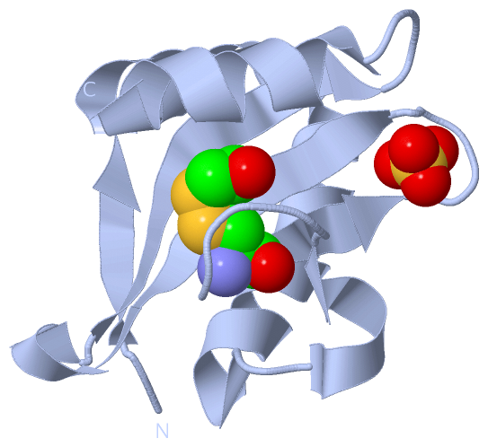
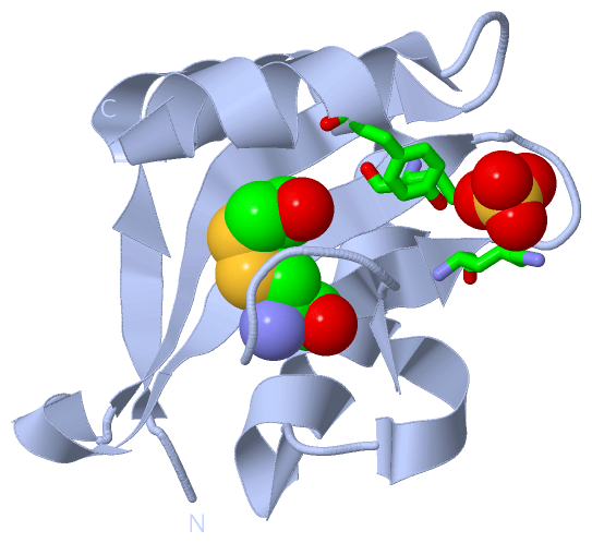
Show PDB file:   
         Plain Text   HTML   (compressed file size)
QuickSearch:   
by PDB,NDB,UniProt,PROSITE Code or Search Term(s)  
(-)Asymmetric Unit
(-)Asym. Unit - sites
(-)Biological Unit 1
(-)Biol. Unit 1 - sites
(-)Biological Unit 2
(-)Biological Unit 3
(-)Biological Unit 4
collapse expand < >
Image Asymmetric Unit
Asymmetric Unit  (Jmol Viewer)
Image Asym. Unit - sites
Asym. Unit - sites  (Jmol Viewer)
Image Biological Unit 1
Biological Unit 1  (Jmol Viewer)
Image Biol. Unit 1 - sites
Biol. Unit 1 - sites  (Jmol Viewer)
Image Biological Unit 2
Biological Unit 2  (Jmol Viewer)
Image Biological Unit 3
Biological Unit 3  (Jmol Viewer)
Image Biological Unit 4
Biological Unit 4  (Jmol Viewer)

(-) Description

Title :  CRYSTAL STRUCTURE OF THE ISOLATED E. COLI RELE TOXIN, P21 FORM
 
Authors :  D. E. Brodersen, A. Boggild, N. Sofos
Date :  03 Jul 12  (Deposition) - 29 Aug 12  (Release) - 26 Dec 12  (Revision)
Method :  X-RAY DIFFRACTION
Resolution :  1.80
Chains :  Asym. Unit :  A,B,C
Biol. Unit 1:  A  (1x)
Biol. Unit 2:  B  (1x)
Biol. Unit 3:  C  (1x)
Biol. Unit 4:  B,C  (1x)
Keywords :  Toxin/Antitoxin System, Toxin, Nuclease, Translational Control, Stress Response, Relb, Ribosome, B-Me On Cys50 (Keyword Search: [Gene Ontology, PubMed, Web (Google))
 
Reference :  A. Boggild, N. Sofos, K. R. Andersen, A. Feddersen, A. D. Easter, L. A. Passmore, D. E. Brodersen
The Crystal Structure Of The Intact E. Coli Relbe Toxin-Antitoxin Complex Provides The Structural Basis For Conditional Cooperativity.
Structure V. 20 1641 2012
PubMed-ID: 22981948  |  Reference-DOI: 10.1016/J.STR.2012.08.017
(for further references see the PDB file header)

(-) Compounds

Molecule 1 - MRNA INTERFERASE RELE
    ChainsA, B, C
    EC Number3.1.-.-
    EngineeredYES
    Expression SystemESCHERICHIA COLI
    Expression System PlasmidPSC2524HE
    Expression System StrainBL21(DE3)
    Expression System Taxid469008
    Expression System Vector TypePLASMID
    FragmentRELE
    GeneB1563, JW1555, RELE
    MutationYES
    Organism ScientificESCHERICHIA COLI
    Organism Taxid83333
    StrainK12
    SynonymENDORIBONUCLEASE RELE, TOXIN RELE

 Structural Features

(-) Chains, Units

  123
Asymmetric Unit ABC
Biological Unit 1 (1x)A  
Biological Unit 2 (1x) B 
Biological Unit 3 (1x)  C
Biological Unit 4 (1x) BC

Summary Information (see also Sequences/Alignments below)

(-) Ligands, Modified Residues, Ions  (2, 9)

Asymmetric Unit (2, 9)
No.NameCountTypeFull Name
1CME3Mod. Amino AcidS,S-(2-HYDROXYETHYL)THIOCYSTEINE
2SO46Ligand/IonSULFATE ION
Biological Unit 1 (2, 2)
No.NameCountTypeFull Name
1CME1Mod. Amino AcidS,S-(2-HYDROXYETHYL)THIOCYSTEINE
2SO41Ligand/IonSULFATE ION
Biological Unit 2 (2, 4)
No.NameCountTypeFull Name
1CME1Mod. Amino AcidS,S-(2-HYDROXYETHYL)THIOCYSTEINE
2SO43Ligand/IonSULFATE ION
Biological Unit 3 (2, 3)
No.NameCountTypeFull Name
1CME1Mod. Amino AcidS,S-(2-HYDROXYETHYL)THIOCYSTEINE
2SO42Ligand/IonSULFATE ION
Biological Unit 4 (2, 7)
No.NameCountTypeFull Name
1CME2Mod. Amino AcidS,S-(2-HYDROXYETHYL)THIOCYSTEINE
2SO45Ligand/IonSULFATE ION

(-) Sites  (6, 6)

Asymmetric Unit (6, 6)
No.NameEvidenceResiduesDescription
1AC1SOFTWARELYS A:54 , ARG A:61 , TYR A:87BINDING SITE FOR RESIDUE SO4 A 101
2AC2SOFTWAREARG B:45 , LYS C:54BINDING SITE FOR RESIDUE SO4 B 101
3AC3SOFTWARELYS B:54 , ARG B:61 , SO4 C:101 , SO4 C:102BINDING SITE FOR RESIDUE SO4 B 102
4AC4SOFTWARELYS B:29 , LYS B:54 , LEU B:55 , ARG B:56 , SER B:57 , HOH B:261 , HOH B:271BINDING SITE FOR RESIDUE SO4 B 103
5AC5SOFTWARETYR B:87 , SO4 B:102 , LYS C:52 , LYS C:54 , ARG C:61BINDING SITE FOR RESIDUE SO4 C 101
6AC6SOFTWARELEU B:44 , ARG B:61 , VAL B:63 , SO4 B:102 , SER C:84 , TYR C:87BINDING SITE FOR RESIDUE SO4 C 102

(-) SS Bonds  (0, 0)

(no "SS Bond" information available for 4FXI)

(-) Cis Peptide Bonds  (1, 1)

Asymmetric Unit
No.Residues
1Ser A:58 -Gly A:59

 Sequence-Structure Mapping

(-) SAPs(SNPs)/Variants  (0, 0)

(no "SAP(SNP)/Variant" information available for 4FXI)

(-) PROSITE Motifs  (0, 0)

(no "PROSITE Motif" information available for 4FXI)

(-) Exons   (0, 0)

(no "Exon" information available for 4FXI)

(-) Sequences/Alignments

Asymmetric Unit
   Reformat: Number of residues per line =  ('0' or empty: single-line sequence representation)
  Number of residues per labelling interval =   
  UniProt sequence: complete  aligned part    
   Show mapping: SCOP domains CATH domains Pfam domains Secondary structure (by author)
SAPs(SNPs) PROSITE motifs Exons
(details for a mapped element are shown in a popup box when the mouse pointer rests over it)
Chain A from PDB  Type:PROTEIN  Length:94
                                                                                                                             
               SCOP domains ---------------------------------------------------------------------------------------------- SCOP domains
               CATH domains ---------------------------------------------------------------------------------------------- CATH domains
               Pfam domains ---------------------------------------------------------------------------------------------- Pfam domains
         Sec.struct. author ..eeeehhhhhhhhhhhhhhhhhhhhhhhhhhhhh..hhhhh......eeeee.....eeeeeeeehhh.eeeeeeeee...hhhhhhhhhhhh Sec.struct. author
                 SAPs(SNPs) ---------------------------------------------------------------------------------------------- SAPs(SNPs)
                    PROSITE ---------------------------------------------------------------------------------------------- PROSITE
                 Transcript ---------------------------------------------------------------------------------------------- Transcript
                  4fxi A  2 AYFLDFDERALKEWRKLGSTVREQLKKKLVEVLESPRIEANKLRGMPDcYKIKLRSSGYRLVYQVIDEKVVVFVISVGKAERSEVYSEAVKRIL 95
                                    11        21        31        41        51        61        71        81        91    
                                                                           50-CME                                         

Chain B from PDB  Type:PROTEIN  Length:94
                                                                                                                             
               SCOP domains ---------------------------------------------------------------------------------------------- SCOP domains
               CATH domains ---------------------------------------------------------------------------------------------- CATH domains
               Pfam domains ---------------------------------------------------------------------------------------------- Pfam domains
         Sec.struct. author ..eeeehhhhhhhhhhhhhhhhhhhhhhhhhhhhh..hhhhh......eeeeee....eeeeeeee....eeeeeeeee...hhhhhhhhhhhh Sec.struct. author
                 SAPs(SNPs) ---------------------------------------------------------------------------------------------- SAPs(SNPs)
                    PROSITE ---------------------------------------------------------------------------------------------- PROSITE
                 Transcript ---------------------------------------------------------------------------------------------- Transcript
                  4fxi B  2 AYFLDFDERALKEWRKLGSTVREQLKKKLVEVLESPRIEANKLRGMPDcYKIKLRSSGYRLVYQVIDEKVVVFVISVGKAERSEVYSEAVKRIL 95
                                    11        21        31        41        51        61        71        81        91    
                                                                           50-CME                                         

Chain C from PDB  Type:PROTEIN  Length:94
                                                                                                                             
               SCOP domains ---------------------------------------------------------------------------------------------- SCOP domains
               CATH domains ---------------------------------------------------------------------------------------------- CATH domains
               Pfam domains ---------------------------------------------------------------------------------------------- Pfam domains
         Sec.struct. author ..eeeehhhhhhhhhhhhhhhhhhhhhhhhhhh....hhhhh......eeeeee....eeeeeeee....eeeeeeeee...hhhhhhhhhhhh Sec.struct. author
                 SAPs(SNPs) ---------------------------------------------------------------------------------------------- SAPs(SNPs)
                    PROSITE ---------------------------------------------------------------------------------------------- PROSITE
                 Transcript ---------------------------------------------------------------------------------------------- Transcript
                  4fxi C  2 AYFLDFDERALKEWRKLGSTVREQLKKKLVEVLESPRIEANKLRGMPDcYKIKLRSSGYRLVYQVIDEKVVVFVISVGKAERSEVYSEAVKRIL 95
                                    11        21        31        41        51        61        71        81        91    
                                                                           50-CME                                         

   Legend:   → Mismatch (orange background)
  - → Gap (green background, '-', border residues have a numbering label)
    → Modified Residue (blue background, lower-case, 'x' indicates undefined single-letter code, labelled with number + name)
  x → Chemical Group (purple background, 'x', labelled with number + name, e.g. ACE or NH2)
  extra numbering lines below/above indicate numbering irregularities and modified residue names etc., number ends below/above '|'

 Classification and Annotation

(-) SCOP Domains  (0, 0)

(no "SCOP Domain" information available for 4FXI)

(-) CATH Domains  (0, 0)

(no "CATH Domain" information available for 4FXI)

(-) Pfam Domains  (0, 0)

(no "Pfam Domain" information available for 4FXI)

(-) Gene Ontology  (16, 16)

Asymmetric Unit(hide GO term definitions)

 Visualization

(-) Interactive Views

Asymmetric Unit
  Complete Structure
    Jena3D(integrated viewing of ligand, site, SAP, PROSITE, SCOP information)
    WebMol | AstexViewer[tm]@PDBe
(Java Applets, require no local installation except for Java; loading may be slow)
    STRAP
(Java WebStart application, automatic local installation, requires Java; full application with system access!)
    RasMol
(require local installation)
    Molscript (VRML)
(requires installation of a VRML viewer; select preferred view via VRML and generate a mono or stereo PDF format file)
 
  Ligands, Modified Residues, Ions
    CME  [ RasMol | Jena3D ]  +environment [ RasMol | Jena3D ]
    SO4  [ RasMol | Jena3D ]  +environment [ RasMol | Jena3D ]
 
  Sites
    AC1  [ RasMol ]  +environment [ RasMol ]
    AC2  [ RasMol ]  +environment [ RasMol ]
    AC3  [ RasMol ]  +environment [ RasMol ]
    AC4  [ RasMol ]  +environment [ RasMol ]
    AC5  [ RasMol ]  +environment [ RasMol ]
    AC6  [ RasMol ]  +environment [ RasMol ]
 
  Cis Peptide Bonds
    Ser A:58 - Gly A:59   [ RasMol ]  
 
Biological Units
  Complete Structure
    Biological Unit 1  [ Jena3D ]
    Biological Unit 2  [ Jena3D ]
    Biological Unit 3  [ Jena3D ]
    Biological Unit 4  [ Jena3D ]

(-) Still Images

Jmol
  protein: cartoon or spacefill or dots and stick; nucleic acid: cartoon and stick; ligands: spacefill; active site: stick
Molscript
  protein, nucleic acid: cartoon; ligands: spacefill; active site: ball and stick

 Databases and Analysis Tools

(-) Databases

Access by PDB/NDB ID
  4fxi
    Family and Domain InformationProDom | SYSTERS
    General Structural InformationGlycoscienceDB | MMDB | NDB | OCA | PDB | PDBe | PDBj | PDBsum | PDBWiki | PQS | PROTEOPEDIA
    Orientation in MembranesOPM
    Protein SurfaceSURFACE
    Secondary StructureDSSP (structure derived) | HSSP (homology derived)
    Structural GenomicsGeneCensus
    Structural NeighboursCE | VAST
    Structure ClassificationCATH | Dali | SCOP
    Validation and Original DataBMRB Data View | BMRB Restraints Grid | EDS | PROCHECK | RECOORD | WHAT_CHECK
 
Access by UniProt ID/Accession number
  RELE_ECOLI | P0C077
    Comparative Protein Structure ModelsModBase
    Genomic InformationEnsembl
    Protein-protein InteractionDIP
    Sequence, Family and Domain InformationInterPro | Pfam | SMART | UniProtKB/SwissProt
 
Access by Enzyme Classificator   (EC Number)
  3.1.-.-
    General Enzyme InformationBRENDA | EC-PDB | Enzyme | IntEnz
    PathwayKEGG | MetaCyc
 
Access by Disease Identifier   (MIM ID)
  (no 'MIM ID' available)
    Disease InformationOMIM
 
Access by GenAge ID
  (no 'GenAge ID' available)
    Age Related InformationGenAge

(-) Analysis Tools

Access by PDB/NDB ID
    Domain InformationXDom
    Interatomic Contacts of Structural UnitsCSU
    Ligand-protein ContactsLPC
    Protein CavitiescastP
    Sequence and Secondary StructurePDBCartoon
    Structure AlignmentSTRAP(Java WebStart application, automatic local installation, requires Java; full application with system access!)
    Structure and Sequence BrowserSTING
 
Access by UniProt ID/Accession number
  RELE_ECOLI | P0C077
    Protein Disorder PredictionDisEMBL | FoldIndex | GLOBPLOT (for more information see DisProt)

 Related Entries

(-) Entries Sharing at Least One Protein Chain (UniProt ID)

UniProtKB/Swiss-Prot
        RELE_ECOLI | P0C0772kc8 2kc9 4fxe 4fxh 4v7j 4v7k

(-) Related Entries Specified in the PDB File

2kc8 NMR STRUCTURE OF RELE MONOMER
2kc9 NMR STRUCTURE OF RELE BOUND TO A FRAGMENT OF RELB
3kha LOWER RESOLUTION CRYSTAL STRUCTURE OF THE SAME CRYSTAL FORM OF RELE.