Show PDB file:   
         Plain Text   HTML   (compressed file size)
QuickSearch:   
by PDB,NDB,UniProt,PROSITE Code or Search Term(s)  
(-)Asymmetric Unit
(-)Asym. Unit - sites
(-)Biological Unit 1
(-)Biol. Unit 1 - sites
(-)Biological Unit 2
collapse expand < >
Image Asymmetric Unit
Asymmetric Unit  (Jmol Viewer)
Image Asym. Unit - sites
Asym. Unit - sites  (Jmol Viewer)
Image Biological Unit 1
Biological Unit 1  (Jmol Viewer)
Image Biol. Unit 1 - sites
Biol. Unit 1 - sites  (Jmol Viewer)
Image Biological Unit 2
Biological Unit 2  (Jmol Viewer)

(-) Description

Title :  CRYSTAL STRUCTURE OF THE C-TERMINAL DOMAIN OF YPBH
 
Authors :  F. Wang, Z. Mei, Y. Qi, C. Yan, J. Wang, Y. Shi
Date :  14 Sep 09  (Deposition) - 29 Sep 09  (Release) - 29 Sep 09  (Revision)
Method :  X-RAY DIFFRACTION
Resolution :  2.09
Chains :  Asym. Unit :  A,B
Biol. Unit 1:  A  (1x)
Biol. Unit 2:  B  (1x)
Keywords :  Ypbh, Adaptor Protein, Competence, Sporulation, Protein Binding (Keyword Search: [Gene Ontology, PubMed, Web (Google))
 
Reference :  F. Wang, Z. Mei, Y. Qi, C. Yan, S. Xiang, Z. Zhou, Q. Hu, J. Wang, Y. Shi
Crystal Structure Of The Meca Degradation Tag
To Be Published
PubMed: search
(for further references see the PDB file header)

(-) Compounds

Molecule 1 - ADAPTER PROTEIN MECA 2
    ChainsA, B
    EngineeredYES
    Expression SystemESCHERICHIA COLI
    Expression System PlasmidPGEX-6P-1
    Expression System StrainBL21(DE3)
    Expression System Taxid562
    Expression System Vector TypePLASMID
    FragmentC-TERMINAL DOMAIN, UNP RESIDUES 104-194
    GeneYPBH
    Organism ScientificBACILLUS SUBTILIS
    Organism Taxid1423
    SynonymYPBH

 Structural Features

(-) Chains, Units

  12
Asymmetric Unit AB
Biological Unit 1 (1x)A 
Biological Unit 2 (1x) B

Summary Information (see also Sequences/Alignments below)

(-) Ligands, Modified Residues, Ions  (1, 14)

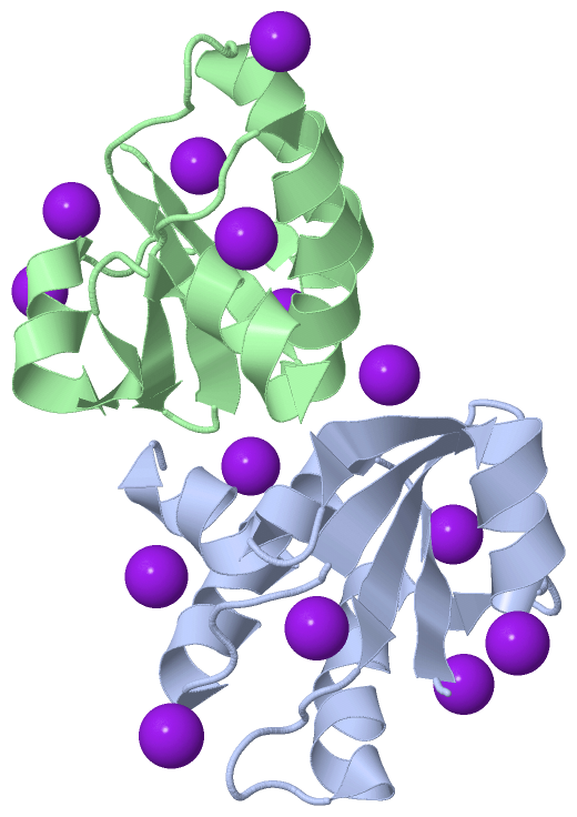
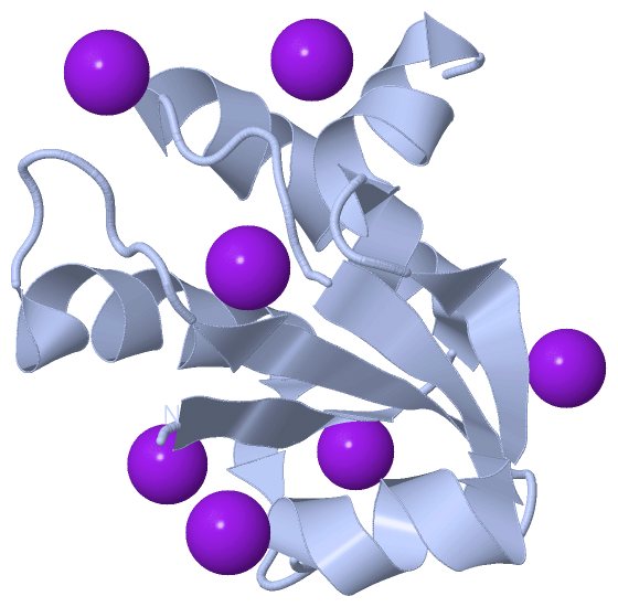
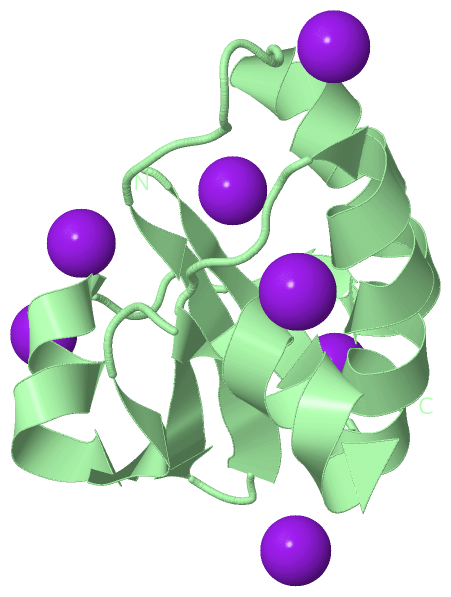
Asymmetric Unit (1, 14)
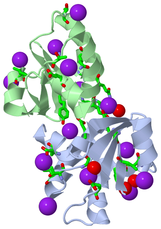
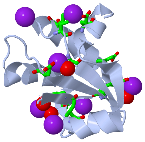
No.NameCountTypeFull Name
1IOD14Ligand/IonIODIDE ION
Biological Unit 1 (1, 7)
No.NameCountTypeFull Name
1IOD7Ligand/IonIODIDE ION
Biological Unit 2 (1, 7)
No.NameCountTypeFull Name
1IOD7Ligand/IonIODIDE ION

(-) Sites  (12, 12)

Asymmetric Unit (12, 12)
No.NameEvidenceResiduesDescription
01AC1SOFTWARETYR A:5 , HOH A:118 , HOH A:122BINDING SITE FOR RESIDUE IOD A 92
02AC2SOFTWAREILE A:67BINDING SITE FOR RESIDUE IOD A 93
03AC3SOFTWAREGLN A:21 , THR A:26 , GLU A:84BINDING SITE FOR RESIDUE IOD A 94
04AC4SOFTWAREGLY A:28 , GLU A:42 , HOH A:125BINDING SITE FOR RESIDUE IOD A 95
05AC5SOFTWAREHIS A:8 , ASN A:61BINDING SITE FOR RESIDUE IOD A 97
06AC6SOFTWARETYR A:37 , HOH A:129 , GLU B:11 , GLN B:15BINDING SITE FOR RESIDUE IOD A 98
07AC7SOFTWARETHR B:66BINDING SITE FOR RESIDUE IOD B 92
08AC8SOFTWAREGLU A:11 , TYR B:37BINDING SITE FOR RESIDUE IOD B 93
09AC9SOFTWAREILE B:67BINDING SITE FOR RESIDUE IOD B 94
10BC1SOFTWAREHIS B:8 , ASN B:61BINDING SITE FOR RESIDUE IOD B 95
11BC2SOFTWAREGLN B:21 , THR B:26 , GLU B:84BINDING SITE FOR RESIDUE IOD B 96
12BC3SOFTWARETHR B:48BINDING SITE FOR RESIDUE IOD B 97

(-) SS Bonds  (0, 0)

(no "SS Bond" information available for 3JTN)

(-) Cis Peptide Bonds  (0, 0)

(no "Cis Peptide Bond" information available for 3JTN)

 Sequence-Structure Mapping

(-) SAPs(SNPs)/Variants  (0, 0)

(no "SAP(SNP)/Variant" information available for 3JTN)

(-) PROSITE Motifs  (0, 0)

(no "PROSITE Motif" information available for 3JTN)

(-) Exons   (0, 0)

(no "Exon" information available for 3JTN)

(-) Sequences/Alignments

Asymmetric Unit
   Reformat: Number of residues per line =  ('0' or empty: single-line sequence representation)
  Number of residues per labelling interval =   
  UniProt sequence: complete  aligned part    
   Show mapping: SCOP domains CATH domains Pfam domains Secondary structure (by author)
SAPs(SNPs) PROSITE motifs Exons
(details for a mapped element are shown in a popup box when the mouse pointer rests over it)
Chain A from PDB  Type:PROTEIN  Length:89
 aligned with MECA2_BACSU | P50734 from UniProtKB/Swiss-Prot  Length:194

    Alignment length:89
                                   114       124       134       144       154       164       174       184         
          MECA2_BACSU   105 DIIYQFHSFEDIIQLSESLQRIGITGGTVYHYDGQYFLSLEDLGSHTAEGVVAVLAEYGNPTTLTIYRLQEYGKLIMDGNAVETIQTHF 193
               SCOP domains ----------------------------------------------------------------------------------------- SCOP domains
               CATH domains ----------------------------------------------------------------------------------------- CATH domains
               Pfam domains ----------------------------------------------------------------------------------------- Pfam domains
         Sec.struct. author .eeeee.hhhhhhhhhhhhhhh.....eeeee..eeeeee......hhhhhhhhhhhhhee...hhhhhhhhheeee..hhhhhhhh.. Sec.struct. author
                 SAPs(SNPs) ----------------------------------------------------------------------------------------- SAPs(SNPs)
                    PROSITE ----------------------------------------------------------------------------------------- PROSITE
                 Transcript ----------------------------------------------------------------------------------------- Transcript
                 3jtn A   2 DIIYQFHSFEDIIQLSESLQRIGITGGTVYHYDGQYFLSLEDLGSHTAEGVVAVLAEYGNPTTLTIYRLQEYGKLIMDGNAVETIQTHF  90
                                    11        21        31        41        51        61        71        81         

Chain B from PDB  Type:PROTEIN  Length:90
 aligned with MECA2_BACSU | P50734 from UniProtKB/Swiss-Prot  Length:194

    Alignment length:90
                                   114       124       134       144       154       164       174       184       194
          MECA2_BACSU   105 DIIYQFHSFEDIIQLSESLQRIGITGGTVYHYDGQYFLSLEDLGSHTAEGVVAVLAEYGNPTTLTIYRLQEYGKLIMDGNAVETIQTHFS 194
               SCOP domains ------------------------------------------------------------------------------------------ SCOP domains
               CATH domains ------------------------------------------------------------------------------------------ CATH domains
           Pfam domains (1) MecA-3jtnB01 B:2-88                                                                    --- Pfam domains (1)
           Pfam domains (2) MecA-3jtnB02 B:2-88                                                                    --- Pfam domains (2)
         Sec.struct. author .eeeee.hhhhhhhhhhhhhhh.....eeeee..eeeeee......hhhhhhhhhhhhhee...hhhhhhhhheeee..hhhhhhhhhh. Sec.struct. author
                 SAPs(SNPs) ------------------------------------------------------------------------------------------ SAPs(SNPs)
                    PROSITE ------------------------------------------------------------------------------------------ PROSITE
                 Transcript ------------------------------------------------------------------------------------------ Transcript
                 3jtn B   2 DIIYQFHSFEDIIQLSESLQRIGITGGTVYHYDGQYFLSLEDLGSHTAEGVVAVLAEYGNPTTLTIYRLQEYGKLIMDGNAVETIQTHFS  91
                                    11        21        31        41        51        61        71        81        91

   Legend:   → Mismatch (orange background)
  - → Gap (green background, '-', border residues have a numbering label)
    → Modified Residue (blue background, lower-case, 'x' indicates undefined single-letter code, labelled with number + name)
  x → Chemical Group (purple background, 'x', labelled with number + name, e.g. ACE or NH2)
  extra numbering lines below/above indicate numbering irregularities and modified residue names etc., number ends below/above '|'

 Classification and Annotation

(-) SCOP Domains  (0, 0)

(no "SCOP Domain" information available for 3JTN)

(-) CATH Domains  (0, 0)

(no "CATH Domain" information available for 3JTN)

(-) Pfam Domains  (1, 2)

Asymmetric Unit

(-) Gene Ontology  (5, 5)

Asymmetric Unit(hide GO term definitions)
Chain A,B   (MECA2_BACSU | P50734)
molecular function
    GO:0030674    protein binding, bridging    The binding activity of a molecule that brings together two or more protein molecules, or a protein and another macromolecule or complex, through a selective, non-covalent, often stoichiometric interaction, permitting those molecules to function in a coordinated way.
biological process
    GO:0030420    establishment of competence for transformation    The process in which a naturally transformable bacterium acquires the ability to take up exogenous DNA. This term should be applied only to naturally transformable bacteria, and should not be used in the context of artificially induced bacterial transformation.
    GO:0045808    negative regulation of establishment of competence for transformation    Any process that stops, prevents, or reduces the frequency, rate or extent of establishment of competence for transformation.
    GO:0042174    negative regulation of sporulation resulting in formation of a cellular spore    Any process that stops, prevents, or reduces the frequency, rate or extent of sporulation.
    GO:0030435    sporulation resulting in formation of a cellular spore    The process in which a relatively unspecialized cell acquires the specialized features of a cellular spore, a cell form that can be used for dissemination, for survival of adverse conditions because of its heat and dessication resistance, and/or for reproduction.

 Visualization

(-) Interactive Views

Asymmetric Unit
  Complete Structure
    Jena3D(integrated viewing of ligand, site, SAP, PROSITE, SCOP information)
    WebMol | AstexViewer[tm]@PDBe
(Java Applets, require no local installation except for Java; loading may be slow)
    STRAP
(Java WebStart application, automatic local installation, requires Java; full application with system access!)
    RasMol
(require local installation)
    Molscript (VRML)
(requires installation of a VRML viewer; select preferred view via VRML and generate a mono or stereo PDF format file)
 
  Ligands, Modified Residues, Ions
    IOD  [ RasMol | Jena3D ]  +environment [ RasMol | Jena3D ]
 
  Sites
    AC1  [ RasMol ]  +environment [ RasMol ]
    AC2  [ RasMol ]  +environment [ RasMol ]
    AC3  [ RasMol ]  +environment [ RasMol ]
    AC4  [ RasMol ]  +environment [ RasMol ]
    AC5  [ RasMol ]  +environment [ RasMol ]
    AC6  [ RasMol ]  +environment [ RasMol ]
    AC7  [ RasMol ]  +environment [ RasMol ]
    AC8  [ RasMol ]  +environment [ RasMol ]
    AC9  [ RasMol ]  +environment [ RasMol ]
    BC1  [ RasMol ]  +environment [ RasMol ]
    BC2  [ RasMol ]  +environment [ RasMol ]
    BC3  [ RasMol ]  +environment [ RasMol ]
 
  Cis Peptide Bonds
(no "Cis Peptide Bonds" information available for 3jtn)
 
Biological Units
  Complete Structure
    Biological Unit 1  [ Jena3D ]
    Biological Unit 2  [ Jena3D ]

(-) Still Images

Jmol
  protein: cartoon or spacefill or dots and stick; nucleic acid: cartoon and stick; ligands: spacefill; active site: stick
Molscript
  protein, nucleic acid: cartoon; ligands: spacefill; active site: ball and stick

 Databases and Analysis Tools

(-) Databases

Access by PDB/NDB ID
  3jtn
    Family and Domain InformationProDom | SYSTERS
    General Structural InformationGlycoscienceDB | MMDB | NDB | OCA | PDB | PDBe | PDBj | PDBsum | PDBWiki | PQS | PROTEOPEDIA
    Orientation in MembranesOPM
    Protein SurfaceSURFACE
    Secondary StructureDSSP (structure derived) | HSSP (homology derived)
    Structural GenomicsGeneCensus
    Structural NeighboursCE | VAST
    Structure ClassificationCATH | Dali | SCOP
    Validation and Original DataBMRB Data View | BMRB Restraints Grid | EDS | PROCHECK | RECOORD | WHAT_CHECK
 
Access by UniProt ID/Accession number
  MECA2_BACSU | P50734
    Comparative Protein Structure ModelsModBase
    Genomic InformationEnsembl
    Protein-protein InteractionDIP
    Sequence, Family and Domain InformationInterPro | Pfam | SMART | UniProtKB/SwissProt
 
Access by Enzyme Classificator   (EC Number)
  (no 'Enzyme Classificator' available)
    General Enzyme InformationBRENDA | EC-PDB | Enzyme | IntEnz
    PathwayKEGG | MetaCyc
 
Access by Disease Identifier   (MIM ID)
  (no 'MIM ID' available)
    Disease InformationOMIM
 
Access by GenAge ID
  (no 'GenAge ID' available)
    Age Related InformationGenAge

(-) Analysis Tools

Access by PDB/NDB ID
    Domain InformationXDom
    Interatomic Contacts of Structural UnitsCSU
    Ligand-protein ContactsLPC
    Protein CavitiescastP
    Sequence and Secondary StructurePDBCartoon
    Structure AlignmentSTRAP(Java WebStart application, automatic local installation, requires Java; full application with system access!)
    Structure and Sequence BrowserSTING
 
Access by UniProt ID/Accession number
  MECA2_BACSU | P50734
    Protein Disorder PredictionDisEMBL | FoldIndex | GLOBPLOT (for more information see DisProt)

 Related Entries

(-) Entries Sharing at Least One Protein Chain (UniProt ID)

UniProtKB/Swiss-Prot
        MECA2_BACSU | P507343jto

(-) Related Entries Specified in the PDB File

3jto 3jtp