Show PDB file:   
         Plain Text   HTML   (compressed file size)
QuickSearch:   
by PDB,NDB,UniProt,PROSITE Code or Search Term(s)  
(-)Asym./Biol. Unit
(-)Asym./Biol. Unit - sites
collapse expand < >
Image Asym./Biol. Unit
Asym./Biol. Unit  (Jmol Viewer)
Image Asym./Biol. Unit - sites
Asym./Biol. Unit - sites  (Jmol Viewer)

(-) Description

Title :  CRYSTAL STRUCTURE OF PCYA-BILIVERDIN XIII ALPHA COMPLEX
 
Authors :  Y. Hagiwara, M. Sugishima, K. Fukuyama
Date :  10 Jul 09  (Deposition) - 27 Oct 09  (Release) - 27 Oct 09  (Revision)
Method :  X-RAY DIFFRACTION
Resolution :  1.04
Chains :  Asym./Biol. Unit :  A
Keywords :  Alpha-Beta-Alpha Sandwich, Enzyme-Substrate Analog Complex, Oxidoreductase (Keyword Search: [Gene Ontology, PubMed, Web (Google))
 
Reference :  Y. Hagiwara, M. Sugishima, H. Khawn, H. Kinoshita, K. Inomata, L. Shang, J. C. Lagarias, Y. Takahashi, K. Fukuyama
Structural Insights Into Vinyl Reduction Regiospecificity Of Phycocyanobilin:Ferredoxin Oxidoreductase (Pcya).
To Be Published
PubMed: search
(for further references see the PDB file header)

(-) Compounds

Molecule 1
    ChainsA
    EC Number1.3.7.5
    EngineeredYES
    Expression SystemESCHERICHIA COLI
    Expression System PlasmidPET21A
    Expression System StrainC41(DE3)
    Expression System Taxid562
    Expression System Vector TypePLASMID
    GenePCYA, SLR0116
    Organism ScientificSYNECHOCYSTIS SP. PCC 6803
    Organism Taxid1148

 Structural Features

(-) Chains, Units

  1
Asymmetric/Biological Unit A

Summary Information (see also Sequences/Alignments below)
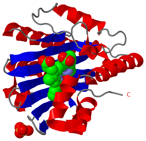
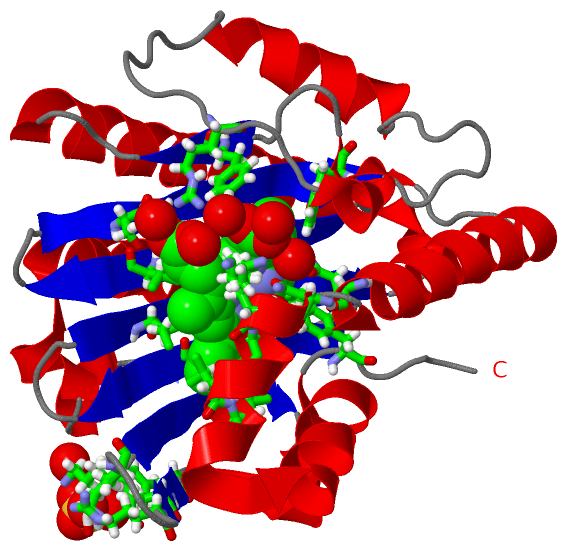
(-) Ligands, Modified Residues, Ions  (2, 2)

Asymmetric/Biological Unit (2, 2)
No.NameCountTypeFull Name
1BL31Ligand/Ion3-[2-[(Z)-[3-(2-CARBOXYETHYL)-5-[(Z)-(3-ETHENYL-4-METHYL-5-OXO-PYRROL-2-YLIDENE)METHYL]-4-METHYL-PYRROL-2-YLIDENE]METHYL]-5-[(Z)-(3-ETHENYL-4-METHYL-5-OXO-PYRROL-2-YLIDENE)METHYL]-4-METHYL-1H-PYRROL-3-YL]PROPANOIC ACID
2SO41Ligand/IonSULFATE ION

(-) Sites  (2, 2)

Asymmetric Unit (2, 2)
No.NameEvidenceResiduesDescription
1AC1SOFTWARESER A:1034 , GLU A:1076 , ILE A:1086 , HIS A:1088 , VAL A:1090 , GLY A:1103 , ASP A:1105 , VAL A:1107 , SER A:1114 , ALA A:1115 , ARG A:1149 , PHE A:1158 , PHE A:1164 , GLN A:1216 , ASN A:1219 , LYS A:1221 , THR A:1222 , VAL A:1225 , LEU A:1226 , PHE A:1244 , HOH A:9027 , HOH A:9042 , HOH A:9053 , HOH A:9080 , HOH A:9147 , HOH A:9201BINDING SITE FOR RESIDUE BL3 A 2130
2AC2SOFTWARETYR A:1048 , GLU A:1050 , ARG A:1052 , LYS A:1057 , HOH A:9182 , HOH A:9314BINDING SITE FOR RESIDUE SO4 A 6610

(-) SS Bonds  (0, 0)

(no "SS Bond" information available for 3I94)

(-) Cis Peptide Bonds  (0, 0)

(no "Cis Peptide Bond" information available for 3I94)

 Sequence-Structure Mapping

(-) SAPs(SNPs)/Variants  (0, 0)

(no "SAP(SNP)/Variant" information available for 3I94)

(-) PROSITE Motifs  (0, 0)

(no "PROSITE Motif" information available for 3I94)

(-) Exons   (0, 0)

(no "Exon" information available for 3I94)

(-) Sequences/Alignments

Asymmetric/Biological Unit
   Reformat: Number of residues per line =  ('0' or empty: single-line sequence representation)
  Number of residues per labelling interval =   
  UniProt sequence: complete  aligned part    
   Show mapping: SCOP domains CATH domains Pfam domains Secondary structure (by author)
SAPs(SNPs) PROSITE motifs Exons
(details for a mapped element are shown in a popup box when the mouse pointer rests over it)
Chain A from PDB  Type:PROTEIN  Length:243
 aligned with PCYA_SYNY3 | Q55891 from UniProtKB/Swiss-Prot  Length:248

    Alignment length:243
                                    15        25        35        45        55        65        75        85        95       105       115       125       135       145       155       165       175       185       195       205       215       225       235       245   
          PCYA_SYNY3      6 LSLTNSSLMPTLNPMIQQLALAIAASWQSLPLKPYQLPEDLGYVEGRLEGEKLVIENRCYQTPQFRKMHLELAKVGKGLDILHCVMFPEPLYGLPLFGCDIVAGPGGVSAAIADLSPTQSDRQLPAAYQKSLAELGQPEFEQQRELPPWGEIFSEYCLFIRPSNVTEEERFVQRVVDFLQIHCHQSIVAEPLSEAQTLEHRQGQIHYCQQQQKNDKTRRVLEKAFGEAWAERYMSQVLFDVIQ  248
               SCOP domains d3i94a_ A: Ferredoxin-dependent bilin reductase                                                                                                                                                                                                     SCOP domains
               CATH domains --------------------------------------------------------------------------------------------------------------------------------------------------------------------------------------------------------------------------------------------------- CATH domains
               Pfam domains --------------------------------------------------------------------------------------------------------------------------------------------------------------------------------------------------------------------------------------------------- Pfam domains
         Sec.struct. author .hhhhh.hhhhhhhhhhhhhhhhhhhhh....ee..hhhhh..eee......eeeeeeeee...eeeeeeeeeee...eeeeeeeeee.hhhh..eeeeeeeee..eeeeeeeeee........hhhhhhhhhhh...........hhhhhhh....eee...hhhhhhhhhhhhhhhhhhhhhhhhhh...hhhhhhhhhhhhhhhhhhhh.hhhhhhhhhhhhhhhhhhhhhhhh...... Sec.struct. author
                 SAPs(SNPs) --------------------------------------------------------------------------------------------------------------------------------------------------------------------------------------------------------------------------------------------------- SAPs(SNPs)
                    PROSITE --------------------------------------------------------------------------------------------------------------------------------------------------------------------------------------------------------------------------------------------------- PROSITE
                 Transcript --------------------------------------------------------------------------------------------------------------------------------------------------------------------------------------------------------------------------------------------------- Transcript
                3i94 A 1006 LSLTNSSLMPTLNPMIQQLALAIAASWQSLPLKPYQLPEDLGYVEGRLEGEKLVIENRCYQTPQFRKMHLELAKVGKGLDILHCVMFPEPLYGLPLFGCDIVAGPGGVSAAIADLSPTQSDRQLPAAYQKSLAELGQPEFEQQRELPPWGEIFSEYCLFIRPSNVTEEERFVQRVVDFLQIHCHQSIVAEPLSEAQTLEHRQGQIHYCQQQQKNDKTRRVLEKAFGEAWAERYMSQVLFDVIQ 1248
                                  1015      1025      1035      1045      1055      1065      1075      1085      1095      1105      1115      1125      1135      1145      1155      1165      1175      1185      1195      1205      1215      1225      1235      1245   

   Legend:   → Mismatch (orange background)
  - → Gap (green background, '-', border residues have a numbering label)
    → Modified Residue (blue background, lower-case, 'x' indicates undefined single-letter code, labelled with number + name)
  x → Chemical Group (purple background, 'x', labelled with number + name, e.g. ACE or NH2)
  extra numbering lines below/above indicate numbering irregularities and modified residue names etc., number ends below/above '|'

 Classification and Annotation

(-) SCOP Domains  (1, 1)

Asymmetric/Biological Unit

(-) CATH Domains  (0, 0)

(no "CATH Domain" information available for 3I94)

(-) Pfam Domains  (0, 0)

(no "Pfam Domain" information available for 3I94)

(-) Gene Ontology  (6, 6)

Asymmetric/Biological Unit(hide GO term definitions)
Chain A   (PCYA_SYNY3 | Q55891)
molecular function
    GO:0050897    cobalt ion binding    Interacting selectively and non-covalently with a cobalt (Co) ion.
    GO:0016491    oxidoreductase activity    Catalysis of an oxidation-reduction (redox) reaction, a reversible chemical reaction in which the oxidation state of an atom or atoms within a molecule is altered. One substrate acts as a hydrogen or electron donor and becomes oxidized, while the other acts as hydrogen or electron acceptor and becomes reduced.
    GO:0016636    oxidoreductase activity, acting on the CH-CH group of donors, iron-sulfur protein as acceptor    Catalysis of an oxidation-reduction (redox) reaction in which a CH-CH group acts as a hydrogen or electron donor and reduces an iron-sulfur protein.
    GO:0050620    phycocyanobilin:ferredoxin oxidoreductase activity    Catalysis of the reaction: (3Z)-phycocyanobilin + oxidized ferredoxin = biliverdin IXa + reduced ferredoxin.
biological process
    GO:0055114    oxidation-reduction process    A metabolic process that results in the removal or addition of one or more electrons to or from a substance, with or without the concomitant removal or addition of a proton or protons.
    GO:0010024    phytochromobilin biosynthetic process    The chemical reactions and pathways resulting in the formation of phytochromobilin, which involves the oxidative cleavage of heme by a heme oxygenase(HO) to form biliverdin IX alpha.

 Visualization

(-) Interactive Views

Asymmetric/Biological Unit
  Complete Structure
    Jena3D(integrated viewing of ligand, site, SAP, PROSITE, SCOP information)
    WebMol | AstexViewer[tm]@PDBe
(Java Applets, require no local installation except for Java; loading may be slow)
    STRAP
(Java WebStart application, automatic local installation, requires Java; full application with system access!)
    RasMol
(require local installation)
    Molscript (VRML)
(requires installation of a VRML viewer; select preferred view via VRML and generate a mono or stereo PDF format file)
 
  Ligands, Modified Residues, Ions
    BL3  [ RasMol | Jena3D ]  +environment [ RasMol | Jena3D ]
    SO4  [ RasMol | Jena3D ]  +environment [ RasMol | Jena3D ]
 
  Sites
    AC1  [ RasMol ]  +environment [ RasMol ]
    AC2  [ RasMol ]  +environment [ RasMol ]
 
  Cis Peptide Bonds
(no "Cis Peptide Bonds" information available for 3i94)
 

(-) Still Images

Jmol
  protein: cartoon or spacefill or dots and stick; nucleic acid: cartoon and stick; ligands: spacefill; active site: stick
Molscript
  protein, nucleic acid: cartoon; ligands: spacefill; active site: ball and stick

 Databases and Analysis Tools

(-) Databases

Access by PDB/NDB ID
  3i94
    Family and Domain InformationProDom | SYSTERS
    General Structural InformationGlycoscienceDB | MMDB | NDB | OCA | PDB | PDBe | PDBj | PDBsum | PDBWiki | PQS | PROTEOPEDIA
    Orientation in MembranesOPM
    Protein SurfaceSURFACE
    Secondary StructureDSSP (structure derived) | HSSP (homology derived)
    Structural GenomicsGeneCensus
    Structural NeighboursCE | VAST
    Structure ClassificationCATH | Dali | SCOP
    Validation and Original DataBMRB Data View | BMRB Restraints Grid | EDS | PROCHECK | RECOORD | WHAT_CHECK
 
Access by UniProt ID/Accession number
  PCYA_SYNY3 | Q55891
    Comparative Protein Structure ModelsModBase
    Genomic InformationEnsembl
    Protein-protein InteractionDIP
    Sequence, Family and Domain InformationInterPro | Pfam | SMART | UniProtKB/SwissProt
 
Access by Enzyme Classificator   (EC Number)
  1.3.7.5
    General Enzyme InformationBRENDA | EC-PDB | Enzyme | IntEnz
    PathwayKEGG | MetaCyc
 
Access by Disease Identifier   (MIM ID)
  (no 'MIM ID' available)
    Disease InformationOMIM
 
Access by GenAge ID
  (no 'GenAge ID' available)
    Age Related InformationGenAge

(-) Analysis Tools

Access by PDB/NDB ID
    Domain InformationXDom
    Interatomic Contacts of Structural UnitsCSU
    Ligand-protein ContactsLPC
    Protein CavitiescastP
    Sequence and Secondary StructurePDBCartoon
    Structure AlignmentSTRAP(Java WebStart application, automatic local installation, requires Java; full application with system access!)
    Structure and Sequence BrowserSTING
 
Access by UniProt ID/Accession number
  PCYA_SYNY3 | Q55891
    Protein Disorder PredictionDisEMBL | FoldIndex | GLOBPLOT (for more information see DisProt)

 Related Entries

(-) Entries Sharing at Least One Protein Chain (UniProt ID)

UniProtKB/Swiss-Prot
        PCYA_SYNY3 | Q558912d1e 2dke 3ajg 3ajh 3f0l 3f0m 3i8u 3i95 3nb8 3nb9 4eoc 4eod 4eoe 4qcd 5b4h 5b4i 5b4j

(-) Related Entries Specified in the PDB File

2d1e THE SAME PROTEIN COMPLEXED WITH BILIVERDIN IX ALPHA
2dke THE SAME PROTEIN OF SUBSTRATE FREE-FORM
3i8u
3i95