Show PDB file:   
         Plain Text   HTML   (compressed file size)
QuickSearch:   
by PDB,NDB,UniProt,PROSITE Code or Search Term(s)  
(-)Asym./Biol. Unit
(-)Asym./Biol. Unit - sites
collapse expand < >
Image Asym./Biol. Unit
Asym./Biol. Unit  (Jmol Viewer)
Image Asym./Biol. Unit - sites
Asym./Biol. Unit - sites  (Jmol Viewer)

(-) Description

Title :  CRYSTAL STRUCTURE OF OXIDIZED H88Q SYNECHOCYSTIS SP. PCYA
 
Authors :  D. D. Gae
Date :  03 Jun 10  (Deposition) - 06 Oct 10  (Release) - 16 Sep 15  (Revision)
Method :  X-RAY DIFFRACTION
Resolution :  1.30
Chains :  Asym./Biol. Unit :  A
Keywords :  Alpha-Beta-Alpha Sandwich, Radical Chemistry, Oxidoreductase (Keyword Search: [Gene Ontology, PubMed, Web (Google))
 
Reference :  A. C. Kohler, D. D. Gae, M. A. Richley, S. Stoll, A. Gunn, S. Lim, S. S. Martin, T. I. Doukov, R. D. Britt, J. B. Ames, J. C. Lagarias, A. J. Fisher
Structural Basis For Hydration Dynamics In Radical Stabilization Of Bilin Reductase Mutants.
Biochemistry V. 49 6206 2010
PubMed-ID: 20557110  |  Reference-DOI: 10.1021/BI100728Q

(-) Compounds

Molecule 1
    ChainsA
    EC Number1.3.7.5
    EngineeredYES
    Expression SystemESCHERICHIA COLI
    Expression System Taxid562
    GenePCYA, SLR0116
    MutationYES
    Organism ScientificSYNECHOCYSTIS SP.
    Organism Taxid1148
    StrainPCC 6803

 Structural Features

(-) Chains, Units

  1
Asymmetric/Biological Unit A

Summary Information (see also Sequences/Alignments below)

(-) Ligands, Modified Residues, Ions  (2, 15)

Asymmetric/Biological Unit (2, 15)
No.NameCountTypeFull Name
1BLA1Ligand/IonBILIVERDINE IX ALPHA
2EDO14Ligand/Ion1,2-ETHANEDIOL

(-) Sites  (15, 15)

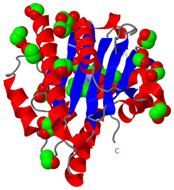
Asymmetric Unit (15, 15)
No.NameEvidenceResiduesDescription
01AC1SOFTWAREGLN A:185 , ARG A:224 , GLU A:227BINDING SITE FOR RESIDUE EDO A 401
02AC2SOFTWAREMET A:20 , GLU A:173 , GLU A:174 , EDO A:410BINDING SITE FOR RESIDUE EDO A 402
03AC3SOFTWAREGLY A:47 , TYR A:48 , GLU A:61 , ASN A:62 , EDO A:411 , HOH A:1236 , HOH A:1237 , HOH A:1374BINDING SITE FOR RESIDUE EDO A 403
04AC4SOFTWAREALA A:132 , TYR A:133 , GLN A:190 , HOH A:1117 , HOH A:1249 , HOH A:1261BINDING SITE FOR RESIDUE EDO A 404
05AC5SOFTWARELEU A:151 , CYS A:162 , PHE A:164 , HOH A:1040 , HOH A:1048 , HOH A:1080BINDING SITE FOR RESIDUE EDO A 405
06AC6SOFTWAREASN A:169 , VAL A:170 , THR A:171 , HOH A:1089 , HOH A:1139BINDING SITE FOR RESIDUE EDO A 406
07AC7SOFTWARESER A:31 , LEU A:184 , GLN A:185 , CYS A:188 , LYS A:221 , HOH A:1043 , HOH A:1055 , HOH A:1112 , HOH A:1292BINDING SITE FOR RESIDUE EDO A 407
08AC8SOFTWAREGLN A:124 , GLN A:201 , GLU A:204 , HOH A:1073 , HOH A:1085 , HOH A:1182 , HOH A:1263BINDING SITE FOR RESIDUE EDO A 408
09AC9SOFTWAREASN A:10 , SER A:12 , TYR A:48 , GLU A:61BINDING SITE FOR RESIDUE EDO A 409
10BC1SOFTWAREMET A:20 , GLN A:23 , GLU A:173 , GLU A:174 , VAL A:177 , EDO A:402 , HOH A:1062BINDING SITE FOR RESIDUE EDO A 410
11BC2SOFTWAREASP A:45 , LEU A:46 , GLY A:47 , GLU A:54 , EDO A:403BINDING SITE FOR RESIDUE EDO A 411
12BC3SOFTWARETHR A:171 , ARG A:175 , HOH A:1074BINDING SITE FOR RESIDUE EDO A 412
13BC4SOFTWAREASN A:18 , PRO A:19 , GLU A:173 , HOH A:1024 , HOH A:1046 , HOH A:1138 , HOH A:1275BINDING SITE FOR RESIDUE EDO A 414
14BC5SOFTWAREGLN A:41 , LEU A:42 , GLU A:44 , HOH A:1134BINDING SITE FOR RESIDUE EDO A 415
15BC6SOFTWARESER A:34 , GLU A:76 , ILE A:86 , GLN A:88 , VAL A:90 , GLY A:103 , CYS A:104 , ASP A:105 , VAL A:107 , SER A:114 , ALA A:115 , ARG A:149 , PHE A:158 , PHE A:164 , ILE A:192 , GLN A:216 , LYS A:221 , THR A:222 , VAL A:225 , PHE A:244 , HOH A:1042 , HOH A:1067 , HOH A:1146 , HOH A:1151 , HOH A:1171 , HOH A:1231 , HOH A:1400BINDING SITE FOR RESIDUE BLA A 249

(-) SS Bonds  (0, 0)

(no "SS Bond" information available for 3NB8)

(-) Cis Peptide Bonds  (0, 0)

(no "Cis Peptide Bond" information available for 3NB8)

 Sequence-Structure Mapping

(-) SAPs(SNPs)/Variants  (0, 0)

(no "SAP(SNP)/Variant" information available for 3NB8)

(-) PROSITE Motifs  (0, 0)

(no "PROSITE Motif" information available for 3NB8)

(-) Exons   (0, 0)

(no "Exon" information available for 3NB8)

(-) Sequences/Alignments

Asymmetric/Biological Unit
   Reformat: Number of residues per line =  ('0' or empty: single-line sequence representation)
  Number of residues per labelling interval =   
  UniProt sequence: complete  aligned part    
   Show mapping: SCOP domains CATH domains Pfam domains Secondary structure (by author)
SAPs(SNPs) PROSITE motifs Exons
(details for a mapped element are shown in a popup box when the mouse pointer rests over it)
Chain A from PDB  Type:PROTEIN  Length:242
 aligned with PCYA_SYNY3 | Q55891 from UniProtKB/Swiss-Prot  Length:248

    Alignment length:242
                                    15        25        35        45        55        65        75        85        95       105       115       125       135       145       155       165       175       185       195       205       215       225       235       245  
           PCYA_SYNY3     6 LSLTNSSLMPTLNPMIQQLALAIAASWQSLPLKPYQLPEDLGYVEGRLEGEKLVIENRCYQTPQFRKMHLELAKVGKGLDILHCVMFPEPLYGLPLFGCDIVAGPGGVSAAIADLSPTQSDRQLPAAYQKSLAELGQPEFEQQRELPPWGEIFSEYCLFIRPSNVTEEERFVQRVVDFLQIHCHQSIVAEPLSEAQTLEHRQGQIHYCQQQQKNDKTRRVLEKAFGEAWAERYMSQVLFDVI 247
               SCOP domains d3nb8a_ A: Ferredoxin-dependent bilin reductase                                                                                                                                                                                                    SCOP domains
               CATH domains -------------------------------------------------------------------------------------------------------------------------------------------------------------------------------------------------------------------------------------------------- CATH domains
               Pfam domains -------------------Fe_bilin_red-3nb8A01 A:25-245                                                                                                                                                                                                -- Pfam domains
         Sec.struct. author .......hhhhhhhhhhhhhhhhhhhhh....ee..hhhhh.eeee......eeeeeeeee...eeeeeeeeeee...eeeeeeeeee.hhhh..eeeeeeeee..eeeeeeeeee........hhhhhhhhhh............hhhhhhh....eee...hhhhhhhhhhhhhhhhhhhhhhhhhh...hhhhhhhhhhhhhhhhhhhh.hhhhhhhhhhhhhhhhhhhhhhhh..... Sec.struct. author
                 SAPs(SNPs) -------------------------------------------------------------------------------------------------------------------------------------------------------------------------------------------------------------------------------------------------- SAPs(SNPs)
                    PROSITE -------------------------------------------------------------------------------------------------------------------------------------------------------------------------------------------------------------------------------------------------- PROSITE
                 Transcript -------------------------------------------------------------------------------------------------------------------------------------------------------------------------------------------------------------------------------------------------- Transcript
                 3nb8 A   6 LSLTNSSLMPTLNPMIQQLALAIAASWQSLPLKPYQLPEDLGYVEGRLEGEKLVIENRCYQTPQFRKMHLELAKVGKGLDILQCVMFPEPLYGLPLFGCDIVAGPGGVSAAIADLSPTQSDRQLPAAYQKSLAELGQPEFEQQRELPPWGEIFSEYCLFIRPSNVTEEERFVQRVVDFLQIHCHQSIVAEPLSEAQTLEHRQGQIHYCQQQQKNDKTRRVLEKAFGEAWAERYMSQVLFDVI 247
                                    15        25        35        45        55        65        75        85        95       105       115       125       135       145       155       165       175       185       195       205       215       225       235       245  

   Legend:   → Mismatch (orange background)
  - → Gap (green background, '-', border residues have a numbering label)
    → Modified Residue (blue background, lower-case, 'x' indicates undefined single-letter code, labelled with number + name)
  x → Chemical Group (purple background, 'x', labelled with number + name, e.g. ACE or NH2)
  extra numbering lines below/above indicate numbering irregularities and modified residue names etc., number ends below/above '|'

 Classification and Annotation

(-) SCOP Domains  (1, 1)

Asymmetric/Biological Unit

(-) CATH Domains  (0, 0)

(no "CATH Domain" information available for 3NB8)

(-) Pfam Domains  (1, 1)

Asymmetric/Biological Unit

(-) Gene Ontology  (6, 6)

Asymmetric/Biological Unit(hide GO term definitions)
Chain A   (PCYA_SYNY3 | Q55891)
molecular function
    GO:0050897    cobalt ion binding    Interacting selectively and non-covalently with a cobalt (Co) ion.
    GO:0016491    oxidoreductase activity    Catalysis of an oxidation-reduction (redox) reaction, a reversible chemical reaction in which the oxidation state of an atom or atoms within a molecule is altered. One substrate acts as a hydrogen or electron donor and becomes oxidized, while the other acts as hydrogen or electron acceptor and becomes reduced.
    GO:0016636    oxidoreductase activity, acting on the CH-CH group of donors, iron-sulfur protein as acceptor    Catalysis of an oxidation-reduction (redox) reaction in which a CH-CH group acts as a hydrogen or electron donor and reduces an iron-sulfur protein.
    GO:0050620    phycocyanobilin:ferredoxin oxidoreductase activity    Catalysis of the reaction: (3Z)-phycocyanobilin + oxidized ferredoxin = biliverdin IXa + reduced ferredoxin.
biological process
    GO:0055114    oxidation-reduction process    A metabolic process that results in the removal or addition of one or more electrons to or from a substance, with or without the concomitant removal or addition of a proton or protons.
    GO:0010024    phytochromobilin biosynthetic process    The chemical reactions and pathways resulting in the formation of phytochromobilin, which involves the oxidative cleavage of heme by a heme oxygenase(HO) to form biliverdin IX alpha.

 Visualization

(-) Interactive Views

Asymmetric/Biological Unit
  Complete Structure
    Jena3D(integrated viewing of ligand, site, SAP, PROSITE, SCOP information)
    WebMol | AstexViewer[tm]@PDBe
(Java Applets, require no local installation except for Java; loading may be slow)
    STRAP
(Java WebStart application, automatic local installation, requires Java; full application with system access!)
    RasMol
(require local installation)
    Molscript (VRML)
(requires installation of a VRML viewer; select preferred view via VRML and generate a mono or stereo PDF format file)
 
  Ligands, Modified Residues, Ions
    BLA  [ RasMol | Jena3D ]  +environment [ RasMol | Jena3D ]
    EDO  [ RasMol | Jena3D ]  +environment [ RasMol | Jena3D ]
 
  Sites
    AC1  [ RasMol ]  +environment [ RasMol ]
    AC2  [ RasMol ]  +environment [ RasMol ]
    AC3  [ RasMol ]  +environment [ RasMol ]
    AC4  [ RasMol ]  +environment [ RasMol ]
    AC5  [ RasMol ]  +environment [ RasMol ]
    AC6  [ RasMol ]  +environment [ RasMol ]
    AC7  [ RasMol ]  +environment [ RasMol ]
    AC8  [ RasMol ]  +environment [ RasMol ]
    AC9  [ RasMol ]  +environment [ RasMol ]
    BC1  [ RasMol ]  +environment [ RasMol ]
    BC2  [ RasMol ]  +environment [ RasMol ]
    BC3  [ RasMol ]  +environment [ RasMol ]
    BC4  [ RasMol ]  +environment [ RasMol ]
    BC5  [ RasMol ]  +environment [ RasMol ]
    BC6  [ RasMol ]  +environment [ RasMol ]
 
  Cis Peptide Bonds
(no "Cis Peptide Bonds" information available for 3nb8)
 

(-) Still Images

Jmol
  protein: cartoon or spacefill or dots and stick; nucleic acid: cartoon and stick; ligands: spacefill; active site: stick
Molscript
  protein, nucleic acid: cartoon; ligands: spacefill; active site: ball and stick

 Databases and Analysis Tools

(-) Databases

Access by PDB/NDB ID
  3nb8
    Family and Domain InformationProDom | SYSTERS
    General Structural InformationGlycoscienceDB | MMDB | NDB | OCA | PDB | PDBe | PDBj | PDBsum | PDBWiki | PQS | PROTEOPEDIA
    Orientation in MembranesOPM
    Protein SurfaceSURFACE
    Secondary StructureDSSP (structure derived) | HSSP (homology derived)
    Structural GenomicsGeneCensus
    Structural NeighboursCE | VAST
    Structure ClassificationCATH | Dali | SCOP
    Validation and Original DataBMRB Data View | BMRB Restraints Grid | EDS | PROCHECK | RECOORD | WHAT_CHECK
 
Access by UniProt ID/Accession number
  PCYA_SYNY3 | Q55891
    Comparative Protein Structure ModelsModBase
    Genomic InformationEnsembl
    Protein-protein InteractionDIP
    Sequence, Family and Domain InformationInterPro | Pfam | SMART | UniProtKB/SwissProt
 
Access by Enzyme Classificator   (EC Number)
  1.3.7.5
    General Enzyme InformationBRENDA | EC-PDB | Enzyme | IntEnz
    PathwayKEGG | MetaCyc
 
Access by Disease Identifier   (MIM ID)
  (no 'MIM ID' available)
    Disease InformationOMIM
 
Access by GenAge ID
  (no 'GenAge ID' available)
    Age Related InformationGenAge

(-) Analysis Tools

Access by PDB/NDB ID
    Domain InformationXDom
    Interatomic Contacts of Structural UnitsCSU
    Ligand-protein ContactsLPC
    Protein CavitiescastP
    Sequence and Secondary StructurePDBCartoon
    Structure AlignmentSTRAP(Java WebStart application, automatic local installation, requires Java; full application with system access!)
    Structure and Sequence BrowserSTING
 
Access by UniProt ID/Accession number
  PCYA_SYNY3 | Q55891
    Protein Disorder PredictionDisEMBL | FoldIndex | GLOBPLOT (for more information see DisProt)

 Related Entries

(-) Entries Sharing at Least One Protein Chain (UniProt ID)

UniProtKB/Swiss-Prot
        PCYA_SYNY3 | Q558912d1e 2dke 3ajg 3ajh 3f0l 3f0m 3i8u 3i94 3i95 3nb9 4eoc 4eod 4eoe 4qcd 5b4h 5b4i 5b4j

(-) Related Entries Specified in the PDB File

3f0j SIDE CHAIN ROTATION OF AMIDE N AND O ATOMS OF GLN88
3nb9