|
|
|
|
 Description
|
|
Compounds
|
||||||||||||||||||||||||||||||||||||||||||||||||||||
Chains, Units
Summary Information (see also Sequences/Alignments below) |
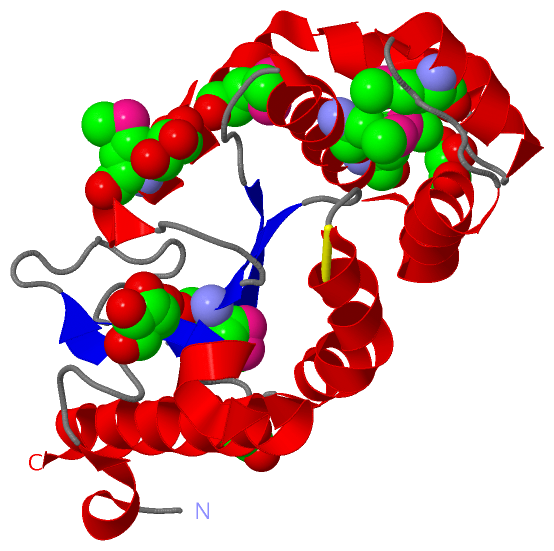
Ligands, Modified Residues, Ions (2, 10)| Asymmetric/Biological Unit (2, 10) |
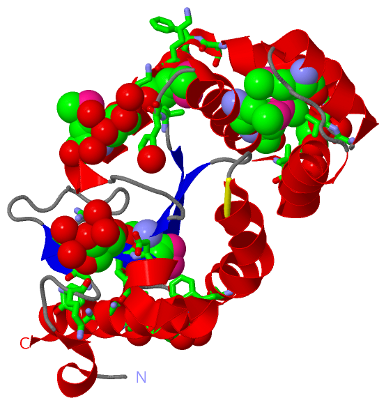
Sites (4, 4)
Asymmetric Unit (4, 4)
|
SS Bonds (1, 1)
Asymmetric/Biological Unit
|
||||||||
Cis Peptide Bonds (1, 1)
Asymmetric/Biological Unit
|
||||||||
SAPs(SNPs)/Variants (0, 0)| (no "SAP(SNP)/Variant" information available for 3H93) |
PROSITE Motifs (1, 1)
Asymmetric/Biological Unit (1, 1)
|
||||||||||||||||||||||||
Exons (0, 0)| (no "Exon" information available for 3H93) |
Sequences/Alignments
Asymmetric/Biological UnitChain A from PDB Type:PROTEIN Length:192 aligned with DSBA_PSEAE | P0C2B2 from UniProtKB/Swiss-Prot Length:211 Alignment length:192 29 39 49 59 69 79 89 99 109 119 129 139 149 159 169 179 189 199 209 DSBA_PSEAE 20 AQADDYTAGKEYVELSSPVPVSQPGKIEVVELFWYGCPHCYAFEPTIVPWSEKLPADVHFVRLPALFGGIWNVHGQMFLTLESMGVEHDVHNAVFEAIHKEHKKLATPEEMADFLAGKGVDKEKFLSTYNSFAIKGQMEKAKKLAMAYQVTGVPTMVVNGKYRFDIGSAGGPEETLKLADYLIEKERAAAKK 211 SCOP domains d3h93a_ A: automated matches SCOP domains CATH domains ------------------------------------------------------------------------------------------------------------------------------------------------------------------------------------------------ CATH domains Pfam domains ------------------------------------------------------------------------------------------------------------------------------------------------------------------------------------------------ Pfam domains SAPs(SNPs) ------------------------------------------------------------------------------------------------------------------------------------------------------------------------------------------------ SAPs(SNPs) PROSITE ----------------------------THIOREDOXIN_1 ------------------------------------------------------------------------------------------------------------------------------------------------- PROSITE Transcript ------------------------------------------------------------------------------------------------------------------------------------------------------------------------------------------------ Transcript 3h93 A 1 SNADDYTAGKEYVELSSPVPVSQPGKIEVVELFWYGCPHCYAFEPTIVPWSEKLPADVHFVRLPALFGGIWNVHGQmFLTLESmGVEHDVHNAVFEAIHKEHKKLATPEEmADFLAGKGVDKEKFLSTYNSFAIKGQmEKAKKLAmAYQVTGVPTmVVNGKYRFDIGSAGGPEETLKLADYLIEKERAAAKK 192 10 20 30 40 50 60 70 | 80 | 90 100 110| 120 130 140 | 150 | 160 170 180 190 77-MSE 84-MSE 111-MSE 138-MSE 146-MSE 156-MSE
|
||||||||||||||||||||
SCOP Domains (1, 1)
Asymmetric/Biological Unit
 |
CATH Domains (0, 0)| (no "CATH Domain" information available for 3H93) |
Pfam Domains (0, 0)| (no "Pfam Domain" information available for 3H93) |
Gene Ontology (4, 4)|
Asymmetric/Biological Unit(hide GO term definitions) Chain A (DSBA_PSEAE | P0C2B2)
|
||||||||||||||||||||||||||||||||||||||||||
Interactive Views
|
|||||||||||||||||||||||||||||||||||||||||||||||||||||||||||||||||||||||||||||||||||||||||||||||||||||||||||||||||||||||||||||||||||||||||||||||||||
Still Images
|
||||||||||||||||
Databases
|
||||||||||||||||||||||||||||||||||||||||||||||||||||||||||||||||||||||||||||||||||||||||||||||||||||||||||||||||||||||||||||||||||||||||||||||||||||||||||||||||
Analysis Tools
|
|||||||||||||||||||||||||||||||||||||||||||||||||||||||||||||
Entries Sharing at Least One Protein Chain (UniProt ID)
Related Entries Specified in the PDB File
|
|