Show PDB file:   
         Plain Text   HTML   (compressed file size)
QuickSearch:   
by PDB,NDB,UniProt,PROSITE Code or Search Term(s)  
(-)Asym./Biol. Unit
(-)Asym./Biol. Unit - sites
collapse expand < >
Image Asym./Biol. Unit
Asym./Biol. Unit  (Jmol Viewer)
Image Asym./Biol. Unit - sites
Asym./Biol. Unit - sites  (Jmol Viewer)

(-) Description

Title :  THE STRUCTURE OF FLUORESCENT PROTEIN FP480
 
Authors :  S. Pletnev, K. S. Morozova, V. V. Verkhusha, Z. Dauter
Date :  13 Apr 09  (Deposition) - 08 Sep 09  (Release) - 13 Jul 11  (Revision)
Method :  X-RAY DIFFRACTION
Resolution :  2.00
Chains :  Asym./Biol. Unit :  A,B
Keywords :  Od-Structure, Order-Disorder Structure, Fluorescent Protein (Keyword Search: [Gene Ontology, PubMed, Web (Google))
 
Reference :  S. Pletnev, K. S. Morozova, V. V. Verkhusha, Z. Dauter
Rotational Order-Disorder Structure Of Fluorescent Protein Fp480
Acta Crystallogr. , Sect. D V. 65 906 2009
PubMed-ID: 19690368  |  Reference-DOI: 10.1107/S0907444909020927

(-) Compounds

Molecule 1 - FLUORESCENT PROTEIN FP480
    ChainsA, B
    EngineeredYES
    Expression SystemESCHERICHIA COLI
    Expression System Taxid562
    Organism ScientificENTACMAEA QUADRICOLOR
    Organism Taxid6118

 Structural Features

(-) Chains, Units

  12
Asymmetric/Biological Unit AB

Summary Information (see also Sequences/Alignments below)
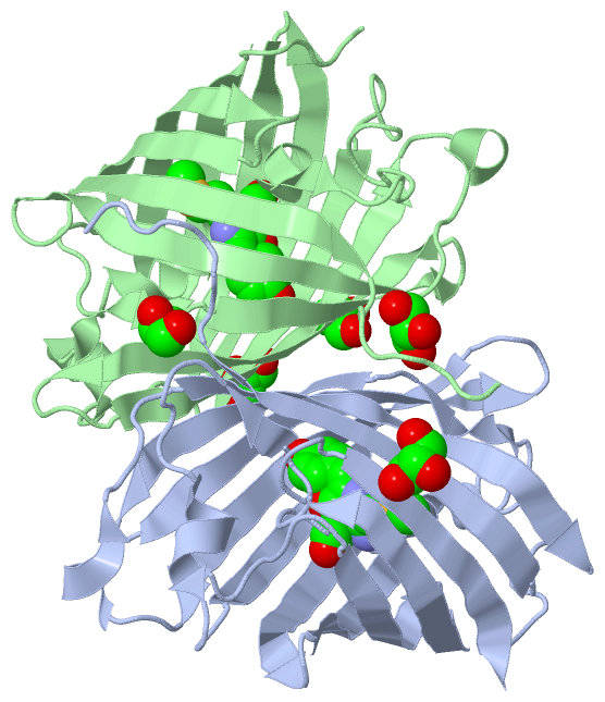
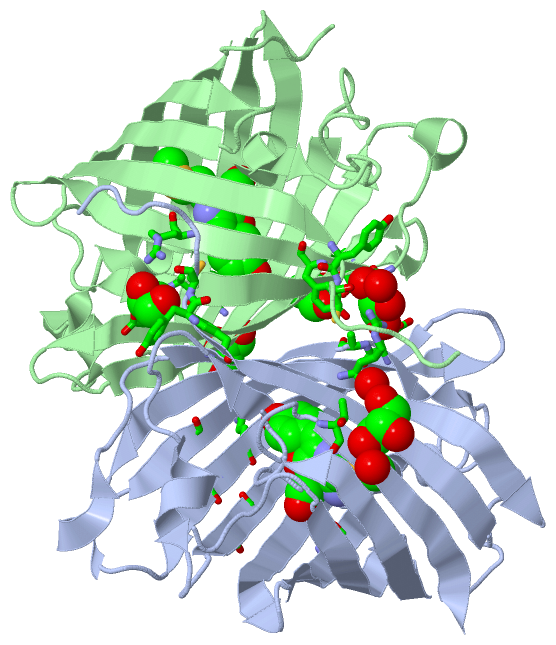
(-) Ligands, Modified Residues, Ions  (2, 7)

Asymmetric/Biological Unit (2, 7)
No.NameCountTypeFull Name
1GOL5Ligand/IonGLYCEROL
2NRQ2Mod. Amino Acid{(4Z)-4-(4-HYDROXYBENZYLIDENE)-2-[3-(METHYLTHIO)PROPANIMIDOYL]-5-OXO-4,5-DIHYDRO-1H-IMIDAZOL-1-YL}ACETIC ACID

(-) Sites  (5, 5)

Asymmetric Unit (5, 5)
No.NameEvidenceResiduesDescription
1AC1SOFTWAREGLU A:141 , ALA A:142 , ARG A:198 , HOH A:278 , VAL B:192 , TYR B:194 , TYR B:221 , CYS B:222BINDING SITE FOR RESIDUE GOL A 234
2AC2SOFTWARETHR A:38 , HIS A:214 , HOH A:302 , HOH A:324BINDING SITE FOR RESIDUE GOL A 235
3AC3SOFTWAREGLU A:91 , GLU A:155 , LYS A:175 , THR A:177 , ARG B:122BINDING SITE FOR RESIDUE GOL B 234
4AC4SOFTWAREARG A:122 , GLU B:155 , THR B:177BINDING SITE FOR RESIDUE GOL B 235
5AC5SOFTWARETYR A:194 , TYR A:221 , CYS A:222 , GLU B:141 , ALA B:142 , ARG B:198BINDING SITE FOR RESIDUE GOL B 236

(-) SS Bonds  (0, 0)

(no "SS Bond" information available for 3H1O)

(-) Cis Peptide Bonds  (4, 4)

Asymmetric/Biological Unit
No.Residues
1Gly A:49 -Pro A:50
2Phe A:84 -Pro A:85
3Gly B:49 -Pro B:50
4Phe B:84 -Pro B:85

 Sequence-Structure Mapping

(-) SAPs(SNPs)/Variants  (0, 0)

(no "SAP(SNP)/Variant" information available for 3H1O)

(-) PROSITE Motifs  (0, 0)

(no "PROSITE Motif" information available for 3H1O)

(-) Exons   (0, 0)

(no "Exon" information available for 3H1O)

(-) Sequences/Alignments

Asymmetric/Biological Unit
   Reformat: Number of residues per line =  ('0' or empty: single-line sequence representation)
  Number of residues per labelling interval =   
  UniProt sequence: complete  aligned part    
   Show mapping: SCOP domains CATH domains Pfam domains Secondary structure (by author)
SAPs(SNPs) PROSITE motifs Exons
(details for a mapped element are shown in a popup box when the mouse pointer rests over it)
Chain A from PDB  Type:PROTEIN  Length:224
 aligned with D0VX33_ENTQU | D0VX33 from UniProtKB/TrEMBL  Length:233

    Alignment length:226
                                    12        22        32        42        52        62        72        82        92       102       112       122       132       142       152       162       172       182       192       202       212       222      
         D0VX33_ENTQU     3 ELITENMHMKLYMEGTVNNHHFKCTSEGEGKPYEGTQTQRIKVVEGGPLPFAFDILATSFMYGSHTFINHTQGIPDFWKQSFPEGFTWERVTTYEDGGVLTATQDTSLQDGCLIYNVKIRGVNFPSNGPVMQKKTLGWEAHTEMLYPADGGLEGRADLALKLVGGGHLICNFKTTYRSKKPAKNLKMPGVYYVDYRLERIKEADKETYVEQHEVAVARYCDLPSKL 228
               SCOP domains d3h1oa_ A: automated matches                                                                                                                                                                                                       SCOP domains
               CATH domains ---------------------------------------------------------------------------------------------------------------------------------------------------------------------------------------------------------------------------------- CATH domains
               Pfam domains ---------------------------------------------------------------------------------------------------------------------------------------------------------------------------------------------------------------------------------- Pfam domains
         Sec.struct. author ......eeeeeeeeeee..eeeeeeeeeeee....eeeeeeeeee.......hhhhhh...--.....ee.....hhhhhh....eeeeeeeee....eeeeeeeeeee..eeeeeeeeeee............eeee..eeeeeeee..eeeeeeeeeeee....eeeeeeeeeeee..hhhhh.....eeeeeeeeeeeee....eeeeeeeeeee........ Sec.struct. author
                 SAPs(SNPs) ---------------------------------------------------------------------------------------------------------------------------------------------------------------------------------------------------------------------------------- SAPs(SNPs)
                    PROSITE ---------------------------------------------------------------------------------------------------------------------------------------------------------------------------------------------------------------------------------- PROSITE
                 Transcript ---------------------------------------------------------------------------------------------------------------------------------------------------------------------------------------------------------------------------------- Transcript
                 3h1o A   3 ELITENMHMKLYMEGTVNNHHFKCTSEGEGKPYEGTQTQRIKVVEGGPLPFAFDILATSFm--SHTFINHTQGIPDFWKQSFPEGFTWERVTTYEDGGVLTATQDTSLQDGCLIYNVKIRGVNFPSNGPVMQKKTLGWEAHTEMLYPADGGLEGRADLALKLVGGGHLICNFKTTYRSKKPAKNLKMPGVYYVDYRLERIKEADKETYVEQHEVAVARYCDLPSKL 228
                                    12        22        32        42        52        62|  |    72        82        92       102       112       122       132       142       152       162       172       182       192       202       212       222      
                                                                                       63-NRQ                                                                                                                                                                 

Chain B from PDB  Type:PROTEIN  Length:224
 aligned with D0VX33_ENTQU | D0VX33 from UniProtKB/TrEMBL  Length:233

    Alignment length:226
                                    12        22        32        42        52        62        72        82        92       102       112       122       132       142       152       162       172       182       192       202       212       222      
         D0VX33_ENTQU     3 ELITENMHMKLYMEGTVNNHHFKCTSEGEGKPYEGTQTQRIKVVEGGPLPFAFDILATSFMYGSHTFINHTQGIPDFWKQSFPEGFTWERVTTYEDGGVLTATQDTSLQDGCLIYNVKIRGVNFPSNGPVMQKKTLGWEAHTEMLYPADGGLEGRADLALKLVGGGHLICNFKTTYRSKKPAKNLKMPGVYYVDYRLERIKEADKETYVEQHEVAVARYCDLPSKL 228
               SCOP domains d3h1ob_ B: automated matches                                                                                                                                                                                                       SCOP domains
               CATH domains ---------------------------------------------------------------------------------------------------------------------------------------------------------------------------------------------------------------------------------- CATH domains
               Pfam domains ---------------------------------------------------------------------------------------------------------------------------------------------------------------------------------------------------------------------------------- Pfam domains
         Sec.struct. author ......eeeeeeeeeee..eeeeeeeeeeee....eeeeeeeeee.......hhhhhhhh.--hhhhhee...............eeeeeeeee....eeeeeeeeeee..eeeeeeeeeee.....hhhhhh.eeee..eeeeeeee..eeeeeeeeeeee....eeeeeeeeeeee..hhhhh.....eeeeeeeeeeeee....eeeeeeeeeee........ Sec.struct. author
                 SAPs(SNPs) ---------------------------------------------------------------------------------------------------------------------------------------------------------------------------------------------------------------------------------- SAPs(SNPs)
                    PROSITE ---------------------------------------------------------------------------------------------------------------------------------------------------------------------------------------------------------------------------------- PROSITE
                 Transcript ---------------------------------------------------------------------------------------------------------------------------------------------------------------------------------------------------------------------------------- Transcript
                 3h1o B   3 ELITENMHMKLYMEGTVNNHHFKCTSEGEGKPYEGTQTQRIKVVEGGPLPFAFDILATSFm--SHTFINHTQGIPDFWKQSFPEGFTWERVTTYEDGGVLTATQDTSLQDGCLIYNVKIRGVNFPSNGPVMQKKTLGWEAHTEMLYPADGGLEGRADLALKLVGGGHLICNFKTTYRSKKPAKNLKMPGVYYVDYRLERIKEADKETYVEQHEVAVARYCDLPSKL 228
                                    12        22        32        42        52        62|  |    72        82        92       102       112       122       132       142       152       162       172       182       192       202       212       222      
                                                                                       63-NRQ                                                                                                                                                                 

   Legend:   → Mismatch (orange background)
  - → Gap (green background, '-', border residues have a numbering label)
    → Modified Residue (blue background, lower-case, 'x' indicates undefined single-letter code, labelled with number + name)
  x → Chemical Group (purple background, 'x', labelled with number + name, e.g. ACE or NH2)
  extra numbering lines below/above indicate numbering irregularities and modified residue names etc., number ends below/above '|'

 Classification and Annotation

(-) SCOP Domains  (1, 2)

Asymmetric/Biological Unit

(-) CATH Domains  (0, 0)

(no "CATH Domain" information available for 3H1O)

(-) Pfam Domains  (0, 0)

(no "Pfam Domain" information available for 3H1O)

(-) Gene Ontology  (1, 1)

Asymmetric/Biological Unit(hide GO term definitions)
Chain A,B   (D0VX33_ENTQU | D0VX33)
biological process
    GO:0008218    bioluminescence    The production of light by certain enzyme-catalyzed reactions in cells.

 Visualization

(-) Interactive Views

Asymmetric/Biological Unit
  Complete Structure
    Jena3D(integrated viewing of ligand, site, SAP, PROSITE, SCOP information)
    WebMol | AstexViewer[tm]@PDBe
(Java Applets, require no local installation except for Java; loading may be slow)
    STRAP
(Java WebStart application, automatic local installation, requires Java; full application with system access!)
    RasMol
(require local installation)
    Molscript (VRML)
(requires installation of a VRML viewer; select preferred view via VRML and generate a mono or stereo PDF format file)
 
  Ligands, Modified Residues, Ions
    GOL  [ RasMol | Jena3D ]  +environment [ RasMol | Jena3D ]
    NRQ  [ RasMol | Jena3D ]  +environment [ RasMol | Jena3D ]
 
  Sites
    AC1  [ RasMol ]  +environment [ RasMol ]
    AC2  [ RasMol ]  +environment [ RasMol ]
    AC3  [ RasMol ]  +environment [ RasMol ]
    AC4  [ RasMol ]  +environment [ RasMol ]
    AC5  [ RasMol ]  +environment [ RasMol ]
 
  Cis Peptide Bonds
    Gly A:49 - Pro A:50   [ RasMol ]  
    Gly B:49 - Pro B:50   [ RasMol ]  
    Phe A:84 - Pro A:85   [ RasMol ]  
    Phe B:84 - Pro B:85   [ RasMol ]  
 

(-) Still Images

Jmol
  protein: cartoon or spacefill or dots and stick; nucleic acid: cartoon and stick; ligands: spacefill; active site: stick
Molscript
  protein, nucleic acid: cartoon; ligands: spacefill; active site: ball and stick

 Databases and Analysis Tools

(-) Databases

Access by PDB/NDB ID
  3h1o
    Family and Domain InformationProDom | SYSTERS
    General Structural InformationGlycoscienceDB | MMDB | NDB | OCA | PDB | PDBe | PDBj | PDBsum | PDBWiki | PQS | PROTEOPEDIA
    Orientation in MembranesOPM
    Protein SurfaceSURFACE
    Secondary StructureDSSP (structure derived) | HSSP (homology derived)
    Structural GenomicsGeneCensus
    Structural NeighboursCE | VAST
    Structure ClassificationCATH | Dali | SCOP
    Validation and Original DataBMRB Data View | BMRB Restraints Grid | EDS | PROCHECK | RECOORD | WHAT_CHECK
 
Access by UniProt ID/Accession number
  D0VX33_ENTQU | D0VX33
    Comparative Protein Structure ModelsModBase
    Genomic InformationEnsembl
    Protein-protein InteractionDIP
    Sequence, Family and Domain InformationInterPro | Pfam | SMART | UniProtKB/TrEMBL
 
Access by Enzyme Classificator   (EC Number)
  (no 'Enzyme Classificator' available)
    General Enzyme InformationBRENDA | EC-PDB | Enzyme | IntEnz
    PathwayKEGG | MetaCyc
 
Access by Disease Identifier   (MIM ID)
  (no 'MIM ID' available)
    Disease InformationOMIM
 
Access by GenAge ID
  (no 'GenAge ID' available)
    Age Related InformationGenAge

(-) Analysis Tools

Access by PDB/NDB ID
    Domain InformationXDom
    Interatomic Contacts of Structural UnitsCSU
    Ligand-protein ContactsLPC
    Protein CavitiescastP
    Sequence and Secondary StructurePDBCartoon
    Structure AlignmentSTRAP(Java WebStart application, automatic local installation, requires Java; full application with system access!)
    Structure and Sequence BrowserSTING
 
Access by UniProt ID/Accession number
  D0VX33_ENTQU | D0VX33
    Protein Disorder PredictionDisEMBL | FoldIndex | GLOBPLOT (for more information see DisProt)

 Related Entries

(-) Entries Sharing at Least One Protein Chain (UniProt ID)

UniProtKB/TrEMBL
        D0VX33_ENTQU | D0VX333h1r 3rwa 3rwt 3t6h 4kpi 4nws 4oj0 4oqw

(-) Related Entries Specified in the PDB File

3h1r