|
|
|
|
 Description
|
|
Compounds
|
||||||||||||||||||||||||||||||||||||||||||||
Chains, Units
Summary Information (see also Sequences/Alignments below) |
Ligands, Modified Residues, Ions (0, 0)| (no "Ligand,Modified Residues,Ions" information available for 3FPR) |
Sites (0, 0)| (no "Site" information available for 3FPR) |
SS Bonds (8, 8)
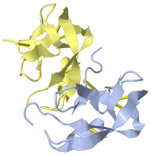
Asymmetric Unit
|
||||||||||||||||||||||||||||||||||||
Cis Peptide Bonds (1, 1)
Asymmetric Unit
|
||||||||
SAPs(SNPs)/Variants (0, 0)| (no "SAP(SNP)/Variant" information available for 3FPR) |
PROSITE Motifs (0, 0)| (no "PROSITE Motif" information available for 3FPR) |
Exons (0, 0)| (no "Exon" information available for 3FPR) |
Sequences/Alignments
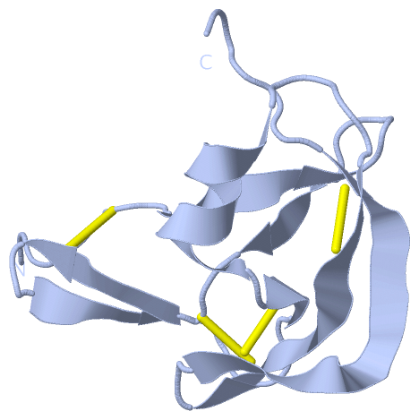
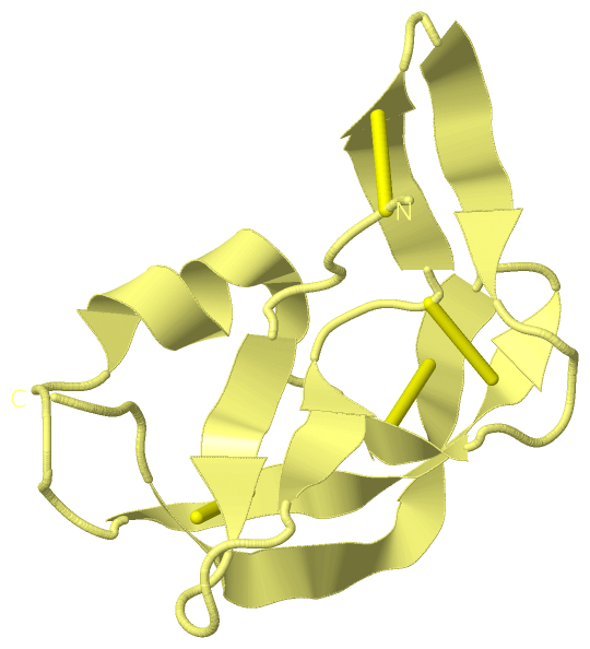
Asymmetric UnitChain A from PDB Type:PROTEIN Length:85 aligned with EVA1_RHISA | P0C8E7 from UniProtKB/Swiss-Prot Length:114 Alignment length:85 36 46 56 66 76 86 96 106 EVA1_RHISA 27 GDLGGCPFLVAENKTGYPTIVACKQDCNGTTETAPNGTRCFSIGDEGLRRMTANLPYDCPLGQCSNGDCIPKETYEVCYRRNWRD 111 SCOP domains ------------------------------------------------------------------------------------- SCOP domains CATH domains ------------------------------------------------------------------------------------- CATH domains Pfam domains ------------------------------------------------------------------------------------- Pfam domains SAPs(SNPs) ------------------------------------------------------------------------------------- SAPs(SNPs) PROSITE ------------------------------------------------------------------------------------- PROSITE Transcript ------------------------------------------------------------------------------------- Transcript 3fpr A 7 GDLGGCPFLVAENKTGYPTIVACKQDCNGTTETAPNGTRCFSIGDEGLRRMTANLPYDCPLGQCSNGDCIPKETYEVCYRRNWRD 91 16 26 36 46 56 66 76 86 Chain D from PDB Type:PROTEIN Length:78 aligned with EVA1_RHISA | P0C8E7 from UniProtKB/Swiss-Prot Length:114 Alignment length:78 40 50 60 70 80 90 100 EVA1_RHISA 31 GCPFLVAENKTGYPTIVACKQDCNGTTETAPNGTRCFSIGDEGLRRMTANLPYDCPLGQCSNGDCIPKETYEVCYRRN 108 SCOP domains ------------------------------------------------------------------------------ SCOP domains CATH domains ------------------------------------------------------------------------------ CATH domains Pfam domains ------------------------------------------------------------------------------ Pfam domains SAPs(SNPs) ------------------------------------------------------------------------------ SAPs(SNPs) PROSITE ------------------------------------------------------------------------------ PROSITE Transcript ------------------------------------------------------------------------------ Transcript 3fpr D 11 GCPFLVAENKTGYPTIVACKQDCNGTTETAPNGTRCFSIGDEGLRRMTANLPYDCPLGQCSNGDCIPKETYEVCYRRN 88 20 30 40 50 60 70 80
|
||||||||||||||||||||
SCOP Domains (0, 0)| (no "SCOP Domain" information available for 3FPR) |
CATH Domains (0, 0)| (no "CATH Domain" information available for 3FPR) |
Pfam Domains (0, 0)| (no "Pfam Domain" information available for 3FPR) |
Gene Ontology (1, 1)|
Asymmetric Unit(hide GO term definitions) Chain A,D (EVA1_RHISA | P0C8E7)
|
||||||||||||
Interactive Views
|
||||||||||||||||||||||||||||||||||||||||||||||||||||||||||||||||||||||||||||||||||||||||||||||||||||||||||||||||||||||||||||||||||||||||||||
Still Images
|
||||||||||||||||
Databases
|
||||||||||||||||||||||||||||||||||||||||||||||||||||||||||||||||||||||||||||||||||||||||||||||||||||||||||||||||||||||||||||||||||||||||||||||||||||||||||||||||
Analysis Tools
|
|||||||||||||||||||||||||||||||||||||||||||||||||||||||||||||
Entries Sharing at Least One Protein Chain (UniProt ID)
Related Entries Specified in the PDB File
|
|