Show PDB file:   
         Plain Text   HTML   (compressed file size)
QuickSearch:   
by PDB,NDB,UniProt,PROSITE Code or Search Term(s)  
(-)Asymmetric Unit
(-)Asym. Unit - sites
(-)Biological Unit 1
(-)Biol. Unit 1 - sites
(-)Biological Unit 2
(-)Biological Unit 3
collapse expand < >
Image Asymmetric Unit
Asymmetric Unit  (Jmol Viewer)
Image Asym. Unit - sites
Asym. Unit - sites  (Jmol Viewer)
Image Biological Unit 1
Biological Unit 1  (Jmol Viewer)
Image Biol. Unit 1 - sites
Biol. Unit 1 - sites  (Jmol Viewer)
Image Biological Unit 2
Biological Unit 2  (Jmol Viewer)
Image Biological Unit 3
Biological Unit 3  (Jmol Viewer)

(-) Description

Title :  THE CRYSTAL STRUCTURE OF THE COMPLEX BETWEEN EVASIN-1 AND CCL3
 
Authors :  J. P. Shaw, J. M. Dias
Date :  06 Jan 09  (Deposition) - 26 Jan 10  (Release) - 26 Jan 10  (Revision)
Method :  X-RAY DIFFRACTION
Resolution :  2.70
Chains :  Asym. Unit :  A,B,C
Biol. Unit 1:  A  (1x)
Biol. Unit 2:  B  (1x)
Biol. Unit 3:  C  (1x)
Keywords :  Novel Fold, Glycosylated Protein, Glycoprotein, Secreted, Immune System (Keyword Search: [Gene Ontology, PubMed, Web (Google))
 
Reference :  J. M. Dias, C. Losberger, M. Deruaz, C. A. Power, A. E. I. Proudfoot, J. P. Shaw
Structural Basis Of Chemokine Sequestration By A Tick Chemokine Binding Protein: The Crystal Structure Of The Complex Between Evasin-1 And Ccl3
Plos One V. 4 2009
PubMed-ID: 20041127  |  Reference-DOI: 10.1371/JOURNAL.PONE.0008514
(for further references see the PDB file header)

(-) Compounds

Molecule 1 - EVASIN-1
    ChainsA, B, C
    EngineeredYES
    Expression SystemSPODOPTERA FRUGIPERDA
    Expression System PlasmidPFASTBAC
    Expression System StrainSF9
    Expression System Taxid7108
    Expression System Vector TypeBACULOVIRUS
    Organism CommonBROWN DOG TICK
    Organism ScientificRHIPICEPHALUS SANGUINEUS
    Organism Taxid34632

 Structural Features

(-) Chains, Units

  123
Asymmetric Unit ABC
Biological Unit 1 (1x)A  
Biological Unit 2 (1x) B 
Biological Unit 3 (1x)  C

Summary Information (see also Sequences/Alignments below)

(-) Ligands, Modified Residues, Ions  (2, 6)

Asymmetric Unit (2, 6)
No.NameCountTypeFull Name
1NAG4Ligand/IonN-ACETYL-D-GLUCOSAMINE
2NDG2Ligand/Ion2-(ACETYLAMINO)-2-DEOXY-A-D-GLUCOPYRANOSE
Biological Unit 1 (1, 2)
No.NameCountTypeFull Name
1NAG2Ligand/IonN-ACETYL-D-GLUCOSAMINE
2NDG-1Ligand/Ion2-(ACETYLAMINO)-2-DEOXY-A-D-GLUCOPYRANOSE
Biological Unit 2 (1, 1)
No.NameCountTypeFull Name
1NAG1Ligand/IonN-ACETYL-D-GLUCOSAMINE
2NDG-1Ligand/Ion2-(ACETYLAMINO)-2-DEOXY-A-D-GLUCOPYRANOSE
Biological Unit 3 (2, 3)
No.NameCountTypeFull Name
1NAG1Ligand/IonN-ACETYL-D-GLUCOSAMINE
2NDG2Ligand/Ion2-(ACETYLAMINO)-2-DEOXY-A-D-GLUCOPYRANOSE

(-) Sites  (6, 6)

Asymmetric Unit (6, 6)
No.NameEvidenceResiduesDescription
1AC1SOFTWAREASN A:19 , THR A:21 , HOH A:113 , GLU C:79BINDING SITE FOR RESIDUE NAG A 119
2AC2SOFTWAREASN A:42BINDING SITE FOR RESIDUE NAG A 142
3AC3SOFTWAREASN B:19 , HOH B:109BINDING SITE FOR RESIDUE NAG B 119
4AC4SOFTWARELYS A:78 , ASN C:19 , THR C:21 , TYR C:23 , ARG C:45 , GLU C:82BINDING SITE FOR RESIDUE NDG C 119
5AC5SOFTWAREASN C:34BINDING SITE FOR RESIDUE NDG C 134
6AC6SOFTWAREASN C:42BINDING SITE FOR RESIDUE NAG C 142

(-) SS Bonds  (12, 12)

Asymmetric Unit
No.Residues
1A:12 -A:33
2A:29 -A:70
3A:46 -A:75
4A:65 -A:84
5B:12 -B:33
6B:29 -B:70
7B:46 -B:75
8B:65 -B:84
9C:12 -C:33
10C:29 -C:70
11C:46 -C:75
12C:65 -C:84

(-) Cis Peptide Bonds  (3, 3)

Asymmetric Unit
No.Residues
1Leu A:9 -Gly A:10
2Gly B:11 -Cys B:12
3Thr B:36 -Thr B:37

 Sequence-Structure Mapping

(-) SAPs(SNPs)/Variants  (0, 0)

(no "SAP(SNP)/Variant" information available for 3FPT)

(-) PROSITE Motifs  (0, 0)

(no "PROSITE Motif" information available for 3FPT)

(-) Exons   (0, 0)

(no "Exon" information available for 3FPT)

(-) Sequences/Alignments

Asymmetric Unit
   Reformat: Number of residues per line =  ('0' or empty: single-line sequence representation)
  Number of residues per labelling interval =   
  UniProt sequence: complete  aligned part    
   Show mapping: SCOP domains CATH domains Pfam domains Secondary structure (by author)
SAPs(SNPs) PROSITE motifs Exons
(details for a mapped element are shown in a popup box when the mouse pointer rests over it)
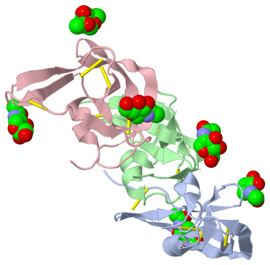
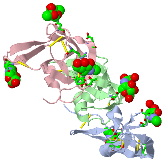
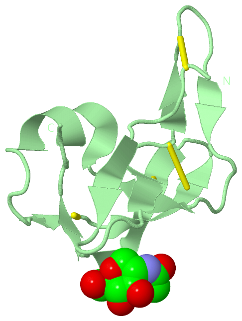
Chain A from PDB  Type:PROTEIN  Length:88
 aligned with EVA1_RHISA | P0C8E7 from UniProtKB/Swiss-Prot  Length:114

    Alignment length:88
                                                                                                                114 
                                    37        47        57        67        77        87        97       107      | 
           EVA1_RHISA    28 DLGGCPFLVAENKTGYPTIVACKQDCNGTTETAPNGTRCFSIGDEGLRRMTANLPYDCPLGQCSNGDCIPKETYEVCYRRNWRDKKN-   -
               SCOP domains ---------------------------------------------------------------------------------------- SCOP domains
               CATH domains ---------------------------------------------------------------------------------------- CATH domains
               Pfam domains ---------------------------------------------------------------------------------------- Pfam domains
         Sec.struct. author ........eee.....eee...eeee..eeee.....eee..hhhhhhh......eeeeeeeee..eeeeeeeeeee...hhhhh... Sec.struct. author
                 SAPs(SNPs) ---------------------------------------------------------------------------------------- SAPs(SNPs)
                    PROSITE ---------------------------------------------------------------------------------------- PROSITE
                 Transcript ---------------------------------------------------------------------------------------- Transcript
                 3fpt A   8 DLGGCPFLVAENKTGYPTIVACKQDCNGTTETAPNGTRCFSIGDEGLRRMTANLPYDCPLGQCSNGDCIPKETYEVCYRRNWRDKKNH  95
                                    17        27        37        47        57        67        77        87        

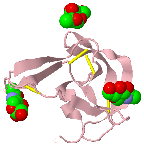
Chain B from PDB  Type:PROTEIN  Length:79
 aligned with EVA1_RHISA | P0C8E7 from UniProtKB/Swiss-Prot  Length:114

    Alignment length:79
                                    40        50        60        70        80        90       100         
           EVA1_RHISA    31 GCPFLVAENKTGYPTIVACKQDCNGTTETAPNGTRCFSIGDEGLRRMTANLPYDCPLGQCSNGDCIPKETYEVCYRRNW 109
               SCOP domains ------------------------------------------------------------------------------- SCOP domains
               CATH domains ------------------------------------------------------------------------------- CATH domains
               Pfam domains ------------------------------------------------------------------------------- Pfam domains
         Sec.struct. author .....eee.....eee...ee......ee.....eee.hhhhhhhhh.....ee..eeeee..eeeeeee..ee..... Sec.struct. author
                 SAPs(SNPs) ------------------------------------------------------------------------------- SAPs(SNPs)
                    PROSITE ------------------------------------------------------------------------------- PROSITE
                 Transcript ------------------------------------------------------------------------------- Transcript
                 3fpt B  11 GCPFLVAENKTGYPTIVACKQDCNGTTETAPNGTRCFSIGDEGLRRMTANLPYDCPLGQCSNGDCIPKETYEVCYRRNW  89
                                    20        30        40        50        60        70        80         

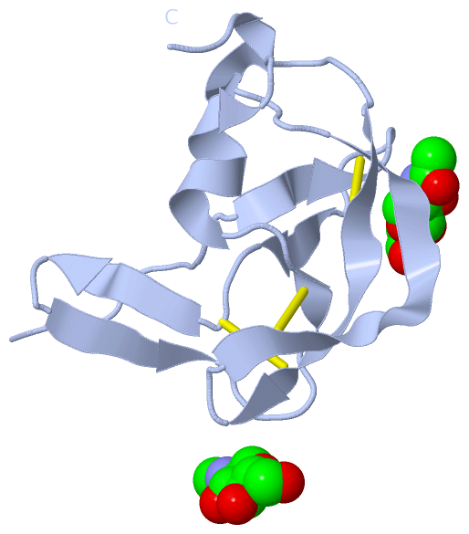
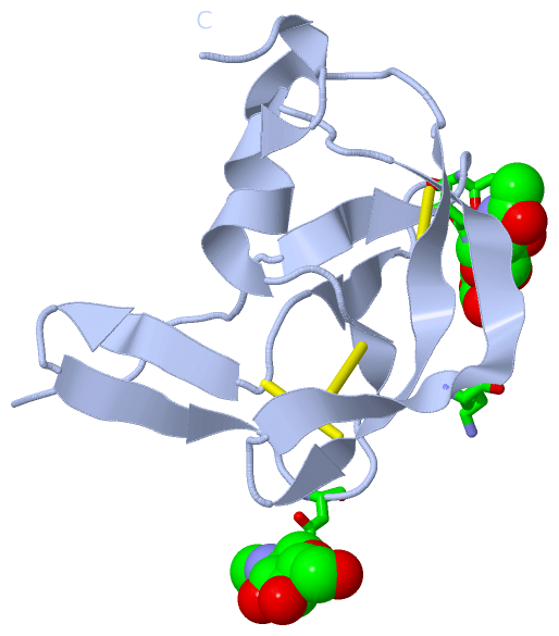
Chain C from PDB  Type:PROTEIN  Length:82
 aligned with EVA1_RHISA | P0C8E7 from UniProtKB/Swiss-Prot  Length:114

    Alignment length:82
                                    37        47        57        67        77        87        97       107  
           EVA1_RHISA    28 DLGGCPFLVAENKTGYPTIVACKQDCNGTTETAPNGTRCFSIGDEGLRRMTANLPYDCPLGQCSNGDCIPKETYEVCYRRNW 109
               SCOP domains ---------------------------------------------------------------------------------- SCOP domains
               CATH domains ---------------------------------------------------------------------------------- CATH domains
               Pfam domains ---------------------------------------------------------------------------------- Pfam domains
         Sec.struct. author ........eee.....eee...eeee..eeee.....eee.hhhhhhhhh.....eeeeeeeee..eeeeeeeeeee..... Sec.struct. author
                 SAPs(SNPs) ---------------------------------------------------------------------------------- SAPs(SNPs)
                    PROSITE ---------------------------------------------------------------------------------- PROSITE
                 Transcript ---------------------------------------------------------------------------------- Transcript
                 3fpt C   8 DLGGCPFLVAENKTGYPTIVACKQDCNGTTETAPNGTRCFSIGDEGLRRMTANLPYDCPLGQCSNGDCIPKETYEVCYRRNW  89
                                    17        27        37        47        57        67        77        87  

   Legend:   → Mismatch (orange background)
  - → Gap (green background, '-', border residues have a numbering label)
    → Modified Residue (blue background, lower-case, 'x' indicates undefined single-letter code, labelled with number + name)
  x → Chemical Group (purple background, 'x', labelled with number + name, e.g. ACE or NH2)
  extra numbering lines below/above indicate numbering irregularities and modified residue names etc., number ends below/above '|'

 Classification and Annotation

(-) SCOP Domains  (0, 0)

(no "SCOP Domain" information available for 3FPT)

(-) CATH Domains  (0, 0)

(no "CATH Domain" information available for 3FPT)

(-) Pfam Domains  (0, 0)

(no "Pfam Domain" information available for 3FPT)

(-) Gene Ontology  (1, 1)

Asymmetric Unit(hide GO term definitions)
Chain A,B,C   (EVA1_RHISA | P0C8E7)
cellular component
    GO:0005576    extracellular region    The space external to the outermost structure of a cell. For cells without external protective or external encapsulating structures this refers to space outside of the plasma membrane. This term covers the host cell environment outside an intracellular parasite.

 Visualization

(-) Interactive Views

Asymmetric Unit
  Complete Structure
    Jena3D(integrated viewing of ligand, site, SAP, PROSITE, SCOP information)
    WebMol | AstexViewer[tm]@PDBe
(Java Applets, require no local installation except for Java; loading may be slow)
    STRAP
(Java WebStart application, automatic local installation, requires Java; full application with system access!)
    RasMol
(require local installation)
    Molscript (VRML)
(requires installation of a VRML viewer; select preferred view via VRML and generate a mono or stereo PDF format file)
 
  Ligands, Modified Residues, Ions
    NAG  [ RasMol | Jena3D ]  +environment [ RasMol | Jena3D ]
    NDG  [ RasMol | Jena3D ]  +environment [ RasMol | Jena3D ]
 
  Sites
    AC1  [ RasMol ]  +environment [ RasMol ]
    AC2  [ RasMol ]  +environment [ RasMol ]
    AC3  [ RasMol ]  +environment [ RasMol ]
    AC4  [ RasMol ]  +environment [ RasMol ]
    AC5  [ RasMol ]  +environment [ RasMol ]
    AC6  [ RasMol ]  +environment [ RasMol ]
 
  Cis Peptide Bonds
    Gly B:11 - Cys B:12   [ RasMol ]  
    Leu A:9 - Gly A:10   [ RasMol ]  
    Thr B:36 - Thr B:37   [ RasMol ]  
 
Biological Units
  Complete Structure
    Biological Unit 1  [ Jena3D ]
    Biological Unit 2  [ Jena3D ]
    Biological Unit 3  [ Jena3D ]

(-) Still Images

Jmol
  protein: cartoon or spacefill or dots and stick; nucleic acid: cartoon and stick; ligands: spacefill; active site: stick
Molscript
  protein, nucleic acid: cartoon; ligands: spacefill; active site: ball and stick

 Databases and Analysis Tools

(-) Databases

Access by PDB/NDB ID
  3fpt
    Family and Domain InformationProDom | SYSTERS
    General Structural InformationGlycoscienceDB | MMDB | NDB | OCA | PDB | PDBe | PDBj | PDBsum | PDBWiki | PQS | PROTEOPEDIA
    Orientation in MembranesOPM
    Protein SurfaceSURFACE
    Secondary StructureDSSP (structure derived) | HSSP (homology derived)
    Structural GenomicsGeneCensus
    Structural NeighboursCE | VAST
    Structure ClassificationCATH | Dali | SCOP
    Validation and Original DataBMRB Data View | BMRB Restraints Grid | EDS | PROCHECK | RECOORD | WHAT_CHECK
 
Access by UniProt ID/Accession number
  EVA1_RHISA | P0C8E7
    Comparative Protein Structure ModelsModBase
    Genomic InformationEnsembl
    Protein-protein InteractionDIP
    Sequence, Family and Domain InformationInterPro | Pfam | SMART | UniProtKB/SwissProt
 
Access by Enzyme Classificator   (EC Number)
  (no 'Enzyme Classificator' available)
    General Enzyme InformationBRENDA | EC-PDB | Enzyme | IntEnz
    PathwayKEGG | MetaCyc
 
Access by Disease Identifier   (MIM ID)
  (no 'MIM ID' available)
    Disease InformationOMIM
 
Access by GenAge ID
  (no 'GenAge ID' available)
    Age Related InformationGenAge

(-) Analysis Tools

Access by PDB/NDB ID
    Domain InformationXDom
    Interatomic Contacts of Structural UnitsCSU
    Ligand-protein ContactsLPC
    Protein CavitiescastP
    Sequence and Secondary StructurePDBCartoon
    Structure AlignmentSTRAP(Java WebStart application, automatic local installation, requires Java; full application with system access!)
    Structure and Sequence BrowserSTING
 
Access by UniProt ID/Accession number
  EVA1_RHISA | P0C8E7
    Protein Disorder PredictionDisEMBL | FoldIndex | GLOBPLOT (for more information see DisProt)

 Related Entries

(-) Entries Sharing at Least One Protein Chain (UniProt ID)

UniProtKB/Swiss-Prot
        EVA1_RHISA | P0C8E73fpr 3fpu

(-) Related Entries Specified in the PDB File

3fpr UNCOMPLEXED NON-GLYCOSYLATED EVASIN-1
3fpu COMPLEX BETWEEN EVASIN-1 AND CCL3