|
|
|
|
 Description
|
|
Compounds
|
||||||||||||||||||||||||||||||||||||||||||||||||||||||||
Chains, Units
Summary Information (see also Sequences/Alignments below) |
Ligands, Modified Residues, Ions (1, 2)
Asymmetric/Biological Unit (1, 2)
|
Sites (2, 2)
Asymmetric Unit (2, 2)
|
SS Bonds (1, 1)
Asymmetric/Biological Unit
|
||||||||
Cis Peptide Bonds (0, 0)| (no "Cis Peptide Bond" information available for 2ZA9) |
SAPs(SNPs)/Variants (0, 0)| (no "SAP(SNP)/Variant" information available for 2ZA9) |
PROSITE Motifs (0, 0)| (no "PROSITE Motif" information available for 2ZA9) |
Exons (0, 0)| (no "Exon" information available for 2ZA9) |
Sequences/Alignments
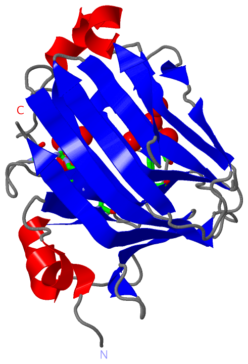
Asymmetric/Biological UnitChain A from PDB Type:PROTEIN Length:227 aligned with Q75WP3_9SPHN | Q75WP3 from UniProtKB/TrEMBL Length:308 Alignment length:227 90 100 110 120 130 140 150 160 170 180 190 200 210 220 230 240 250 260 270 280 290 300 Q75WP3_9SPHN 81 PAAAPGKNFDLSHWKLQLPDANTTEISSANLGLGYTSQYFYTDTDGAMTFWAPTTGGTTANSSYPRSELREMLDPSNSKVNWGWQGTHTMKLSGKTVQLPSSGKIIVAQIHGIMDDGTNAPPLVKAVFQDGQLDMQVKQNSDGTGSDVHNYFTGIKLGDLYNMEIRVTDGVAYVTMNGDTRSVDFVGKDAGWKNLKYYFKAGNYVQDNTSTGGSAIAKLYSLSVSHS 307 SCOP domains d2za9a_ A: automated matches SCOP domains CATH domains ----------------------------------------------------------------------------------------------------------------------------------------------------------------------------------------------------------------------------------- CATH domains Pfam domains --------Alginate_lyase2-2za9A01 A:89-306 - Pfam domains SAPs(SNPs) ----------------------------------------------------------------------------------------------------------------------------------------------------------------------------------------------------------------------------------- SAPs(SNPs) PROSITE ----------------------------------------------------------------------------------------------------------------------------------------------------------------------------------------------------------------------------------- PROSITE Transcript ----------------------------------------------------------------------------------------------------------------------------------------------------------------------------------------------------------------------------------- Transcript 2za9 A 81 PAAAPGKNFDLSHWKLQLPDANTTEISSANLGLGYTSQYFYTDTDGAMTFWAPTTGGTTACSSYPRSELREMLDPSNSKVNWGWQGTHTMKLSGKTVQLPSSGKIIVAQIHGIMDDGTCAPPLVKAVFQDGQLDMQVKQNSDGTGSDVHNYFTGIKLGDLYNMEIRVTDGVAYVTMNGDTRSVDFVGKDAGWKNLKYYFKAGNYVQDNTSTGGSAIAKLYSLSVSHS 307 90 100 110 120 130 140 150 160 170 180 190 200 210 220 230 240 250 260 270 280 290 300
|
||||||||||||||||||||
SCOP Domains (1, 1)
Asymmetric/Biological Unit

|
CATH Domains (0, 0)| (no "CATH Domain" information available for 2ZA9) |
Pfam Domains (1, 1)
Asymmetric/Biological Unit
|
Gene Ontology (1, 1)|
Asymmetric/Biological Unit(hide GO term definitions) Chain A (Q75WP3_9SPHN | Q75WP3)
|
||||||||||||
Interactive Views
|
|||||||||||||||||||||||||||||||||||||||||||||||||||||||||||||||||||||||||||||||||||||||||||||||||||||||||||||||||||||||||||||
Still Images
|
||||||||||||||||
Databases
|
||||||||||||||||||||||||||||||||||||||||||||||||||||||||||||||||||||||||||||||||||||||||||||||||||||||||||||||||||||||||||||||||||||||||||||||||||||||||||||||||
Analysis Tools
|
|||||||||||||||||||||||||||||||||||||||||||||||||||||||||||||
Entries Sharing at Least One Protein Chain (UniProt ID)
Related Entries Specified in the PDB File
|
|