|
|
|
|
 Description
|
|
Compounds
|
||||||||||||||||||||||||||||||||||||||||||||
Chains, Units
Summary Information (see also Sequences/Alignments below) |
Ligands, Modified Residues, Ions (2, 3)| Asymmetric/Biological Unit (2, 3) |
Sites (3, 3)
Asymmetric Unit (3, 3)
|
SS Bonds (0, 0)| (no "SS Bond" information available for 2CWS) |
Cis Peptide Bonds (0, 0)| (no "Cis Peptide Bond" information available for 2CWS) |
SAPs(SNPs)/Variants (0, 0)| (no "SAP(SNP)/Variant" information available for 2CWS) |
PROSITE Motifs (0, 0)| (no "PROSITE Motif" information available for 2CWS) |
Exons (0, 0)| (no "Exon" information available for 2CWS) |
Sequences/Alignments
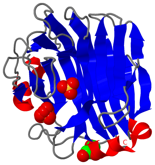
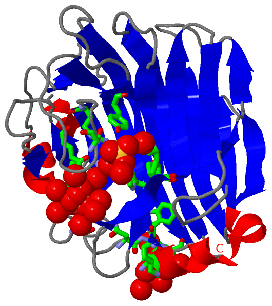
Asymmetric/Biological UnitChain A from PDB Type:PROTEIN Length:227 aligned with Q75WP3_9SPHN | Q75WP3 from UniProtKB/TrEMBL Length:308 Alignment length:227 308 92 102 112 122 132 142 152 162 172 182 192 202 212 222 232 242 252 262 272 282 292 302 | Q75WP3_9SPHN 83 AAPGKNFDLSHWKLQLPDANTTEISSANLGLGYTSQYFYTDTDGAMTFWAPTTGGTTANSSYPRSELREMLDPSNSKVNWGWQGTHTMKLSGKTVQLPSSGKIIVAQIHGIMDDGTNAPPLVKAVFQDGQLDMQVKQNSDGTGSDVHNYFTGIKLGDLYNMEIRVTDGVAYVTMNGDTRSVDFVGKDAGWKNLKYYFKAGNYVQDNTSTGGSAIAKLYSLSVSHSN- - SCOP domains d2cwsa_ A: automated matches SCOP domains CATH domains ----------------------------------------------------------------------------------------------------------------------------------------------------------------------------------------------------------------------------------- CATH domains Pfam domains ----------------------------------------------------------------------------------------------------------------------------------------------------------------------------------------------------------------------------------- Pfam domains SAPs(SNPs) ----------------------------------------------------------------------------------------------------------------------------------------------------------------------------------------------------------------------------------- SAPs(SNPs) PROSITE ----------------------------------------------------------------------------------------------------------------------------------------------------------------------------------------------------------------------------------- PROSITE Transcript ----------------------------------------------------------------------------------------------------------------------------------------------------------------------------------------------------------------------------------- Transcript 2cws A 83 AAPGKNFDLSHWKLQLPDANTTEISSANLGLGYTSQYFYTDTDGAMTFWAPTTGGTTANSSYPRSELREMLDPSNSKVNWGWQGTHTMKLSGKTVQLPSSGKIIVAQIHGIMDDGTNAPPLVKAVFQDGQLDMQVKQNSDGTGSDVHNYFTGIKLGDLYNMEIRVTDGVAYVTMNGDTRSVDFVGKDAGWKNLKYYFKAGNYVQDNTSTGGSAIAKLYSLSVSHSNL 309 92 102 112 122 132 142 152 162 172 182 192 202 212 222 232 242 252 262 272 282 292 302
|
||||||||||||||||||||
SCOP Domains (1, 1)
Asymmetric/Biological Unit
 |
CATH Domains (0, 0)| (no "CATH Domain" information available for 2CWS) |
Pfam Domains (0, 0)| (no "Pfam Domain" information available for 2CWS) |
Gene Ontology (1, 1)|
Asymmetric/Biological Unit(hide GO term definitions) Chain A (Q75WP3_9SPHN | Q75WP3)
|
||||||||||||
Interactive Views
|
|||||||||||||||||||||||||||||||||||||||||||||||||||||||||||||||||||||||||||||||||||||||||||||||||||||||||||||||||||||||||||||||||||||||||||
Still Images
|
||||||||||||||||
Databases
|
||||||||||||||||||||||||||||||||||||||||||||||||||||||||||||||||||||||||||||||||||||||||||||||||||||||||||||||||||||||||||||||||||||||||||||||||||||||||||||||||
Analysis Tools
|
|||||||||||||||||||||||||||||||||||||||||||||||||||||||||||||
Entries Sharing at Least One Protein Chain (UniProt ID)
Related Entries Specified in the PDB File
|
|