Show PDB file:   
         Plain Text   HTML   (compressed file size)
QuickSearch:   
by PDB,NDB,UniProt,PROSITE Code or Search Term(s)  
(-)Biol.Unit 1 - manually
(-)Asymmetric Unit
(-)Asym. Unit - sites
(-)Biological Unit 1
(-)Biol. Unit 1 - sites
collapse expand < >
Image Biol.Unit 1 - manually
Biol.Unit 1 - manually  (Jmol Viewer)
Image Asymmetric Unit
Asymmetric Unit  (Jmol Viewer)
Image Asym. Unit - sites
Asym. Unit - sites  (Jmol Viewer)
Image Biological Unit 1
Biological Unit 1  (Jmol Viewer)
Image Biol. Unit 1 - sites
Biol. Unit 1 - sites  (Jmol Viewer)

(-) Description

Title :  CRYSTAL STRUCTURE OF NEAT DOMAIN FROM STAPHYLOCOCCUS AUREUS IN COMPLEX WITH HEME
 
Authors :  Y. Tanaka, A. Suenaga, K. Tsumoto
Date :  31 Jul 07  (Deposition) - 29 Jul 08  (Release) - 09 Dec 08  (Revision)
Method :  X-RAY DIFFRACTION
Resolution :  1.90
Chains :  Asym. Unit :  A
Biol. Unit 1:  A  (2x)
Keywords :  Igg-Like Fold, Cell Wall, Peptidoglycan-Anchor, Secreted, Heme Binding Protein (Keyword Search: [Gene Ontology, PubMed, Web (Google))
 
Reference :  M. Watanabe, Y. Tanaka, A. Suenaga, M. Kuroda, M. Yao, N. Watanabe, F. Arisaka, T. Ohta, I. Tanaka, K. Tsumoto
Structural Basis For Multimeric Heme Complexation Through A Specific Protein-Heme Interaction: The Case Of The Third Neat Domain Of Isdh From Staphylococcus Aureus
J. Biol. Chem. V. 283 28649 2008
PubMed-ID: 18667422  |  Reference-DOI: 10.1074/JBC.M803383200
(for further references see the PDB file header)

(-) Compounds

Molecule 1 - IRON-REGULATED SURFACE DETERMINANT PROTEIN H
    ChainsA
    EngineeredYES
    Expression SystemESCHERICHIA COLI
    Expression System PlasmidPDBHT3
    Expression System StrainBL21(DE3)
    Expression System Taxid469008
    Expression System Vector TypePLASMID
    FragmentNEAT 3 DOMAIN, RESIDUE 539-664
    GeneISDH
    Organism ScientificSTAPHYLOCOCCUS AUREUS
    Organism Taxid158878
    StrainMU50
    SynonymHAPTOGLOBIN RECEPTOR A, STAPHYLOCOCCUS AUREUS SURFACE PROTEIN I

 Structural Features

(-) Chains, Units

  1
Asymmetric Unit A
Biological Unit 1 (2x)A

Summary Information (see also Sequences/Alignments below)

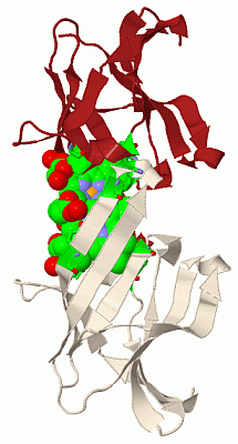
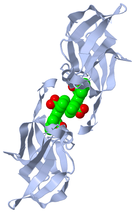
(-) Ligands, Modified Residues, Ions  (1, 1)

Asymmetric Unit (1, 1)
No.NameCountTypeFull Name
1HEM1Ligand/IonPROTOPORPHYRIN IX CONTAINING FE
Biological Unit 1 (1, 2)
No.NameCountTypeFull Name
1HEM2Ligand/IonPROTOPORPHYRIN IX CONTAINING FE

(-) Sites  (1, 1)

Asymmetric Unit (1, 1)
No.NameEvidenceResiduesDescription
1AC1SOFTWAREHOH A:2 , HOH A:33 , HOH A:76 , SER A:563 , VAL A:564 , PHE A:568 , TRP A:594 , VAL A:635 , ILE A:640 , TYR A:642 , TYR A:646 , VAL A:648BINDING SITE FOR RESIDUE HEM A3747

(-) SS Bonds  (0, 0)

(no "SS Bond" information available for 2Z6F)

(-) Cis Peptide Bonds  (1, 1)

Asymmetric Unit
No.Residues
1His A:571 -Pro A:572

 Sequence-Structure Mapping

(-) SAPs(SNPs)/Variants  (0, 0)

(no "SAP(SNP)/Variant" information available for 2Z6F)

(-) PROSITE Motifs  (0, 0)

(no "PROSITE Motif" information available for 2Z6F)

(-) Exons   (0, 0)

(no "Exon" information available for 2Z6F)

(-) Sequences/Alignments

Asymmetric Unit
   Reformat: Number of residues per line =  ('0' or empty: single-line sequence representation)
  Number of residues per labelling interval =   
  UniProt sequence: complete  aligned part    
   Show mapping: SCOP domains CATH domains Pfam domains Secondary structure (by author)
SAPs(SNPs) PROSITE motifs Exons
(details for a mapped element are shown in a popup box when the mouse pointer rests over it)
Chain A from PDB  Type:PROTEIN  Length:112
 aligned with ISDH_STAAM | Q931P4 from UniProtKB/Swiss-Prot  Length:891

    Alignment length:112
                                   553       563       573       583       593       603       613       623       633       643       653  
           ISDH_STAAM   544 LTDLQEAHFVVFESEENSESVMDGFVEHPFYTATLNGQKYVVMKTKDDSYWKDLIVEGKRVTTVSKDPKNNSRTLIFPYIPDKAVYNAIVKVVVANIGYEGQYHVRIINQDI 655
               SCOP domains d2z6fa_ A: automated matches                                                                                     SCOP domains
               CATH domains ---------------------------------------------------------------------------------------------------------------- CATH domains
               Pfam domains NEAT-2z6fA01 A:544-655                                                                                           Pfam domains
         Sec.struct. author .eeeeee.eeeee......hhhhhh....eeeeee..eeeeeee..hhh.eeeeee..ee.eeeeeehhhheeeeeee......eeeeeeeeeehhhheeeeeeeeeee... Sec.struct. author
                 SAPs(SNPs) ---------------------------------------------------------------------------------------------------------------- SAPs(SNPs)
                    PROSITE ---------------------------------------------------------------------------------------------------------------- PROSITE
                 Transcript ---------------------------------------------------------------------------------------------------------------- Transcript
                 2z6f A 544 LTDLQEAHFVVFESEENSESVMDGFVEHPFYTATLNGQKYVVMKTKDDSYWKDLIVEGKRVTTVSKDPKNNSRTLIFPYIPDKAVYNAIVKVVVANIGYEGQYHVRIINQDI 655
                                   553       563       573       583       593       603       613       623       633       643       653  
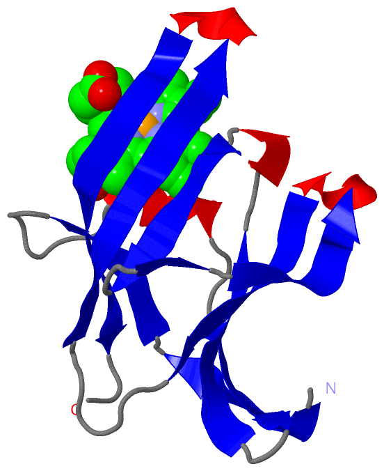
   Legend:   → Mismatch (orange background)
  - → Gap (green background, '-', border residues have a numbering label)
    → Modified Residue (blue background, lower-case, 'x' indicates undefined single-letter code, labelled with number + name)
  x → Chemical Group (purple background, 'x', labelled with number + name, e.g. ACE or NH2)
  extra numbering lines below/above indicate numbering irregularities and modified residue names etc., number ends below/above '|'

 Classification and Annotation

(-) SCOP Domains  (1, 1)

Asymmetric Unit

(-) CATH Domains  (0, 0)

(no "CATH Domain" information available for 2Z6F)
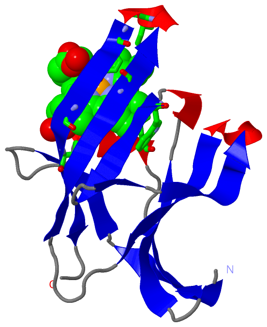
(-) Pfam Domains  (1, 1)

Asymmetric Unit

(-) Gene Ontology  (3, 3)

Asymmetric Unit(hide GO term definitions)
Chain A   (ISDH_STAAM | Q931P4)
cellular component
    GO:0005618    cell wall    The rigid or semi-rigid envelope lying outside the cell membrane of plant, fungal, most prokaryotic cells and some protozoan parasites, maintaining their shape and protecting them from osmotic lysis. In plants it is made of cellulose and, often, lignin; in fungi it is composed largely of polysaccharides; in bacteria it is composed of peptidoglycan; in protozoan parasites such as Giardia species, it's made of carbohydrates and proteins.
    GO:0005576    extracellular region    The space external to the outermost structure of a cell. For cells without external protective or external encapsulating structures this refers to space outside of the plasma membrane. This term covers the host cell environment outside an intracellular parasite.
    GO:0016020    membrane    A lipid bilayer along with all the proteins and protein complexes embedded in it an attached to it.

 Visualization

(-) Interactive Views

Asymmetric Unit
  Complete Structure
    Jena3D(integrated viewing of ligand, site, SAP, PROSITE, SCOP information)
    WebMol | AstexViewer[tm]@PDBe
(Java Applets, require no local installation except for Java; loading may be slow)
    STRAP
(Java WebStart application, automatic local installation, requires Java; full application with system access!)
    RasMol
(require local installation)
    Molscript (VRML)
(requires installation of a VRML viewer; select preferred view via VRML and generate a mono or stereo PDF format file)
 
  Ligands, Modified Residues, Ions
    HEM  [ RasMol | Jena3D ]  +environment [ RasMol | Jena3D ]
 
  Sites
    AC1  [ RasMol ]  +environment [ RasMol ]
 
  Cis Peptide Bonds
    His A:571 - Pro A:572   [ RasMol ]  
 
Biological Unit
  Complete Structure
    Biological Unit 1  [ Jena3D ]

(-) Still Images

Jmol
  protein: cartoon or spacefill or dots and stick; nucleic acid: cartoon and stick; ligands: spacefill; active site: stick
Molscript
  protein, nucleic acid: cartoon; ligands: spacefill; active site: ball and stick

 Databases and Analysis Tools

(-) Databases

Access by PDB/NDB ID
  2z6f
    Family and Domain InformationProDom | SYSTERS
    General Structural InformationGlycoscienceDB | MMDB | NDB | OCA | PDB | PDBe | PDBj | PDBsum | PDBWiki | PQS | PROTEOPEDIA
    Orientation in MembranesOPM
    Protein SurfaceSURFACE
    Secondary StructureDSSP (structure derived) | HSSP (homology derived)
    Structural GenomicsGeneCensus
    Structural NeighboursCE | VAST
    Structure ClassificationCATH | Dali | SCOP
    Validation and Original DataBMRB Data View | BMRB Restraints Grid | EDS | PROCHECK | RECOORD | WHAT_CHECK
 
Access by UniProt ID/Accession number
  ISDH_STAAM | Q931P4
    Comparative Protein Structure ModelsModBase
    Genomic InformationEnsembl
    Protein-protein InteractionDIP
    Sequence, Family and Domain InformationInterPro | Pfam | SMART | UniProtKB/SwissProt
 
Access by Enzyme Classificator   (EC Number)
  (no 'Enzyme Classificator' available)
    General Enzyme InformationBRENDA | EC-PDB | Enzyme | IntEnz
    PathwayKEGG | MetaCyc
 
Access by Disease Identifier   (MIM ID)
  (no 'MIM ID' available)
    Disease InformationOMIM
 
Access by GenAge ID
  (no 'GenAge ID' available)
    Age Related InformationGenAge

(-) Analysis Tools

Access by PDB/NDB ID
    Domain InformationXDom
    Interatomic Contacts of Structural UnitsCSU
    Ligand-protein ContactsLPC
    Protein CavitiescastP
    Sequence and Secondary StructurePDBCartoon
    Structure AlignmentSTRAP(Java WebStart application, automatic local installation, requires Java; full application with system access!)
    Structure and Sequence BrowserSTING
 
Access by UniProt ID/Accession number
  ISDH_STAAM | Q931P4
    Protein Disorder PredictionDisEMBL | FoldIndex | GLOBPLOT (for more information see DisProt)

 Related Entries

(-) Entries Sharing at Least One Protein Chain (UniProt ID)

UniProtKB/Swiss-Prot
        ISDH_STAAM | Q931P42e7d 3qug 3quh 3vtm 3vua

(-) Related Entries Specified in the PDB File

2e7d CRYSTAL STRUCTURE OF THE PROTEIN IN APO FORM
2h3k SOLUTION STRUCTURE OF THE PROTEIN WHICH HAS A SIMILAR MOTIF
2ite CRYSTAL STRUCTURE OF THE PROTEIN WHICH HAS A SIMILAR MOTIF
2itf CRYSTAL STRUCTURE OF THE PROTEIN WHICH HAS A SIMILAR MOTIF
2o1a CRYSTAL STRUCTURE OF THE PROTEIN WHICH HAS A SIMILAR MOTIF
2o6p CRYSTAL STRUCTURE OF THE PROTEIN WHICH HAS A SIMILAR MOTIF