|
|
|
|
 Description
|
|
Compounds
|
||||||||||||||||||||||||||||||||||||||||||||||||||||||||
Chains, Units
Summary Information (see also Sequences/Alignments below) |
Ligands, Modified Residues, Ions (3, 6)
Asymmetric Unit (3, 6)
|
Sites (6, 6)
Asymmetric Unit (6, 6)
|
SS Bonds (0, 0)| (no "SS Bond" information available for 2E7D) |
Cis Peptide Bonds (2, 2)
Asymmetric Unit
|
||||||||||||
SAPs(SNPs)/Variants (0, 0)| (no "SAP(SNP)/Variant" information available for 2E7D) |
PROSITE Motifs (1, 1)
Asymmetric Unit (1, 1)
|
||||||||||||||||||||||||||||||||||||||||||||||||||||||||||||||||||||||||
Exons (0, 0)| (no "Exon" information available for 2E7D) |
Sequences/Alignments
Asymmetric UnitChain A from PDB Type:PROTEIN Length:116 aligned with ISDH_STAAM | Q931P4 from UniProtKB/Swiss-Prot Length:891 Alignment length:116 551 561 571 581 591 601 611 621 631 641 651 ISDH_STAAM 542 DQLTDLQEAHFVVFESEENSESVMDGFVEHPFYTATLNGQKYVVMKTKDDSYWKDLIVEGKRVTTVSKDPKNNSRTLIFPYIPDKAVYNAIVKVVVANIGYEGQYHVRIINQDINT 657 SCOP domains d2e7da_ A: automated matches SCOP domains CATH domains -------------------------------------------------------------------------------------------------------------------- CATH domains Pfam domains -------------------------------------------------------------------------------------------------------------------- Pfam domains SAPs(SNPs) -------------------------------------------------------------------------------------------------------------------- SAPs(SNPs) PROSITE -NEAT PDB: A:10-124 UniProt: 543-660 PROSITE Transcript -------------------------------------------------------------------------------------------------------------------- Transcript 2e7d A 9 DQLTDLQEAHFVVFESEENSESVMDGFVEHPFYTATLNGQKYVVMKTKDDSYWKDLIVEGKRVTTVSKDPKNNSRTLIFPYIPDKAVYNAIVKVVVANIGYEGQYHVRIINQDINT 124 18 28 38 48 58 68 78 88 98 108 118 Chain B from PDB Type:PROTEIN Length:112 aligned with ISDH_STAAM | Q931P4 from UniProtKB/Swiss-Prot Length:891 Alignment length:112 553 563 573 583 593 603 613 623 633 643 653 ISDH_STAAM 544 LTDLQEAHFVVFESEENSESVMDGFVEHPFYTATLNGQKYVVMKTKDDSYWKDLIVEGKRVTTVSKDPKNNSRTLIFPYIPDKAVYNAIVKVVVANIGYEGQYHVRIINQDI 655 SCOP domains d2e7db_ B: automated matches SCOP domains CATH domains ---------------------------------------------------------------------------------------------------------------- CATH domains Pfam domains ---------------------------------------------------------------------------------------------------------------- Pfam domains SAPs(SNPs) ---------------------------------------------------------------------------------------------------------------- SAPs(SNPs) PROSITE NEAT PDB: - UniProt: 543-660 PROSITE Transcript ---------------------------------------------------------------------------------------------------------------- Transcript 2e7d B 11 LTDLQEAHFVVFESEENSESVMDGFVEHPFYTATLNGQKYVVMKTKDDSYWKDLIVEGKRVTTVSKDPKNNSRTLIFPYIPDKAVYNAIVKVVVANIGYEGQYHVRIINQDI 122 20 30 40 50 60 70 80 90 100 110 120
|
||||||||||||||||||||
SCOP Domains (1, 2)
Asymmetric Unit
 |
CATH Domains (0, 0)| (no "CATH Domain" information available for 2E7D) |
Pfam Domains (0, 0)| (no "Pfam Domain" information available for 2E7D) |
Gene Ontology (3, 3)|
Asymmetric Unit(hide GO term definitions) Chain A,B (ISDH_STAAM | Q931P4)
|
||||||||||||||||||||||||
Interactive Views
|
||||||||||||||||||||||||||||||||||||||||||||||||||||||||||||||||||||||||||||||||||||||||||||||||||||||||||||||||||||||||||||||||||||||||||||||||||||||||||||||||||||||||||||||||||||||||||||||||||||||
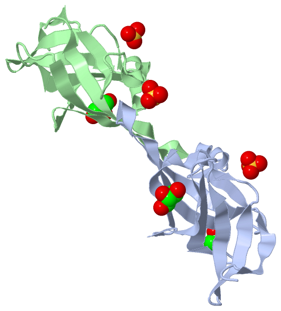
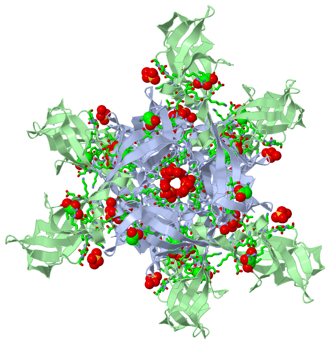
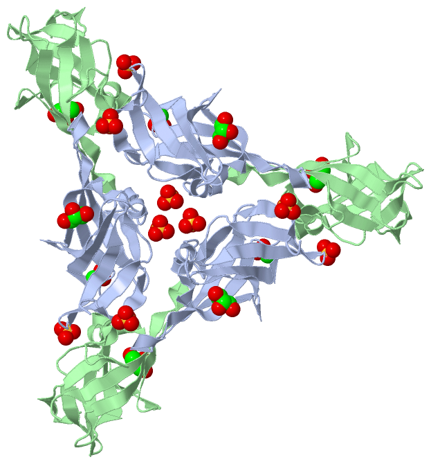
Still Images
|
||||||||||||||||
Databases
|
||||||||||||||||||||||||||||||||||||||||||||||||||||||||||||||||||||||||||||||||||||||||||||||||||||||||||||||||||||||||||||||||||||||||||||||||||||||||||||||||
Analysis Tools
|
|||||||||||||||||||||||||||||||||||||||||||||||||||||||||||||
Entries Sharing at Least One Protein Chain (UniProt ID)
Related Entries Specified in the PDB File
|
|