|
|
|
|
 Description
|
|
Compounds
|
||||||||||||||||||||||||||||||||||||||||||||||||||||||||
Chains, Units
Summary Information (see also Sequences/Alignments below) |
Ligands, Modified Residues, Ions (2, 3)| Asymmetric/Biological Unit (2, 3) |
Sites (3, 3)
Asymmetric Unit (3, 3)
|
SS Bonds (4, 4)
Asymmetric/Biological Unit
|
||||||||||||||||||||
Cis Peptide Bonds (0, 0)| (no "Cis Peptide Bond" information available for 2Z2F) |
SAPs(SNPs)/Variants (0, 0)| (no "SAP(SNP)/Variant" information available for 2Z2F) |
PROSITE Motifs (2, 2)
Asymmetric/Biological Unit (2, 2)
|
||||||||||||||||||||||||||||||||
Exons (0, 0)| (no "Exon" information available for 2Z2F) |
Sequences/Alignments
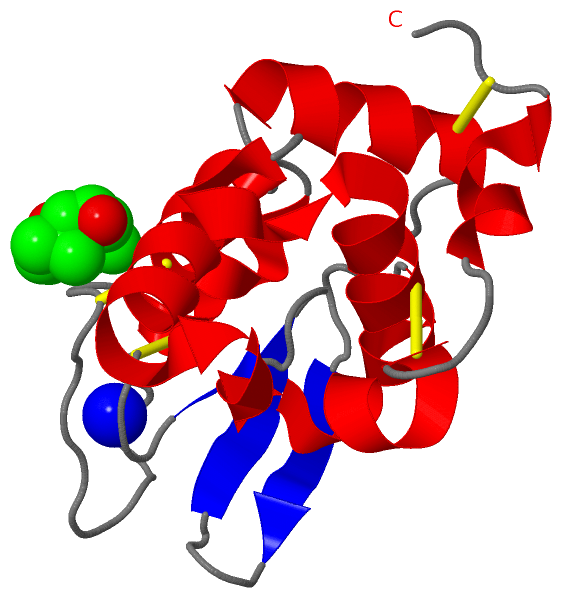
Asymmetric/Biological UnitChain A from PDB Type:PROTEIN Length:129 aligned with LYSC2_BOVIN | Q06283 from UniProtKB/Swiss-Prot Length:147 Alignment length:129 28 38 48 58 68 78 88 98 108 118 128 138 LYSC2_BOVIN 19 KVFERCELARTLKKLGLDGYKGVSLANWLCLTKWESSYNTKATNYNPSSESTDYGIFQINSKWWCNDGKTPNAVDGCHVSCSELMENDIAKAVACAKHIVSEQGITAWVAWKSHCRDHDVSSYVEGCTL 147 SCOP domains d2z2fa_ A: automated matches SCOP domains CATH domains 2z2fA00 A:1-129 [code=1.10.530.10, no name defined] CATH domains Pfam domains Lys-2z2fA01 A:1-127 -- Pfam domains SAPs(SNPs) --------------------------------------------------------------------------------------------------------------------------------- SAPs(SNPs) PROSITE (1) LACTALBUMIN_LYSOZYME_2 PDB: A:1-129 UniProt: 19-147 PROSITE (1) PROSITE (2) ----------------------------------------------------------------------------LACTALBUMIN_LYSOZYM---------------------------------- PROSITE (2) Transcript --------------------------------------------------------------------------------------------------------------------------------- Transcript 2z2f A 1 KVFERCELARTLKKLGLDGYKGVSLANWLCLTKWESSYNTKATNYNPSSESTDYGIFQINSKWWCNDGKTPNAVDGCHVSCSELMENDIAKAVACAKHIVSEQGITAWVAWKSHCRDHDVSSYVEGCTL 129 10 20 30 40 50 60 70 80 90 100 110 120
|
||||||||||||||||||||
SCOP Domains (1, 1)
Asymmetric/Biological Unit
 |
CATH Domains (1, 1)
Asymmetric/Biological Unit
|
Pfam Domains (1, 1)
Asymmetric/Biological Unit
|
Gene Ontology (8, 8)|
Asymmetric/Biological Unit(hide GO term definitions) Chain A (LYSC2_BOVIN | Q06283)
|
||||||||||||||||||||||||||||||||||||||||||||||||||||||||||||
Interactive Views
|
|||||||||||||||||||||||||||||||||||||||||||||||||||||||||||||||||||||||||||||||||||||||||||||||||||||||||||||||||||||||||||||||||||||||||||
Still Images
|
||||||||||||||||
Databases
|
||||||||||||||||||||||||||||||||||||||||||||||||||||||||||||||||||||||||||||||||||||||||||||||||||||||||||||||||||||||||||||||||||||||||||||||||||||||||||||||||
Analysis Tools
|
|||||||||||||||||||||||||||||||||||||||||||||||||||||||||||||
Entries Sharing at Least One Protein Chain (UniProt ID)
Related Entries Specified in the PDB File
|
|