|
|
|
|
 Description
|
|
Compounds
|
||||||||||||||||||||||||||||||||||||||||||||
Chains, Units
Summary Information (see also Sequences/Alignments below) |
Ligands, Modified Residues, Ions (0, 0)| (no "Ligand,Modified Residues,Ions" information available for 2YXM) |
Sites (0, 0)| (no "Site" information available for 2YXM) |
SS Bonds (0, 0)| (no "SS Bond" information available for 2YXM) |
Cis Peptide Bonds (0, 0)| (no "Cis Peptide Bond" information available for 2YXM) |
SAPs(SNPs)/Variants (0, 0)| (no "SAP(SNP)/Variant" information available for 2YXM) |
PROSITE Motifs (0, 0)| (no "PROSITE Motif" information available for 2YXM) |
Exons (0, 0)| (no "Exon" information available for 2YXM) |
Sequences/Alignments
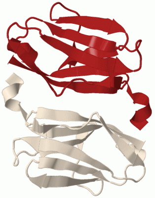
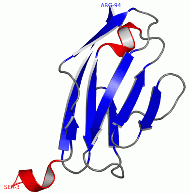
Asymmetric UnitChain A from PDB Type:PROTEIN Length:92 aligned with MYPC1_HUMAN | Q00872 from UniProtKB/Swiss-Prot Length:1141 Alignment length:161 187 197 207 217 227 237 247 257 267 277 287 297 307 317 327 337 MYPC1_HUMAN 178 SGEGQEDAGELDFSGLLKRREVKQQEEEPQVDVWELLKNAKPSEYEKIAFQYGITDLRGMLKRLKRMRREEKKSAAFAKILDPAYQVDKGGRVRFVVELADPKLEVKWYKNGQEIRPSTKYIFEHKGCQRILFINNCQMTDDSEYYVTAGDEKCSTELFVR 338 SCOP domains d2y xm a_ A: automated matches SCOP domains CATH domains ----------------------------------------------------------------------------------------------------------------------------------------------------------------- CATH domains Pfam domains ---------------------------------------------------------------------------I-set-2yxmA01 A:9-93 - Pfam domains SAPs(SNPs) ----------------------------------------------------------------------------------------------------------------------------------------------------------------- SAPs(SNPs) PROSITE ----------------------------------------------------------------------------------------------------------------------------------------------------------------- PROSITE Transcript ----------------------------------------------------------------------------------------------------------------------------------------------------------------- Transcript 2yxm A 3 SGS----------SG-----------------------------------------------------------AAFAKILDPAYQVDKGGRVRFVVELADPKLEVKWYKNGQEIRPSTKYIFEHKGCQRILFINNCQMTDDSEYYVTAGDEKCSTELFVR 94 | - || - - - - - - | 13 23 33 43 53 63 73 83 93 5 6| 8 7
|
||||||||||||||||||||
SCOP Domains (1, 1)
Asymmetric Unit

|
CATH Domains (0, 0)| (no "CATH Domain" information available for 2YXM) |
Pfam Domains (1, 1)
Asymmetric Unit
|
Gene Ontology (11, 11)|
Asymmetric Unit(hide GO term definitions) Chain A (MYPC1_HUMAN | Q00872)
|
||||||||||||||||||||||||||||||||||||||||||||||||||||||||||||||||||||||||||||||||||||
Interactive Views
|
|||||||||||||||||||||||||||||||||||||||||||||||||||||||||||||||||||||||||||||||||||||||||||||||||||||||||||||||||||||||||||||||||||||||||||
Still Images
|
||||||||||||||||
Databases
|
||||||||||||||||||||||||||||||||||||||||||||||||||||||||||||||||||||||||||||||||||||||||||||||||||||||||||||||||||||||||||||||||||||||||||||||||||||||||||||||||
Analysis Tools
|
|||||||||||||||||||||||||||||||||||||||||||||||||||||||||||||
Entries Sharing at Least One Protein Chain (UniProt ID)
Related Entries Specified in the PDB File
|
|