| molecular function |
|---|
| | GO:0051400 | | BH domain binding | | Interacting selectively and non-covalently with the Bcl-2 homology (BH) domain of a protein. Bcl-2-related proteins share homology in one to four conserved regions designated the Bcl-2 homology (BH) domains BH1, BH2, BH3 and BH4. These domains contribute at multiple levels to the function of these proteins in cell death and survival. Anti-apoptotic members of the Bcl-2 family have four BH domains (BH1-BH4). Pro-apoptotic members have fewer BH domains. |
| | GO:0005515 | | protein binding | | Interacting selectively and non-covalently with any protein or protein complex (a complex of two or more proteins that may include other nonprotein molecules). |
| | GO:0046982 | | protein heterodimerization activity | | Interacting selectively and non-covalently with a nonidentical protein to form a heterodimer. |
| | GO:0042803 | | protein homodimerization activity | | Interacting selectively and non-covalently with an identical protein to form a homodimer. |
| biological process |
|---|
| | GO:0060011 | | Sertoli cell proliferation | | The multiplication or reproduction of Sertoli cells, resulting in the expansion of the Sertoli cell population. A Sertoli cell is a supporting cell projecting inward from the basement membrane of seminiferous tubules. |
| | GO:0006915 | | apoptotic process | | A programmed cell death process which begins when a cell receives an internal (e.g. DNA damage) or external signal (e.g. an extracellular death ligand), and proceeds through a series of biochemical events (signaling pathway phase) which trigger an execution phase. The execution phase is the last step of an apoptotic process, and is typically characterized by rounding-up of the cell, retraction of pseudopodes, reduction of cellular volume (pyknosis), chromatin condensation, nuclear fragmentation (karyorrhexis), plasma membrane blebbing and fragmentation of the cell into apoptotic bodies. When the execution phase is completed, the cell has died. |
| | GO:0097192 | | extrinsic apoptotic signaling pathway in absence of ligand | | A series of molecular signals in which a signal is conveyed from the cell surface to trigger the apoptotic death of a cell. The pathway starts with withdrawal of a ligand from a cell surface receptor, and ends when the execution phase of apoptosis is triggered. |
| | GO:0008630 | | intrinsic apoptotic signaling pathway in response to DNA damage | | A series of molecular signals in which an intracellular signal is conveyed to trigger the apoptotic death of a cell. The pathway is induced by the detection of DNA damage, and ends when the execution phase of apoptosis is triggered. |
| | GO:0043066 | | negative regulation of apoptotic process | | Any process that stops, prevents, or reduces the frequency, rate or extent of cell death by apoptotic process. |
| | GO:2001243 | | negative regulation of intrinsic apoptotic signaling pathway | | Any process that stops, prevents or reduces the frequency, rate or extent of intrinsic apoptotic signaling pathway. |
| | GO:0042981 | | regulation of apoptotic process | | Any process that modulates the occurrence or rate of cell death by apoptotic process. |
| | GO:0007283 | | spermatogenesis | | The process of formation of spermatozoa, including spermatocytogenesis and spermiogenesis. |
| cellular component |
|---|
| | GO:0005737 | | cytoplasm | | All of the contents of a cell excluding the plasma membrane and nucleus, but including other subcellular structures. |
| | GO:0005829 | | cytosol | | The part of the cytoplasm that does not contain organelles but which does contain other particulate matter, such as protein complexes. |
| | GO:0070062 | | extracellular exosome | | A vesicle that is released into the extracellular region by fusion of the limiting endosomal membrane of a multivesicular body with the plasma membrane. Extracellular exosomes, also simply called exosomes, have a diameter of about 40-100 nm. |
| | GO:0016020 | | membrane | | A lipid bilayer along with all the proteins and protein complexes embedded in it an attached to it. |
| | GO:0031966 | | mitochondrial membrane | | Either of the lipid bilayers that surround the mitochondrion and form the mitochondrial envelope. |
| | GO:0005741 | | mitochondrial outer membrane | | The outer, i.e. cytoplasm-facing, lipid bilayer of the mitochondrial envelope. |
| | GO:0005739 | | mitochondrion | | A semiautonomous, self replicating organelle that occurs in varying numbers, shapes, and sizes in the cytoplasm of virtually all eukaryotic cells. It is notably the site of tissue respiration. |
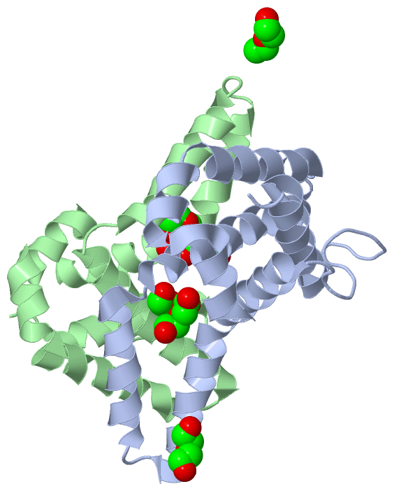
 Description
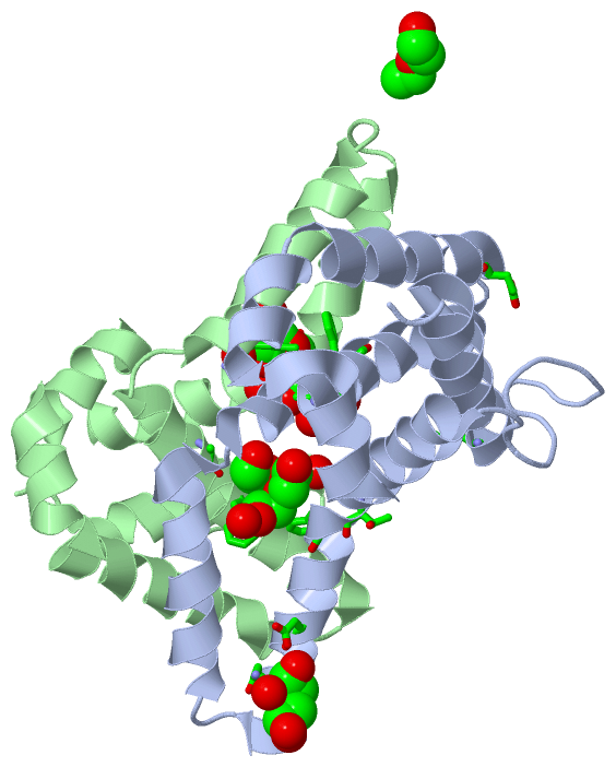