|
|
|
|
 Description
|
|
Compounds
|
||||||||||||||||||||||||||||||||||||||||||||||||||||
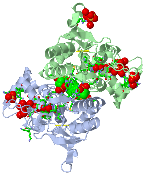
Chains, Units
Summary Information (see also Sequences/Alignments below) |
Ligands, Modified Residues, Ions (3, 8)| Asymmetric/Biological Unit (3, 8) |
Sites (8, 8)
Asymmetric Unit (8, 8)
|
SS Bonds (2, 2)
Asymmetric/Biological Unit
|
||||||||||||
Cis Peptide Bonds (6, 6)
Asymmetric/Biological Unit
|
||||||||||||||||||||||||||||
SAPs(SNPs)/Variants (0, 0)| (no "SAP(SNP)/Variant" information available for 2XHD) |
PROSITE Motifs (0, 0)| (no "PROSITE Motif" information available for 2XHD) |
Exons (0, 0)| (no "Exon" information available for 2XHD) |
Sequences/Alignments
Asymmetric/Biological UnitChain A from PDB Type:PROTEIN Length:262 aligned with GRIA2_HUMAN | P42262 from UniProtKB/Swiss-Prot Length:883 Alignment length:391 422 432 442 452 462 472 482 492 502 512 522 532 542 552 562 572 582 592 602 612 622 632 642 652 662 672 682 692 702 712 722 732 742 752 762 772 782 792 802 GRIA2_HUMAN 413 NKTVVVTTILESPYVMMKKNHEMLEGNERYEGYCVDLAAEIAKHCGFKYKLTIVGDGKYGARDADTKIWNGMVGELVYGKADIAIAPLTITLVREEVIDFSKPFMSLGISIMIKKPQKSKPGVFSFLDPLAYEIWMCIVFAYIGVSVVLFLVSRFSPYEWHTEEFEDGRETQSSESTNEFGIFNSLWFSLGAFMQQGCDISPRSLSGRIVGGVWWFFTLIIISSYTANLAAFLTVERMVSPIESAEDLSKQTEIAYGTLDSGSTKEFFRRSKIAVFDKMWTYMRSAEPSVFVRTTAEGVARVRKSKGKYAYLLESTMNEYIEQRKPCDTMKVGGNLDSKGYGIATPKGSSLRNAVNLAVLKLNEQGLLDKLKNKWWYDKGECGSGGGDSKE 803 SCOP domains d2xhda_ A: automated matches SCOP domains CATH domains -2xhdA01 A:4-109,A:219-258 Periplasmic binding protein-like II ----------------------------------------------------------------------------------------------------------------------------------------------------------------------------------------------------------------------------------------2xhdA01 A:4-109,A:219-258 ------------ CATH domains Pfam domains ------------------------------------------------------------------------------------------------------------------------------------------------------------------------------------------------------------------------------------------------------------------------------------------------------------------------------------------------------------------------------------------------------- Pfam domains SAPs(SNPs) ------------------------------------------------------------------------------------------------------------------------------------------------------------------------------------------------------------------------------------------------------------------------------------------------------------------------------------------------------------------------------------------------------- SAPs(SNPs) PROSITE ------------------------------------------------------------------------------------------------------------------------------------------------------------------------------------------------------------------------------------------------------------------------------------------------------------------------------------------------------------------------------------------------------- PROSITE Transcript ------------------------------------------------------------------------------------------------------------------------------------------------------------------------------------------------------------------------------------------------------------------------------------------------------------------------------------------------------------------------------------------------------- Transcript 2xhd A 3 NKTVVVTTILESPYVMMKKNHEMLEGNERYEGYCVDLAAEIAKHCGFKYKLTIVGDGKYGARDADTKIWNGMVGELVYGKADIAIAPLTITLVREEVIDFSKPFMSLGISIMIKKG---------------------------------------------------------------------------------------------------------------------------TPIESAEDLSKQTEIAYGTLDSGSTKEFFRRSKIAVFDKMWTYMRSAEPSVFVRTTAEGVARVRKSKGKYAYLLESTMNEYIEQRKPCDTMKVGGNLDSKGYGIATPKGSSLRTPVNLAVLKLSEQGVLDKLKNKWWYDKGECGA------E 301 12 22 32 42 52 62 72 82 92 102 112 | - - - - - - - - - - - - 119 129 139 149 159 169 179 189 199 209 219 229 239 249 259 | -| 118 119 263 301 Chain B from PDB Type:PROTEIN Length:257 aligned with GRIA2_HUMAN | P42262 from UniProtKB/Swiss-Prot Length:883 Alignment length:390 423 433 443 453 463 473 483 493 503 513 523 533 543 553 563 573 583 593 603 613 623 633 643 653 663 673 683 693 703 713 723 733 743 753 763 773 783 793 803 GRIA2_HUMAN 414 KTVVVTTILESPYVMMKKNHEMLEGNERYEGYCVDLAAEIAKHCGFKYKLTIVGDGKYGARDADTKIWNGMVGELVYGKADIAIAPLTITLVREEVIDFSKPFMSLGISIMIKKPQKSKPGVFSFLDPLAYEIWMCIVFAYIGVSVVLFLVSRFSPYEWHTEEFEDGRETQSSESTNEFGIFNSLWFSLGAFMQQGCDISPRSLSGRIVGGVWWFFTLIIISSYTANLAAFLTVERMVSPIESAEDLSKQTEIAYGTLDSGSTKEFFRRSKIAVFDKMWTYMRSAEPSVFVRTTAEGVARVRKSKGKYAYLLESTMNEYIEQRKPCDTMKVGGNLDSKGYGIATPKGSSLRNAVNLAVLKLNEQGLLDKLKNKWWYDKGECGSGGGDSKE 803 SCOP domains d2xhdb_ B: automated matches SCOP domains CATH domains 2xhdB01 B:4-109,B:219-262 Periplasmic binding protein-like II --------- ----------------------------------------------------------------------------------------------------2xhdB01 B:4-109,B:219-262 - CATH domains Pfam domains (1) -----------Lig_chan-Glu_bd-2xhdB03 B:15-80 -------------------------------------- -Lig_chan-2xhdB01 B:120-262 - Pfam domains (1) Pfam domains (2) -----------Lig_chan-Glu_bd-2xhdB04 B:15-80 -------------------------------------- -Lig_chan-2xhdB02 B:120-262 - Pfam domains (2) SAPs(SNPs) ------------------------------------------------------------------------------------------------------------------------------------------------------------------------------------------------------------------------------------------------------------------------------------------------------------------------------------------------------------------------------------------------------ SAPs(SNPs) PROSITE ------------------------------------------------------------------------------------------------------------------------------------------------------------------------------------------------------------------------------------------------------------------------------------------------------------------------------------------------------------------------------------------------------ PROSITE Transcript ------------------------------------------------------------------------------------------------------------------------------------------------------------------------------------------------------------------------------------------------------------------------------------------------------------------------------------------------------------------------------------------------------ Transcript 2xhd B 4 KTVVVTTILESPYVMMKKNHEMLEGNERYEGYCVDLAAEIAKHCGFKYKLTIVGDGKYGARDADTKIWNGMVGELVYGKADIAIAPLTITLVREEVIDFSKPFMSLGISIMIKKG---------------------------------------------------------------------------------------------------------------------------TPIESAEDLSKQTEIAYGTLDSGSTKEFFRRSKIAVFDKMWTYMRSAEPSVFVRTTAEGVARVRKSKGKYAYLLESTMNEYIEQRKPCDTMKVGGNLDSKGYGIATPKGSSLRTPVNLAVLKLSEQGVLDKLKNKWWYD---CG-------E 301 13 23 33 43 53 63 73 83 93 103 113 | - - - - - - - - - - - - 120 130 140 150 160 170 180 190 200 210 220 230 240 250 | -|| 301 118 119 257 261| 301 262
|
||||||||||||||||||||
SCOP Domains (1, 2)
Asymmetric/Biological Unit
 |
CATH Domains (1, 2)
Asymmetric/Biological Unit
|
Pfam Domains (2, 4)
Asymmetric/Biological Unit
|
Gene Ontology (24, 24)|
Asymmetric/Biological Unit(hide GO term definitions) Chain A,B (GRIA2_HUMAN | P42262)
|
||||||||||||||||||||||||||||||||||||||||||||||||||||||||||||||||||||||||||||||||||||||||||||||||||||||||||||||||||||||||||||||||||||||||||||||||||||||||||||||||||
Interactive Views
|
|||||||||||||||||||||||||||||||||||||||||||||||||||||||||||||||||||||||||||||||||||||||||||||||||||||||||||||||||||||||||||||||||||||||||||||||||||||||||||||||||||||||||||||||||||||||||||||||||||||||||||||||||||||||||
Still Images
|
||||||||||||||||
Databases
|
||||||||||||||||||||||||||||||||||||||||||||||||||||||||||||||||||||||||||||||||||||||||||||||||||||||||||||||||||||||||||||||||||||||||||||||||||||||||||||||||
Analysis Tools
|
|||||||||||||||||||||||||||||||||||||||||||||||||||||||||||||
Entries Sharing at Least One Protein Chain (UniProt ID)
Related Entries Specified in the PDB File
|
|