Show PDB file:   
         Plain Text   HTML   (compressed file size)
QuickSearch:   
by PDB,NDB,UniProt,PROSITE Code or Search Term(s)  
(-)Asymmetric Unit
(-)Asym. Unit - sites
(-)Biological Unit 1
(-)Biol. Unit 1 - sites
collapse expand < >
Image Asymmetric Unit
Asymmetric Unit  (Jmol Viewer)
Image Asym. Unit - sites
Asym. Unit - sites  (Jmol Viewer)
Image Biological Unit 1
Biological Unit 1  (Jmol Viewer)
Image Biol. Unit 1 - sites
Biol. Unit 1 - sites  (Jmol Viewer)

(-) Description

Title :  CRYSTAL STRUCTURE OF THE NATIVE E. COLI ZINC AMIDASE AMID
 
Authors :  S. Petrella, F. Kerff, R. Herman, C. Genereux, A. Pennartz, E. Sauvage, P. Charlier
Date :  18 Jun 09  (Deposition) - 12 Jan 10  (Release) - 13 Jul 11  (Revision)
Method :  X-RAY DIFFRACTION
Resolution :  1.80
Chains :  Asym. Unit :  A
Biol. Unit 1:  A  (2x)
Keywords :  Outer Membrane, Cell Wall Biogenesis/Degradation, Cell Membrane, Metal-Binding, Hydrolase (Keyword Search: [Gene Ontology, PubMed, Web (Google))
 
Reference :  F. Kerff, S. Petrella, F. Mercier, E. Sauvage, R. Herman, A. Pennartz, A. Zervosen, A. Luxen, J. M. Frere, B. Joris, P. Charlier
Specific Structural Features Of The N- Acetylmuramoyl-L-Alanine Amidase Amid From Escherichia Coli And Mechanistic Implications For Enzymes Of This Family.
J. Mol. Biol. V. 397 249 2010
PubMed-ID: 20036252  |  Reference-DOI: 10.1016/J.JMB.2009.12.038

(-) Compounds

Molecule 1 - N-ACETYLMURAMOYL-L-ALANINE AMIDASE AMID
    ChainsA
    EC Number3.5.1.28
    EngineeredYES
    Expression SystemESCHERICHIA COLI
    Expression System PlasmidPBAD/MYC-HISA
    Expression System StrainLMG194
    Expression System Taxid562
    Expression System Vector TypePLASMID
    FragmentRESIDUES 18-276
    Organism ScientificESCHERICHIA COLI
    Organism Taxid83333
    StrainK-12 MG1655

 Structural Features

(-) Chains, Units

  1
Asymmetric Unit A
Biological Unit 1 (2x)A

Summary Information (see also Sequences/Alignments below)
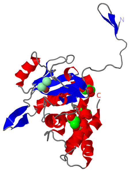
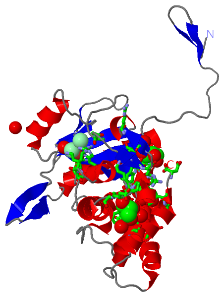
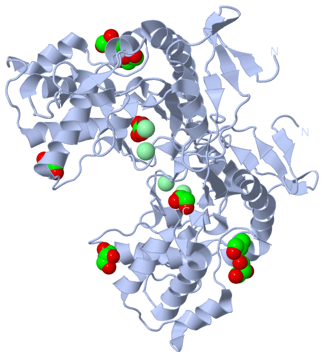
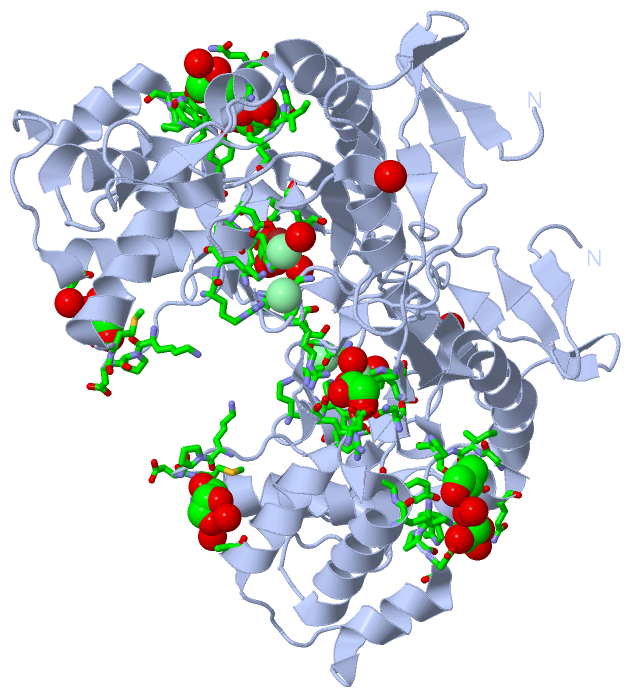
(-) Ligands, Modified Residues, Ions  (3, 7)

Asymmetric Unit (3, 7)
No.NameCountTypeFull Name
1CL2Ligand/IonCHLORIDE ION
2GOL4Ligand/IonGLYCEROL
3ZN1Ligand/IonZINC ION
Biological Unit 1 (1, 8)
No.NameCountTypeFull Name
1CL-1Ligand/IonCHLORIDE ION
2GOL8Ligand/IonGLYCEROL
3ZN-1Ligand/IonZINC ION

(-) Sites  (7, 7)

Asymmetric Unit (7, 7)
No.NameEvidenceResiduesDescription
1AC1SOFTWAREHIS A:35 , HIS A:151 , ASP A:161 , CL A:302BINDING SITE FOR RESIDUE ZN A 300
2AC2SOFTWAREALA A:85 , GLY A:86 , HIS A:151 , ARG A:158BINDING SITE FOR RESIDUE CL A 301
3AC3SOFTWAREHIS A:151 , LYS A:159 , ASP A:161 , ZN A:300 , HOH A:2091BINDING SITE FOR RESIDUE CL A 302
4AC4SOFTWAREALA A:201 , LYS A:215 , PRO A:216 , ASP A:217 , MET A:218 , HOH A:2036 , HOH A:2235 , HOH A:2236BINDING SITE FOR RESIDUE GOL A 303
5AC5SOFTWAREPRO A:145 , ALA A:172 , GLY A:175 , ILE A:176 , GLY A:177 , LYS A:257 , TYR A:258 , GOL A:305 , HOH A:2238BINDING SITE FOR RESIDUE GOL A 304
6AC6SOFTWAREGLN A:173 , TRP A:179 , PRO A:180 , ASP A:181 , LYS A:257 , GOL A:304 , HOH A:2238 , HOH A:2239BINDING SITE FOR RESIDUE GOL A 305
7AC7SOFTWAREVAL A:31 , TRP A:90 , ARG A:91 , ALA A:93 , ARG A:95 , ASN A:147 , ASP A:261 , HOH A:2089BINDING SITE FOR RESIDUE GOL A 306

(-) SS Bonds  (0, 0)

(no "SS Bond" information available for 2WKX)

(-) Cis Peptide Bonds  (1, 1)

Asymmetric Unit
No.Residues
1Asp A:161 -Pro A:162

 Sequence-Structure Mapping

(-) SAPs(SNPs)/Variants  (0, 0)

(no "SAP(SNP)/Variant" information available for 2WKX)

(-) PROSITE Motifs  (0, 0)

(no "PROSITE Motif" information available for 2WKX)

(-) Exons   (0, 0)

(no "Exon" information available for 2WKX)

(-) Sequences/Alignments

Asymmetric Unit
   Reformat: Number of residues per line =  ('0' or empty: single-line sequence representation)
  Number of residues per labelling interval =   
  UniProt sequence: complete  aligned part    
   Show mapping: SCOP domains CATH domains Pfam domains Secondary structure (by author)
SAPs(SNPs) PROSITE motifs Exons
(details for a mapped element are shown in a popup box when the mouse pointer rests over it)
Chain A from PDB  Type:PROTEIN  Length:257
 aligned with AMID_ECOLI | P75820 from UniProtKB/Swiss-Prot  Length:276

    Alignment length:257
                                    29        39        49        59        69        79        89        99       109       119       129       139       149       159       169       179       189       199       209       219       229       239       249       259       269       
           AMID_ECOLI    20 EKGIVEKEGYQLDTRRQAQAAYPRIKVLVIHYTADDFDSSLATLTDKQVSSHYLVPAVPPRYNGKPRIWQLVPEQELAWHAGISAWRGATRLNDTSIGIELENRGWQKSAGVKYFAPFEPAQIQALIPLAKDIIARYHIKPENVVAHADIAPQRKDDPGPLFPWQQLAQQGIGAWPDAQRVNFYLAGRAPHTPVDTASLLELLARYGYDVKPDMTPREQRRVIMAFQMHFRPTLYNGEADAETQAIAEALLEKYGQD 276
               SCOP domains d2wkxa2 A:5-179 Probable N-acetylmuramoyl-L-alanine amidase YbjR, N-terminal domain                                                                                            d2wkxa1 A:180-261                                                                  SCOP domains
               CATH domains ----------------------------------------------------------------------------------------------------------------------------------------------------------------------------------------------------------------------------------------------------------------- CATH domains
               Pfam domains ----------------------------------------------------------------------------------------------------------------------------------------------------------------------------------------------------------------------------------------------------------------- Pfam domains
         Sec.struct. author ...eee....eee.............eeeeee...hhhhhhhhhh.......eee.....ee..ee..ee..............ee..ee.hhh.eeeeee....eeee..eeee...hhhhhhhhhhhhhhhhhhhh.hhh.eeehhhhh...........hhhhhhhh......hhhhhhhhhh........hhhhhhhhhhhhh.......hhhhhhhhhhhhhhhhh........hhhhhhhhhhhhhhhh.. Sec.struct. author
                 SAPs(SNPs) ----------------------------------------------------------------------------------------------------------------------------------------------------------------------------------------------------------------------------------------------------------------- SAPs(SNPs)
                    PROSITE ----------------------------------------------------------------------------------------------------------------------------------------------------------------------------------------------------------------------------------------------------------------- PROSITE
                 Transcript ----------------------------------------------------------------------------------------------------------------------------------------------------------------------------------------------------------------------------------------------------------------- Transcript
                 2wkx A   5 EKGIVEKEGYQLDTRRQAQAAYPRIKVLVIHYTADDFDSSLATLTDKQVSSHYLVPAVPPRYNGKPRIWQLVPEQELAWHAGISAWRGATRLNDTSIGIELENRGWQKSAGVKYFAPFEPAQIQALIPLAKDIIARYHIKPENVVAHADIAPQRKDDPGPLFPWQQLAQQGIGAWPDAQRVNFYLAGRAPHTPVDTASLLELLARYGYDVKPDMTPREQRRVIMAFQMHFRPTLYNGEADAETQAIAEALLEKYGQD 261
                                    14        24        34        44        54        64        74        84        94       104       114       124       134       144       154       164       174       184       194       204       214       224       234       244       254       

   Legend:   → Mismatch (orange background)
  - → Gap (green background, '-', border residues have a numbering label)
    → Modified Residue (blue background, lower-case, 'x' indicates undefined single-letter code, labelled with number + name)
  x → Chemical Group (purple background, 'x', labelled with number + name, e.g. ACE or NH2)
  extra numbering lines below/above indicate numbering irregularities and modified residue names etc., number ends below/above '|'

 Classification and Annotation

(-) SCOP Domains  (2, 2)

Asymmetric Unit

(-) CATH Domains  (0, 0)

(no "CATH Domain" information available for 2WKX)

(-) Pfam Domains  (0, 0)

(no "Pfam Domain" information available for 2WKX)

(-) Gene Ontology  (11, 11)

Asymmetric Unit(hide GO term definitions)
Chain A   (AMID_ECOLI | P75820)
molecular function
    GO:0009392    N-acetyl-anhydromuramoyl-L-alanine amidase activity    Catalysis of the reaction: GlcNAc-1,6-anhMurNAc-L-Ala-gamma-D-Glu-DAP-D-Ala + H2O glcNAc-1,6-anhMurNAc + L-Ala-gamma-D-Glu-DAP-D-Ala.
    GO:0008745    N-acetylmuramoyl-L-alanine amidase activity    Catalysis of the hydrolysis of the link between N-acetylmuramoyl residues and L-amino acid residues in certain bacterial cell-wall glycopeptides.
    GO:0016787    hydrolase activity    Catalysis of the hydrolysis of various bonds, e.g. C-O, C-N, C-C, phosphoric anhydride bonds, etc. Hydrolase is the systematic name for any enzyme of EC class 3.
    GO:0046872    metal ion binding    Interacting selectively and non-covalently with any metal ion.
    GO:0008270    zinc ion binding    Interacting selectively and non-covalently with zinc (Zn) ions.
biological process
    GO:0071555    cell wall organization    A process that results in the assembly, arrangement of constituent parts, or disassembly of the cell wall, the rigid or semi-rigid envelope lying outside the cell membrane of plant, fungal and most prokaryotic cells, maintaining their shape and protecting them from osmotic lysis.
    GO:0009253    peptidoglycan catabolic process    The chemical reactions and pathways resulting in the breakdown of peptidoglycans, any of a class of glycoconjugates found in bacterial cell walls.
    GO:0009254    peptidoglycan turnover    The continual breakdown and regeneration of peptidoglycan required to maintain the cell wall.
cellular component
    GO:0009279    cell outer membrane    A lipid bilayer that forms the outermost membrane of the cell envelope; enriched in polysaccharide and protein; the outer leaflet of the membrane contains specific lipopolysaccharide structures.
    GO:0016020    membrane    A lipid bilayer along with all the proteins and protein complexes embedded in it an attached to it.
    GO:0019867    outer membrane    The external membrane of Gram-negative bacteria or certain organelles such as mitochondria and chloroplasts; freely permeable to most ions and metabolites.

 Visualization

(-) Interactive Views

Asymmetric Unit
  Complete Structure
    Jena3D(integrated viewing of ligand, site, SAP, PROSITE, SCOP information)
    WebMol | AstexViewer[tm]@PDBe
(Java Applets, require no local installation except for Java; loading may be slow)
    STRAP
(Java WebStart application, automatic local installation, requires Java; full application with system access!)
    RasMol
(require local installation)
    Molscript (VRML)
(requires installation of a VRML viewer; select preferred view via VRML and generate a mono or stereo PDF format file)
 
  Ligands, Modified Residues, Ions
    CL  [ RasMol | Jena3D ]  +environment [ RasMol | Jena3D ]
    GOL  [ RasMol | Jena3D ]  +environment [ RasMol | Jena3D ]
    ZN  [ RasMol | Jena3D ]  +environment [ RasMol | Jena3D ]
 
  Sites
    AC1  [ RasMol ]  +environment [ RasMol ]
    AC2  [ RasMol ]  +environment [ RasMol ]
    AC3  [ RasMol ]  +environment [ RasMol ]
    AC4  [ RasMol ]  +environment [ RasMol ]
    AC5  [ RasMol ]  +environment [ RasMol ]
    AC6  [ RasMol ]  +environment [ RasMol ]
    AC7  [ RasMol ]  +environment [ RasMol ]
 
  Cis Peptide Bonds
    Asp A:161 - Pro A:162   [ RasMol ]  
 
Biological Unit
  Complete Structure
    Biological Unit 1  [ Jena3D ]

(-) Still Images

Jmol
  protein: cartoon or spacefill or dots and stick; nucleic acid: cartoon and stick; ligands: spacefill; active site: stick
Molscript
  protein, nucleic acid: cartoon; ligands: spacefill; active site: ball and stick

 Databases and Analysis Tools

(-) Databases

Access by PDB/NDB ID
  2wkx
    Family and Domain InformationProDom | SYSTERS
    General Structural InformationGlycoscienceDB | MMDB | NDB | OCA | PDB | PDBe | PDBj | PDBsum | PDBWiki | PQS | PROTEOPEDIA
    Orientation in MembranesOPM
    Protein SurfaceSURFACE
    Secondary StructureDSSP (structure derived) | HSSP (homology derived)
    Structural GenomicsGeneCensus
    Structural NeighboursCE | VAST
    Structure ClassificationCATH | Dali | SCOP
    Validation and Original DataBMRB Data View | BMRB Restraints Grid | EDS | PROCHECK | RECOORD | WHAT_CHECK
 
Access by UniProt ID/Accession number
  AMID_ECOLI | P75820
    Comparative Protein Structure ModelsModBase
    Genomic InformationEnsembl
    Protein-protein InteractionDIP
    Sequence, Family and Domain InformationInterPro | Pfam | SMART | UniProtKB/SwissProt
 
Access by Enzyme Classificator   (EC Number)
  3.5.1.28
    General Enzyme InformationBRENDA | EC-PDB | Enzyme | IntEnz
    PathwayKEGG | MetaCyc
 
Access by Disease Identifier   (MIM ID)
  (no 'MIM ID' available)
    Disease InformationOMIM
 
Access by GenAge ID
  (no 'GenAge ID' available)
    Age Related InformationGenAge

(-) Analysis Tools

Access by PDB/NDB ID
    Domain InformationXDom
    Interatomic Contacts of Structural UnitsCSU
    Ligand-protein ContactsLPC
    Protein CavitiescastP
    Sequence and Secondary StructurePDBCartoon
    Structure AlignmentSTRAP(Java WebStart application, automatic local installation, requires Java; full application with system access!)
    Structure and Sequence BrowserSTING
 
Access by UniProt ID/Accession number
  AMID_ECOLI | P75820
    Protein Disorder PredictionDisEMBL | FoldIndex | GLOBPLOT (for more information see DisProt)

 Related Entries

(-) Entries Sharing at Least One Protein Chain (UniProt ID)

UniProtKB/Swiss-Prot
        AMID_ECOLI | P758202bh7 3d2y 3d2z

(-) Related Entries Specified in the PDB File

2bgx CRYSTAL STRUCTURE OF NATIVE AMID AT 1.8 ANGSTROM
2bh7 CRYSTAL STRUCTURE OF A SEMET DERIVATIVE OF AMID AT 2.2 ANGSTROMS