|
|
|
|
 Description
|
|
Compounds
|
||||||||||||||||||||||||
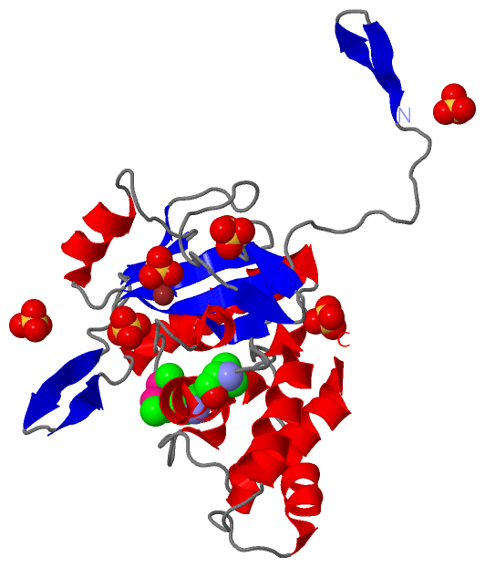
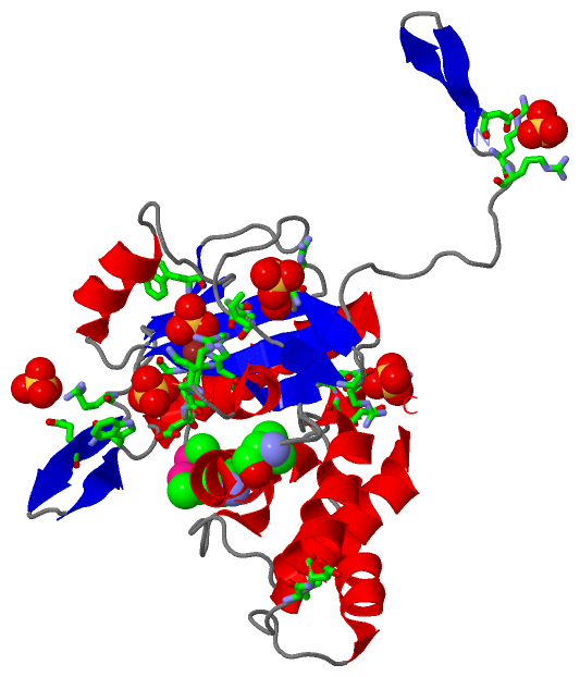
Chains, Units
Summary Information (see also Sequences/Alignments below) |
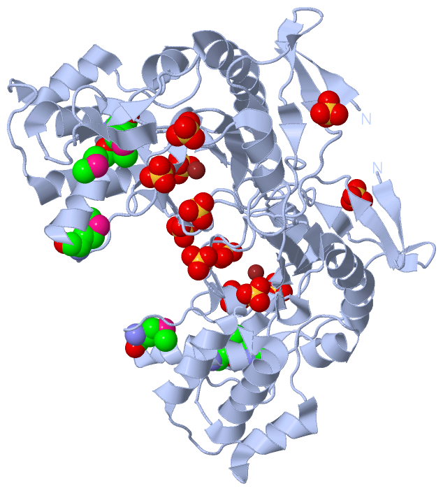
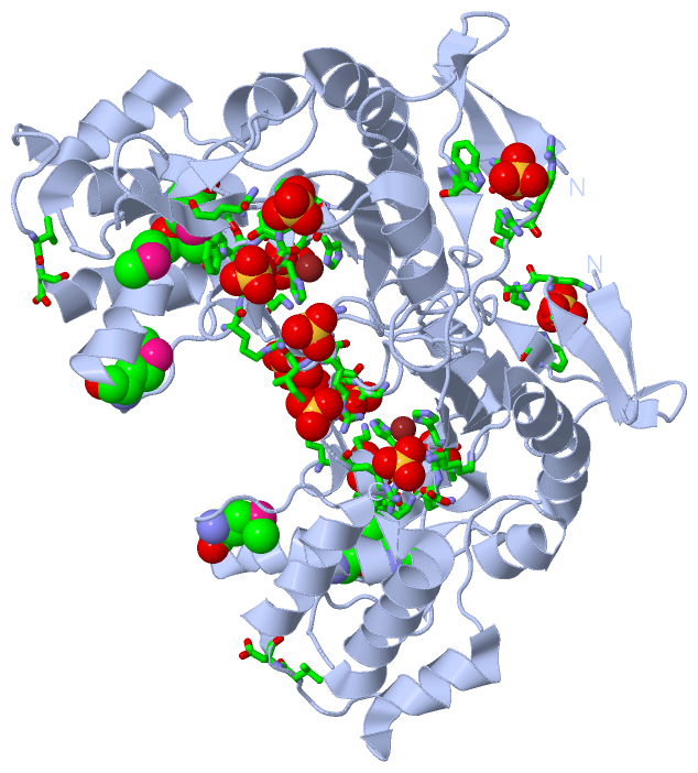
Ligands, Modified Residues, Ions (3, 10)| Asymmetric Unit (3, 10) Biological Unit 1 (2, 18) |
Sites (7, 7)
Asymmetric Unit (7, 7)
|
SS Bonds (0, 0)| (no "SS Bond" information available for 2BH7) |
Cis Peptide Bonds (1, 1)
Asymmetric Unit
|
||||||||
SAPs(SNPs)/Variants (0, 0)| (no "SAP(SNP)/Variant" information available for 2BH7) |
PROSITE Motifs (0, 0)| (no "PROSITE Motif" information available for 2BH7) |
Exons (0, 0)| (no "Exon" information available for 2BH7) |
Sequences/Alignments
Asymmetric UnitChain A from PDB Type:PROTEIN Length:254 aligned with AMID_ECOLI | P75820 from UniProtKB/Swiss-Prot Length:276 Alignment length:254 31 41 51 61 71 81 91 101 111 121 131 141 151 161 171 181 191 201 211 221 231 241 251 261 271 AMID_ECOLI 22 GIVEKEGYQLDTRRQAQAAYPRIKVLVIHYTADDFDSSLATLTDKQVSSHYLVPAVPPRYNGKPRIWQLVPEQELAWHAGISAWRGATRLNDTSIGIELENRGWQKSAGVKYFAPFEPAQIQALIPLAKDIIARYHIKPENVVAHADIAPQRKDDPGPLFPWQQLAQQGIGAWPDAQRVNFYLAGRAPHTPVDTASLLELLARYGYDVKPDMTPREQRRVIMAFQMHFRPTLYNGEADAETQAIAEALLEKYGQ 275 SCOP domains d2bh7a2 A:7-179 Probable N-acetylmuramoyl-L-alanine amidase YbjR, N-terminal domain d2bh7a1 A:180-260 SCOP domains CATH domains -------------------------------------------------------------------------------------------------------------------------------------------------------------------------------------------------------------------------------------------------------------- CATH domains Pfam domains -------------------------------------------------------------------------------------------------------------------------------------------------------------------------------------------------------------------------------------------------------------- Pfam domains SAPs(SNPs) -------------------------------------------------------------------------------------------------------------------------------------------------------------------------------------------------------------------------------------------------------------- SAPs(SNPs) PROSITE -------------------------------------------------------------------------------------------------------------------------------------------------------------------------------------------------------------------------------------------------------------- PROSITE Transcript -------------------------------------------------------------------------------------------------------------------------------------------------------------------------------------------------------------------------------------------------------------- Transcript 2bh7 A 7 GIVEKEGYQLDTRRQAQAAYPRIKVLVIHYTADDFDSSLATLTDKQVSSHYLVPAVPPRYNGKPRIWQLVPEQELAWHAGISAWRGATRLNDTSIGIELENRGWQKSAGVKYFAPFEPAQIQALIPLAKDIIARYHIKPENVVAHADIAPQRKDDPGPLFPWQQLAQQGIGAWPDAQRVNFYLAGRAPHTPVDTASLLELLARYGYDVKPDmTPREQRRVImAFQmHFRPTLYNGEADAETQAIAEALLEKYGQ 260 16 26 36 46 56 66 76 86 96 106 116 126 136 146 156 166 176 186 196 206 216 | 226 | | 236 246 256 218-MSE 228-MSE 232-MSE
|
||||||||||||||||||||
SCOP Domains (2, 2)
Asymmetric Unit
 |
CATH Domains (0, 0)| (no "CATH Domain" information available for 2BH7) |
Pfam Domains (0, 0)| (no "Pfam Domain" information available for 2BH7) |
Gene Ontology (11, 11)|
Asymmetric Unit(hide GO term definitions) Chain A (AMID_ECOLI | P75820)
|
||||||||||||||||||||||||||||||||||||||||||||||||||||||||||||||||||||||||||||||||||||
Interactive Views
|
|||||||||||||||||||||||||||||||||||||||||||||||||||||||||||||||||||||||||||||||||||||||||||||||||||||||||||||||||||||||||||||||||||||||||||||||||||||||||||||||||||||||||||||||||||||||||||||||||
Still Images
|
||||||||||||||||
Databases
|
||||||||||||||||||||||||||||||||||||||||||||||||||||||||||||||||||||||||||||||||||||||||||||||||||||||||||||||||||||||||||||||||||||||||||||||||||||||||||||||||
Analysis Tools
|
|||||||||||||||||||||||||||||||||||||||||||||||||||||||||||||
Entries Sharing at Least One Protein Chain (UniProt ID)
Related Entries Specified in the PDB File
|
|