Asymmetric Unit (11, 11)
| No. | Name | Evidence | Residues | Description |
|---|
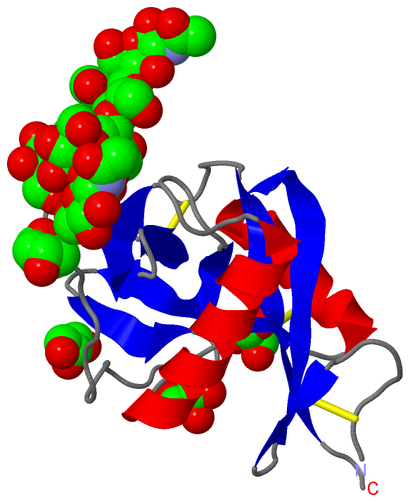
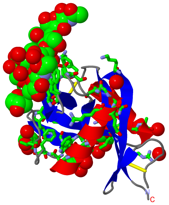
| 01 | AC1 | SOFTWARE | GLU A:93 , ASN A:95 , GLU A:101 , ASN A:112 , ASP A:113 , MAN A:205 | BINDING SITE FOR RESIDUE CA A1130 |
| 02 | AC2 | SOFTWARE | GLU A:55 , NAG A:208 | BINDING SITE FOR RESIDUE NAG A1139 |
| 03 | AC3 | SOFTWARE | LYS A:54 , GLU A:55 , ASN A:56 , GLY A:57 , BMA A:202 , MAN A:205 , NAG A:206 , NAG A:207 , HOH A:378 , HOH A:305 | BINDING SITE FOR RESIDUE NAG A1140 |
| 04 | AC4 | SOFTWARE | GLY A:57 , SER A:61 , PHE A:62 , ASN A:107 , TRP A:108 , MAN A:203 , MAN A:205 , NAG A:208 , HOH A:378 , HOH A:360 | BINDING SITE FOR RESIDUE BMA A1131 |
| 05 | AC5 | SOFTWARE | GLY A:57 , ASP A:58 , BMA A:202 , NAG A:204 , HOH A:325 , HOH A:392 , HOH A:345 , HOH A:355 | BINDING SITE FOR RESIDUE MAN A1132 |
| 06 | AC6 | SOFTWARE | LYS A:20 , TRP A:108 , MAN A:203 , HOH A:344 | BINDING SITE FOR RESIDUE NAG A1133 |
| 07 | AC7 | SOFTWARE | PHE A:62 , GLU A:93 , ASN A:95 , GLU A:101 , ASN A:112 , ASP A:113 , ARG A:115 , CA A:201 , BMA A:202 , NAG A:206 , NAG A:208 , HOH A:305 | BINDING SITE FOR RESIDUE MAN A1134 |
| 08 | AC8 | SOFTWARE | GLU A:93 , ASN A:95 , SER A:97 , MAN A:205 , NAG A:208 , HOH A:378 | BINDING SITE FOR RESIDUE NAG A1135 |
| 09 | AC9 | SOFTWARE | GLU A:39 , ASP A:41 , TRP A:77 , SER A:80 , ALA A:82 , HOH A:320 , HOH A:376 | BINDING SITE FOR RESIDUE GOL A1136 |
| 10 | BC1 | SOFTWARE | ALA A:32 , TRP A:75 , ALA A:83 , PHE A:84 , LEU A:87 , ARG A:126 , HOH A:374 , HOH A:388 , HOH A:309 , HOH A:385 | BINDING SITE FOR RESIDUE GOL A1137 |
| 11 | BC2 | SOFTWARE | GLN A:27 , GLN A:31 , ILE A:36 , VAL A:71 , GLN A:76 , SER A:78 , SER A:79 , HOH A:314 | BINDING SITE FOR RESIDUE GOL A1138 |
|
 Description