|
|
|
|
 Description
|
|
Compounds
|
||||||||||||||||||||
Chains, Units
Summary Information (see also Sequences/Alignments below) |
Ligands, Modified Residues, Ions (4, 5)| Asymmetric/Biological Unit (4, 5) |
Sites (4, 4)
Asymmetric Unit (4, 4)
|
SS Bonds (4, 4)
Asymmetric/Biological Unit
|
||||||||||||||||||||
Cis Peptide Bonds (1, 1)
Asymmetric/Biological Unit
|
||||||||
SAPs(SNPs)/Variants (0, 0)| (no "SAP(SNP)/Variant" information available for 2VUV) |
PROSITE Motifs (0, 0)| (no "PROSITE Motif" information available for 2VUV) |
Exons (0, 0)| (no "Exon" information available for 2VUV) |
Sequences/Alignments
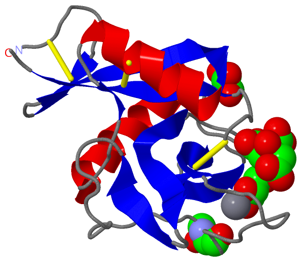
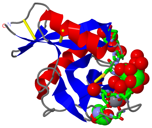
Asymmetric/Biological UnitChain A from PDB Type:PROTEIN Length:129 aligned with Q3KVL7_9BIVA | Q3KVL7 from UniProtKB/TrEMBL Length:148 Alignment length:129 29 39 49 59 69 79 89 99 109 119 129 139 Q3KVL7_9BIVA 20 GCPDGWTQFLDLCYIYQSAKASWASAQSSCQALGGILAEPDTACENEVLIHMCRENGDAGSFGPWLGGQKVGGAWQWSSSGAAFDYLRWGPNEPNNSGGNEDCLHYNWLSWNDLRCHYQASYLCQRAAE 148 SCOP domains --------------------------------------------------------------------------------------------------------------------------------- SCOP domains CATH domains --------------------------------------------------------------------------------------------------------------------------------- CATH domains Pfam domains ------------------Lectin_C-2vuvA01 A:19-126 --- Pfam domains SAPs(SNPs) --------------------------------------------------------------------------------------------------------------------------------- SAPs(SNPs) PROSITE --------------------------------------------------------------------------------------------------------------------------------- PROSITE Transcript --------------------------------------------------------------------------------------------------------------------------------- Transcript 2vuv A 1 GCPDGWTQFLDLCYIYQSAKASWASAQSSCQALGGILAEPDTACENEVLIHMCKENGDAGSFGPWLGGQKVGGAWQWSSSGAAFDYLRWGpNEPNNSGGNEDCLHYNWLSWNDLRCHYQASYLCQRAAE 129 10 20 30 40 50 60 70 80 90| 100 110 120 91-HYP
|
||||||||||||||||||||
SCOP Domains (0, 0)| (no "SCOP Domain" information available for 2VUV) |
CATH Domains (0, 0)| (no "CATH Domain" information available for 2VUV) |
Pfam Domains (1, 1)
Asymmetric/Biological Unit
|
Gene Ontology (1, 1)|
Asymmetric/Biological Unit(hide GO term definitions) Chain A (Q3KVL7_9BIVA | Q3KVL7)
|
||||||||||||
Interactive Views
|
|||||||||||||||||||||||||||||||||||||||||||||||||||||||||||||||||||||||||||||||||||||||||||||||||||||||||||||||||||||||||||||||||||||||||||||||||||||||||||||||||
Still Images
|
||||||||||||||||
Databases
|
||||||||||||||||||||||||||||||||||||||||||||||||||||||||||||||||||||||||||||||||||||||||||||||||||||||||||||||||||||||||||||||||||||||||||||||||||||||||||||||||
Analysis Tools
|
|||||||||||||||||||||||||||||||||||||||||||||||||||||||||||||
Entries Sharing at Least One Protein Chain (UniProt ID)
Related Entries Specified in the PDB File
|
|