|
|
|
|
 Description
|
|
Compounds
|
||||||||||||||||||||||||||||||||||||||||
Chains, Units
Summary Information (see also Sequences/Alignments below) |
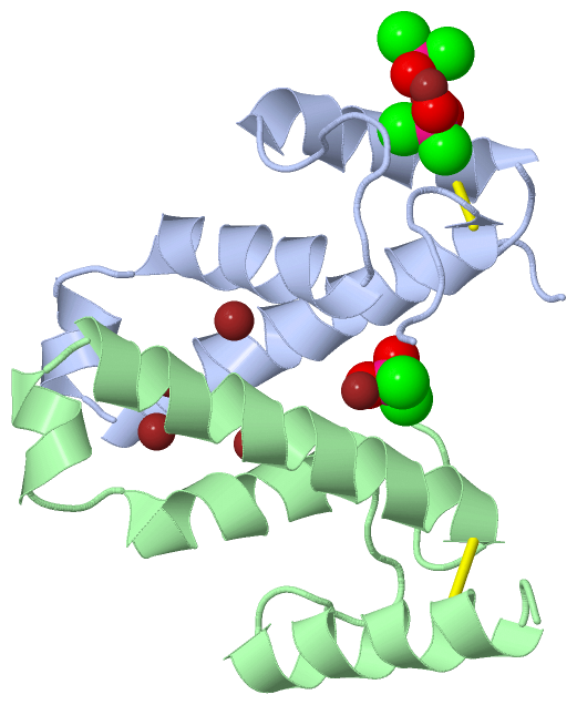
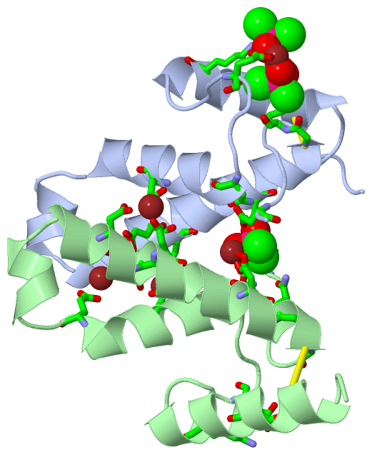
Ligands, Modified Residues, Ions (2, 11)| Asymmetric Unit (2, 11) Biological Unit 1 (1, 2) Biological Unit 2 (1, 1) |
Sites (11, 11)
Asymmetric Unit (11, 11)
|
SS Bonds (2, 2)
Asymmetric Unit
|
||||||||||||
Cis Peptide Bonds (0, 0)| (no "Cis Peptide Bond" information available for 2VQH) |
SAPs(SNPs)/Variants (0, 0)| (no "SAP(SNP)/Variant" information available for 2VQH) |
PROSITE Motifs (0, 0)| (no "PROSITE Motif" information available for 2VQH) |
Exons (0, 0)| (no "Exon" information available for 2VQH) |
Sequences/Alignments
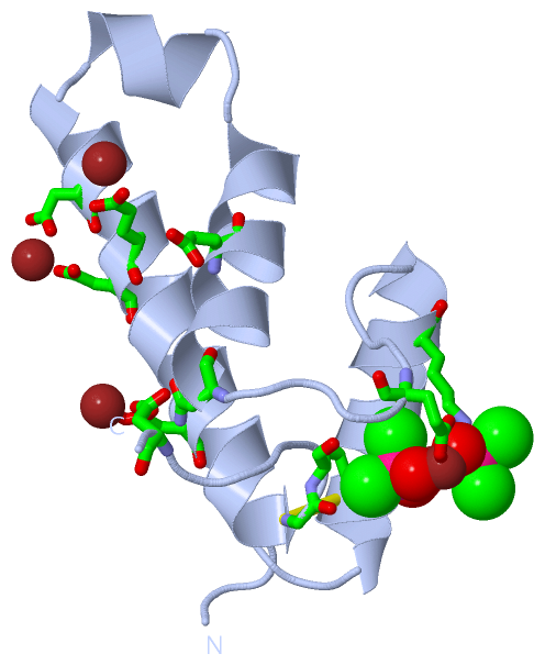
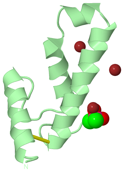
Asymmetric UnitChain A from PDB Type:PROTEIN Length:76 aligned with Q8NRS3_CORGL | Q8NRS3 from UniProtKB/TrEMBL Length:126 Alignment length:76 49 59 69 79 89 99 109 Q8NRS3_CORGL 40 LSPQYNWVACGILEGGLKAAGVLEEGQYNRELAEAIAAKGEGFWTTQFPQIGDWNEDQAAALADRAQTCGLVKADT 115 SCOP domains ---------------------------------------------------------------------------- SCOP domains CATH domains ---------------------------------------------------------------------------- CATH domains Pfam domains ---------------------------------------------------------------------------- Pfam domains SAPs(SNPs) ---------------------------------------------------------------------------- SAPs(SNPs) PROSITE ---------------------------------------------------------------------------- PROSITE Transcript ---------------------------------------------------------------------------- Transcript 2vqh A 13 LSPQYNWVACGILEGGLKAAGVLEEGQYNRELAEAIAAKGEGFWTTQFPQIGDWNEDQAAALADRAQTCGLVKADT 88 22 32 42 52 62 72 82 Chain B from PDB Type:PROTEIN Length:72 aligned with Q8NRS3_CORGL | Q8NRS3 from UniProtKB/TrEMBL Length:126 Alignment length:72 53 63 73 83 93 103 113 Q8NRS3_CORGL 44 YNWVACGILEGGLKAAGVLEEGQYNRELAEAIAAKGEGFWTTQFPQIGDWNEDQAAALADRAQTCGLVKADT 115 SCOP domains ------------------------------------------------------------------------ SCOP domains CATH domains ------------------------------------------------------------------------ CATH domains Pfam domains (1) PorB-2vqhB01 B:17-88 Pfam domains (1) Pfam domains (2) PorB-2vqhB02 B:17-88 Pfam domains (2) SAPs(SNPs) ------------------------------------------------------------------------ SAPs(SNPs) PROSITE ------------------------------------------------------------------------ PROSITE Transcript ------------------------------------------------------------------------ Transcript 2vqh B 17 YNWVACGILEGGLKAAGVLEEGQYNRELAEAIAAKGEGFWTTQFPQIGDWNEDQAAALADRAQTCGLVKADT 88 26 36 46 56 66 76 86
|
||||||||||||||||||||
SCOP Domains (0, 0)| (no "SCOP Domain" information available for 2VQH) |
CATH Domains (0, 0)| (no "CATH Domain" information available for 2VQH) |
Pfam Domains (1, 2)
Asymmetric Unit
|
Gene Ontology (1, 1)|
Asymmetric Unit(hide GO term definitions) Chain A,B (Q8NRS3_CORGL | Q8NRS3)
|
||||||||||||
Interactive Views
|
||||||||||||||||||||||||||||||||||||||||||||||||||||||||||||||||||||||||||||||||||||||||||||||||||||||||||||||||||||||||||||||||||||||||||||||||||||||||||||||||||||||||||||||||||||||||||||||||||||||||||||||||||||||||||
Still Images
|
||||||||||||||||
Databases
|
||||||||||||||||||||||||||||||||||||||||||||||||||||||||||||||||||||||||||||||||||||||||||||||||||||||||||||||||||||||||||||||||||||||||||||||||||||||||||||||||
Analysis Tools
|
|||||||||||||||||||||||||||||||||||||||||||||||||||||||||||||
Entries Sharing at Least One Protein Chain (UniProt ID)
Related Entries Specified in the PDB File
|
|