|
|
|
|
 Description
|
|
Compounds
|
||||||||||||||||||||||||||||||||||||||||||||
Chains, Units
Summary Information (see also Sequences/Alignments below) |
Ligands, Modified Residues, Ions (1, 2)
Asymmetric/Biological Unit (1, 2)
|
Sites (0, 0)| (no "Site" information available for 2VQC) |
SS Bonds (1, 1)
Asymmetric/Biological Unit
|
||||||||
Cis Peptide Bonds (0, 0)| (no "Cis Peptide Bond" information available for 2VQC) |
SAPs(SNPs)/Variants (0, 0)| (no "SAP(SNP)/Variant" information available for 2VQC) |
PROSITE Motifs (0, 0)| (no "PROSITE Motif" information available for 2VQC) |
Exons (0, 0)| (no "Exon" information available for 2VQC) |
Sequences/Alignments
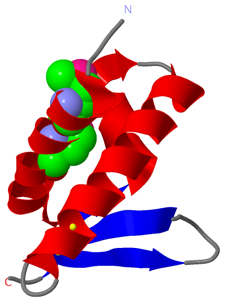
Asymmetric/Biological UnitChain A from PDB Type:PROTEIN Length:70 aligned with F112_SSV1 | P20220 from UniProtKB/Swiss-Prot Length:112 Alignment length:70 13 23 33 43 53 63 73 F112_SSV1 4 TLNSYKMAEIMYKILEKKGELTLEDILAQFEISVPSAYNIQRALKAICERHPDECEVQYKNRKTTFKWIK 73 SCOP domains d2vqca1 A:4-73 F-112 SCOP domains CATH domains ---------------------------------------------------------------------- CATH domains Pfam domains F-112-2vqcA01 A:4-73 Pfam domains SAPs(SNPs) ---------------------------------------------------------------------- SAPs(SNPs) PROSITE ---------------------------------------------------------------------- PROSITE Transcript ---------------------------------------------------------------------- Transcript 2vqc A 4 TLNSYKmAEImYKILEKKGELTLEDILAQFEISVPSAYNIQRALKAICERHPDECEVQYKNRKTTFKWIK 73 | 13| 23 33 43 53 63 73 10-MSE 14-MSE
|
||||||||||||||||||||
SCOP Domains (1, 1)
Asymmetric/Biological Unit
 |
CATH Domains (0, 0)| (no "CATH Domain" information available for 2VQC) |
Pfam Domains (1, 1)
Asymmetric/Biological Unit
|
Gene Ontology (0, 0)|
Asymmetric/Biological Unit(hide GO term definitions)
(no "Gene Ontology" information available for 2VQC)
|
Interactive Views
|
|||||||||||||||||||||||||||||||||||||||||||||||||||||||||||||||||||||||||||||||||||||||||||||||||||||||||||||||||||||
Still Images
|
||||||||||||||||
Databases
|
||||||||||||||||||||||||||||||||||||||||||||||||||||||||||||||||||||||||||||||||||||||||||||||||||||||||||||||||||||||||||||||||||||||||||||||||||||||||||||||||
Analysis Tools
|
|||||||||||||||||||||||||||||||||||||||||||||||||||||||||||||
Entries Sharing at Least One Protein Chain (UniProt ID)
Related Entries Specified in the PDB File
|
|