| molecular function |
|---|
| | GO:0015267 | | channel activity | | Enables the energy-independent facilitated diffusion, mediated by passage of a solute through a transmembrane aqueous pore or channel. Stereospecificity is not exhibited but this transport may be specific for a particular molecular species or class of molecules. |
| | GO:0005515 | | protein binding | | Interacting selectively and non-covalently with any protein or protein complex (a complex of two or more proteins that may include other nonprotein molecules). |
| | GO:0046982 | | protein heterodimerization activity | | Interacting selectively and non-covalently with a nonidentical protein to form a heterodimer. |
| | GO:0042803 | | protein homodimerization activity | | Interacting selectively and non-covalently with an identical protein to form a homodimer. |
| biological process |
|---|
| | GO:0001782 | | B cell homeostasis | | The process of regulating the proliferation and elimination of B cells such that the total number of B cells within a whole or part of an organism is stable over time in the absence of an outside stimulus. |
| | GO:0006915 | | apoptotic process | | A programmed cell death process which begins when a cell receives an internal (e.g. DNA damage) or external signal (e.g. an extracellular death ligand), and proceeds through a series of biochemical events (signaling pathway phase) which trigger an execution phase. The execution phase is the last step of an apoptotic process, and is typically characterized by rounding-up of the cell, retraction of pseudopodes, reduction of cellular volume (pyknosis), chromatin condensation, nuclear fragmentation (karyorrhexis), plasma membrane blebbing and fragmentation of the cell into apoptotic bodies. When the execution phase is completed, the cell has died. |
| | GO:0097192 | | extrinsic apoptotic signaling pathway in absence of ligand | | A series of molecular signals in which a signal is conveyed from the cell surface to trigger the apoptotic death of a cell. The pathway starts with withdrawal of a ligand from a cell surface receptor, and ends when the execution phase of apoptosis is triggered. |
| | GO:0008630 | | intrinsic apoptotic signaling pathway in response to DNA damage | | A series of molecular signals in which an intracellular signal is conveyed to trigger the apoptotic death of a cell. The pathway is induced by the detection of DNA damage, and ends when the execution phase of apoptosis is triggered. |
| | GO:0008053 | | mitochondrial fusion | | Merging of two or more mitochondria within a cell to form a single compartment. |
| | GO:0002903 | | negative regulation of B cell apoptotic process | | Any process that stops, prevents, or reduces the frequency, rate, or extent of B cell apoptotic process. |
| | GO:0043066 | | negative regulation of apoptotic process | | Any process that stops, prevents, or reduces the frequency, rate or extent of cell death by apoptotic process. |
| | GO:0043065 | | positive regulation of apoptotic process | | Any process that activates or increases the frequency, rate or extent of cell death by apoptotic process. |
| | GO:0042981 | | regulation of apoptotic process | | Any process that modulates the occurrence or rate of cell death by apoptotic process. |
| | GO:0001836 | | release of cytochrome c from mitochondria | | The process that results in the movement of cytochrome c from the mitochondrial intermembrane space into the cytosol, which is part of the apoptotic signaling pathway and leads to caspase activation. |
| | GO:0055085 | | transmembrane transport | | The process in which a solute is transported across a lipid bilayer, from one side of a membrane to the other |
| cellular component |
|---|
| | GO:0005737 | | cytoplasm | | All of the contents of a cell excluding the plasma membrane and nucleus, but including other subcellular structures. |
| | GO:0005741 | | mitochondrial outer membrane | | The outer, i.e. cytoplasm-facing, lipid bilayer of the mitochondrial envelope. |
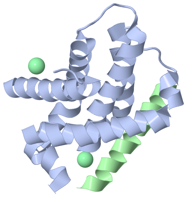
 Description
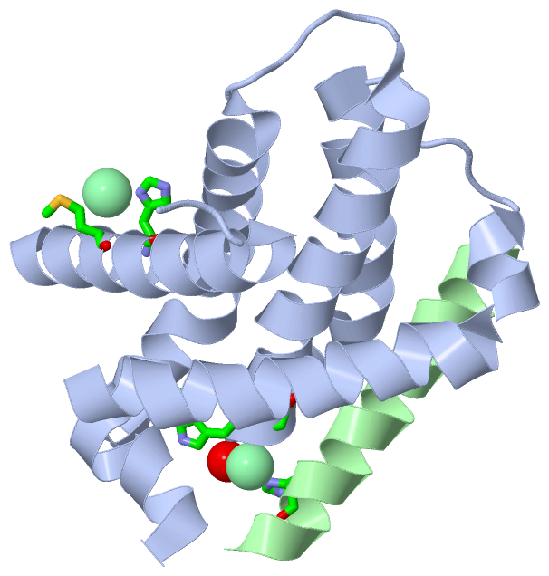