Show PDB file:   
         Plain Text   HTML   (compressed file size)
QuickSearch:   
by PDB,NDB,UniProt,PROSITE Code or Search Term(s)  
(-)Asymmetric Unit
(-)Asym. Unit - sites
(-)Biological Unit 1
(-)Biol. Unit 1 - sites
(-)Biological Unit 2
(-)Biological Unit 3
collapse expand < >
Image Asymmetric Unit
Asymmetric Unit  (Jmol Viewer)
Image Asym. Unit - sites
Asym. Unit - sites  (Jmol Viewer)
Image Biological Unit 1
Biological Unit 1  (Jmol Viewer)
Image Biol. Unit 1 - sites
Biol. Unit 1 - sites  (Jmol Viewer)
Image Biological Unit 2
Biological Unit 2  (Jmol Viewer)
Image Biological Unit 3
Biological Unit 3  (Jmol Viewer)

(-) Description

Title :  STRUCTURE OF MOUSE A1 BOUND TO THE PUMA BH3-DOMAIN
 
Authors :  C. Smits, P. E. Czabotar, M. G. Hinds, C. L. Day
Date :  17 Feb 08  (Deposition) - 04 Mar 08  (Release) - 13 Jul 11  (Revision)
Method :  X-RAY DIFFRACTION
Resolution :  1.80
Chains :  Asym. Unit :  A,B,C,D
Biol. Unit 1:  A,B,C,D  (1x)
Biol. Unit 2:  A,B  (1x)
Biol. Unit 3:  C,D  (1x)
Keywords :  Bh3, Bcl-2, Apoptosis, Pro-Survival, Mitochondrion, Protein-Protein Complex (Keyword Search: [Gene Ontology, PubMed, Web (Google))
 
Reference :  C. Smits, P. E. Czabotar, M. G. Hinds, C. L. Day
Structural Plasticity Underpins Promiscuous Binding Of The Prosurvival Protein A1.
Structure V. 16 818 2008
PubMed-ID: 18462686  |  Reference-DOI: 10.1016/J.STR.2008.02.009

(-) Compounds

Molecule 1 - BCL-2-RELATED PROTEIN A1
    ChainsA, C
    EngineeredYES
    Expression SystemESCHERICHIA COLI
    Expression System PlasmidPGEX 6P-3
    Expression System StrainBL21(DE3)
    Expression System Taxid469008
    Expression System VariantPSJS 1240
    Expression System Vector TypePLASMID
    FragmentRESIDUES 1-152
    MutationYES
    Organism CommonMOUSE
    Organism ScientificMUS MUSCULUS
    Organism Taxid10090
    SynonymPROTEIN BFL-1, HEMOPOIETIC-SPECIFIC EARLY RESPONSE PROTEIN, A1-A
 
Molecule 2 - BCL-2-BINDING COMPONENT 3
    ChainsB, D
    EngineeredYES
    Expression SystemESCHERICHIA COLI
    Expression System PlasmidPET31B PUMA TR
    Expression System StrainBL21(DE3)
    Expression System Taxid469008
    Expression System Vector TypePLASMID
    FragmentBH3-DOMAIN, RESIDUES 130-155
    MutationYES
    Organism CommonMOUSE
    Organism ScientificMUS MUSCULUS
    Organism Taxid10090
    Other DetailsC-TERMINAL HOMOSERINE LACTONE DUE TO CNBR CLEAVAGE
    SynonymP53 UP-REGULATED MODULATOR OF APOPTOSIS

 Structural Features

(-) Chains, Units

  1234
Asymmetric Unit ABCD
Biological Unit 1 (1x)ABCD
Biological Unit 2 (1x)AB  
Biological Unit 3 (1x)  CD

Summary Information (see also Sequences/Alignments below)

(-) Ligands, Modified Residues, Ions  (2, 10)

Asymmetric Unit (2, 10)
No.NameCountTypeFull Name
1CL2Ligand/IonCHLORIDE ION
2MSE8Mod. Amino AcidSELENOMETHIONINE
Biological Unit 1 (1, 8)
No.NameCountTypeFull Name
1CL-1Ligand/IonCHLORIDE ION
2MSE8Mod. Amino AcidSELENOMETHIONINE
Biological Unit 2 (1, 4)
No.NameCountTypeFull Name
1CL-1Ligand/IonCHLORIDE ION
2MSE4Mod. Amino AcidSELENOMETHIONINE
Biological Unit 3 (1, 4)
No.NameCountTypeFull Name
1CL-1Ligand/IonCHLORIDE ION
2MSE4Mod. Amino AcidSELENOMETHIONINE

(-) Sites  (2, 2)

Asymmetric Unit (2, 2)
No.NameEvidenceResiduesDescription
1AC1SOFTWAREHIS A:10 , MSE C:7 , HIS C:10BINDING SITE FOR RESIDUE CL A1151
2AC2SOFTWAREASN C:85 , TRP C:86 , HOH C:2076 , ASN D:149BINDING SITE FOR RESIDUE CL C1150

(-) SS Bonds  (0, 0)

(no "SS Bond" information available for 2VOF)

(-) Cis Peptide Bonds  (0, 0)

(no "Cis Peptide Bond" information available for 2VOF)

 Sequence-Structure Mapping

(-) SAPs(SNPs)/Variants  (0, 0)

(no "SAP(SNP)/Variant" information available for 2VOF)

(-) PROSITE Motifs  (3, 6)

Asymmetric Unit (3, 6)
 PROSITEUniProtKBPDB
No.IDACDescriptionIDLocationCountLocation
1BCL2_FAMILYPS50062 BCL2-like apoptosis inhibitors family profile.B2LA1_MOUSE37-142
 
  2A:37-142
C:37-142
2BH1PS01080 Apoptosis regulator, Bcl-2 family BH1 motif signature.B2LA1_MOUSE78-97
 
  2A:78-97
C:78-97
3BH2PS01258 Apoptosis regulator, Bcl-2 family BH2 motif signature.B2LA1_MOUSE133-144
 
  2A:133-144
C:133-144
Biological Unit 1 (3, 6)
 PROSITEUniProtKBPDB
No.IDACDescriptionIDLocationCountLocation
1BCL2_FAMILYPS50062 BCL2-like apoptosis inhibitors family profile.B2LA1_MOUSE37-142
 
  2A:37-142
C:37-142
2BH1PS01080 Apoptosis regulator, Bcl-2 family BH1 motif signature.B2LA1_MOUSE78-97
 
  2A:78-97
C:78-97
3BH2PS01258 Apoptosis regulator, Bcl-2 family BH2 motif signature.B2LA1_MOUSE133-144
 
  2A:133-144
C:133-144
Biological Unit 2 (3, 3)
 PROSITEUniProtKBPDB
No.IDACDescriptionIDLocationCountLocation
1BCL2_FAMILYPS50062 BCL2-like apoptosis inhibitors family profile.B2LA1_MOUSE37-142
 
  1A:37-142
-
2BH1PS01080 Apoptosis regulator, Bcl-2 family BH1 motif signature.B2LA1_MOUSE78-97
 
  1A:78-97
-
3BH2PS01258 Apoptosis regulator, Bcl-2 family BH2 motif signature.B2LA1_MOUSE133-144
 
  1A:133-144
-
Biological Unit 3 (3, 3)
 PROSITEUniProtKBPDB
No.IDACDescriptionIDLocationCountLocation
1BCL2_FAMILYPS50062 BCL2-like apoptosis inhibitors family profile.B2LA1_MOUSE37-142
 
  1-
C:37-142
2BH1PS01080 Apoptosis regulator, Bcl-2 family BH1 motif signature.B2LA1_MOUSE78-97
 
  1-
C:78-97
3BH2PS01258 Apoptosis regulator, Bcl-2 family BH2 motif signature.B2LA1_MOUSE133-144
 
  1-
C:133-144

(-) Exons   (0, 0)

(no "Exon" information available for 2VOF)

(-) Sequences/Alignments

Asymmetric Unit
   Reformat: Number of residues per line =  ('0' or empty: single-line sequence representation)
  Number of residues per labelling interval =   
  UniProt sequence: complete  aligned part    
   Show mapping: SCOP domains CATH domains Pfam domains Secondary structure (by author)
SAPs(SNPs) PROSITE motifs Exons
(details for a mapped element are shown in a popup box when the mouse pointer rests over it)
Chain A from PDB  Type:PROTEIN  Length:145
 aligned with B2LA1_MOUSE | Q07440 from UniProtKB/Swiss-Prot  Length:172

    Alignment length:152
                              1                                                                                                                                                     
                              |      8        18        28        38        48        58        68        78        88        98       108       118       128       138       148  
          B2LA1_MOUSE     - --MAESELMHIHSLAEHYLQYVLQVPAFESAPSQACRVLQRVAFSVQKEVEKNLKSYLDDFHVESIDTARIIFNQVMEKEFEDGIINWGRIVTIFAFGGVLLKKLPQEQIALDVCAYKQVSSFVAEFIMNNTGEWIRQNGGWEDGFIKKFEP 150
               SCOP domains d2vofa_ A: automated matches                                                                                                                             SCOP domains
               CATH domains -------------------------------------------------------------------------------------------------------------------------------------------------------- CATH domains
               Pfam domains -------------------------------------------------------------------------------------------------------------------------------------------------------- Pfam domains
         Sec.struct. author .hhhhhhhhhhhhhhhhhhhhhhh.....-..hhhhhhhhhhhhhhhhhhhhhhhhhhh.....hhhhhhhhhhhhhhhhhh....hhhhhhhhhhhhhhhhhhhhhh------hhhhhhhhhhhhhhhhhhhhhhhhh....hhhhhhh.. Sec.struct. author
                 SAPs(SNPs) -------------------------------------------------------------------------------------------------------------------------------------------------------- SAPs(SNPs)
                PROSITE (1) --------------------------------------BCL2_FAMILY  PDB: A:37-142 UniProt: 37-142                                                                -------- PROSITE (1)
                PROSITE (2) -------------------------------------------------------------------------------BH1  PDB: A:78-97   -----------------------------------BH2         ------ PROSITE (2)
                 Transcript -------------------------------------------------------------------------------------------------------------------------------------------------------- Transcript
                 2vof A  -1 GSmAESELmHIHSLAEHYLQYVLQVPAFE-APSQACRVLQRVAFSVQKEVEKNLKSYLDDFHVESIDTARIIFNQVmEKEFEDGIINWGRIVTIFAFGGVLLKKLKQE------SAYKQVSSFVAEFImNNTGEWIRQNGGWEDGFIKKFEP 150
                              |     |8        18        |-|       38        48        58        68      | 78        88        98       | -    |  118       128       138       148  
                              |     7-MSE              27 |                                            75-MSE                        106    113           127-MSE                   
                              1-MSE                      29                                                                                                                         

Chain B from PDB  Type:PROTEIN  Length:23
 aligned with BBC3_MOUSE | Q99ML1 from UniProtKB/Swiss-Prot  Length:193

    Alignment length:23
                                   140       150   
           BBC3_MOUSE   131 EEWAREIGAQLRRMADDLNAQYE 153
               SCOP domains ----------------------- SCOP domains
               CATH domains ----------------------- CATH domains
               Pfam domains ----------------------- Pfam domains
         Sec.struct. author hhhhhhhhhhhhhhhhhhhhhh. Sec.struct. author
                 SAPs(SNPs) ----------------------- SAPs(SNPs)
                    PROSITE ----------------------- PROSITE
                 Transcript ----------------------- Transcript
                 2vof B 131 EEWAREIGAQLRRIADDLNAQYE 153
                                   140       150   

Chain C from PDB  Type:PROTEIN  Length:150
 aligned with B2LA1_MOUSE | Q07440 from UniProtKB/Swiss-Prot  Length:172

    Alignment length:152
                               1                                                                                                                                                    
                               |     7        17        27        37        47        57        67        77        87        97       107       117       127       137       147  
          B2LA1_MOUSE     - ---MAESELMHIHSLAEHYLQYVLQVPAFESAPSQACRVLQRVAFSVQKEVEKNLKSYLDDFHVESIDTARIIFNQVMEKEFEDGIINWGRIVTIFAFGGVLLKKLPQEQIALDVCAYKQVSSFVAEFIMNNTGEWIRQNGGWEDGFIKKFE 149
               SCOP domains d2vofc_ C: automated matches                                                                                                                             SCOP domains
               CATH domains -------------------------------------------------------------------------------------------------------------------------------------------------------- CATH domains
           Pfam domains (1) ---------------------------------------Bcl-2-2vofC01 C:37-140                                                                                  --------- Pfam domains (1)
           Pfam domains (2) ---------------------------------------Bcl-2-2vofC02 C:37-140                                                                                  --------- Pfam domains (2)
         Sec.struct. author ..hhhhhhhhhhhhhhhhhhhhhhh.....--.hhhhhhhhhhhhhhhhhhhhhhhhhhh.....hhhhhhhhhhhhhhhhhh....hhhhhhhhhhhhhhhhhhhhhhh.....hhhhhhhhhhhhhhhhhhhhhhhhhhhhhhhhhhhhh Sec.struct. author
                 SAPs(SNPs) -------------------------------------------------------------------------------------------------------------------------------------------------------- SAPs(SNPs)
                PROSITE (1) ---------------------------------------BCL2_FAMILY  PDB: C:37-142 UniProt: 37-142                                                                ------- PROSITE (1)
                PROSITE (2) --------------------------------------------------------------------------------BH1  PDB: C:78-97   -----------------------------------BH2         ----- PROSITE (2)
                 Transcript -------------------------------------------------------------------------------------------------------------------------------------------------------- Transcript
                 2vof C  -2 LGSmAESELmHIHSLAEHYLQYVLQVPAFE--PSQACRVLQRVAFSVQKEVEKNLKSYLDDFHVESIDTARIIFNQVmEKEFEDGIINWGRIVTIFAFGGVLLKKLKQEQIALDVSAYKQVSSFVAEFImNNTGEWIRQNGGWEDGFIKKFE 149
                               |     7        17        27  |     37        47        57        67       |77        87        97       107       117       127       137       147  
                               1-MSE 7-MSE              27 30                                           75-MSE                                             127-MSE                  

Chain D from PDB  Type:PROTEIN  Length:22
 aligned with BBC3_MOUSE | Q99ML1 from UniProtKB/Swiss-Prot  Length:193

    Alignment length:22
                                   141       151  
           BBC3_MOUSE   132 EWAREIGAQLRRMADDLNAQYE 153
               SCOP domains ---------------------- SCOP domains
               CATH domains ---------------------- CATH domains
               Pfam domains ---------------------- Pfam domains
         Sec.struct. author hhhhhhhhhhhhhhhhhhhhh. Sec.struct. author
                 SAPs(SNPs) ---------------------- SAPs(SNPs)
                    PROSITE ---------------------- PROSITE
                 Transcript ---------------------- Transcript
                 2vof D 132 EWAREIGAQLRRIADDLNAQYE 153
                                   141       151  

   Legend:   → Mismatch (orange background)
  - → Gap (green background, '-', border residues have a numbering label)
    → Modified Residue (blue background, lower-case, 'x' indicates undefined single-letter code, labelled with number + name)
  x → Chemical Group (purple background, 'x', labelled with number + name, e.g. ACE or NH2)
  extra numbering lines below/above indicate numbering irregularities and modified residue names etc., number ends below/above '|'

 Classification and Annotation

(-) SCOP Domains  (1, 2)

Asymmetric Unit

(-) CATH Domains  (0, 0)

(no "CATH Domain" information available for 2VOF)

(-) Pfam Domains  (1, 2)

Asymmetric Unit

(-) Gene Ontology  (45, 49)

Asymmetric Unit(hide GO term definitions)
Chain A,C   (B2LA1_MOUSE | Q07440)
molecular function
    GO:0015267    channel activity    Enables the energy-independent facilitated diffusion, mediated by passage of a solute through a transmembrane aqueous pore or channel. Stereospecificity is not exhibited but this transport may be specific for a particular molecular species or class of molecules.
    GO:0005515    protein binding    Interacting selectively and non-covalently with any protein or protein complex (a complex of two or more proteins that may include other nonprotein molecules).
    GO:0046982    protein heterodimerization activity    Interacting selectively and non-covalently with a nonidentical protein to form a heterodimer.
    GO:0042803    protein homodimerization activity    Interacting selectively and non-covalently with an identical protein to form a homodimer.
biological process
    GO:0001782    B cell homeostasis    The process of regulating the proliferation and elimination of B cells such that the total number of B cells within a whole or part of an organism is stable over time in the absence of an outside stimulus.
    GO:0006915    apoptotic process    A programmed cell death process which begins when a cell receives an internal (e.g. DNA damage) or external signal (e.g. an extracellular death ligand), and proceeds through a series of biochemical events (signaling pathway phase) which trigger an execution phase. The execution phase is the last step of an apoptotic process, and is typically characterized by rounding-up of the cell, retraction of pseudopodes, reduction of cellular volume (pyknosis), chromatin condensation, nuclear fragmentation (karyorrhexis), plasma membrane blebbing and fragmentation of the cell into apoptotic bodies. When the execution phase is completed, the cell has died.
    GO:0097192    extrinsic apoptotic signaling pathway in absence of ligand    A series of molecular signals in which a signal is conveyed from the cell surface to trigger the apoptotic death of a cell. The pathway starts with withdrawal of a ligand from a cell surface receptor, and ends when the execution phase of apoptosis is triggered.
    GO:0008630    intrinsic apoptotic signaling pathway in response to DNA damage    A series of molecular signals in which an intracellular signal is conveyed to trigger the apoptotic death of a cell. The pathway is induced by the detection of DNA damage, and ends when the execution phase of apoptosis is triggered.
    GO:0008053    mitochondrial fusion    Merging of two or more mitochondria within a cell to form a single compartment.
    GO:0002903    negative regulation of B cell apoptotic process    Any process that stops, prevents, or reduces the frequency, rate, or extent of B cell apoptotic process.
    GO:0043066    negative regulation of apoptotic process    Any process that stops, prevents, or reduces the frequency, rate or extent of cell death by apoptotic process.
    GO:0043065    positive regulation of apoptotic process    Any process that activates or increases the frequency, rate or extent of cell death by apoptotic process.
    GO:0042981    regulation of apoptotic process    Any process that modulates the occurrence or rate of cell death by apoptotic process.
    GO:0001836    release of cytochrome c from mitochondria    The process that results in the movement of cytochrome c from the mitochondrial intermembrane space into the cytosol, which is part of the apoptotic signaling pathway and leads to caspase activation.
    GO:0055085    transmembrane transport    The process in which a solute is transported across a lipid bilayer, from one side of a membrane to the other
cellular component
    GO:0005737    cytoplasm    All of the contents of a cell excluding the plasma membrane and nucleus, but including other subcellular structures.
    GO:0005741    mitochondrial outer membrane    The outer, i.e. cytoplasm-facing, lipid bilayer of the mitochondrial envelope.

Chain B,D   (BBC3_MOUSE | Q99ML1)
molecular function
    GO:0051117    ATPase binding    Interacting selectively and non-covalently with an ATPase, any enzyme that catalyzes the hydrolysis of ATP.
    GO:0005515    protein binding    Interacting selectively and non-covalently with any protein or protein complex (a complex of two or more proteins that may include other nonprotein molecules).
biological process
    GO:0006919    activation of cysteine-type endopeptidase activity involved in apoptotic process    Any process that initiates the activity of the inactive enzyme cysteine-type endopeptidase in the context of an apoptotic process.
    GO:0006915    apoptotic process    A programmed cell death process which begins when a cell receives an internal (e.g. DNA damage) or external signal (e.g. an extracellular death ligand), and proceeds through a series of biochemical events (signaling pathway phase) which trigger an execution phase. The execution phase is the last step of an apoptotic process, and is typically characterized by rounding-up of the cell, retraction of pseudopodes, reduction of cellular volume (pyknosis), chromatin condensation, nuclear fragmentation (karyorrhexis), plasma membrane blebbing and fragmentation of the cell into apoptotic bodies. When the execution phase is completed, the cell has died.
    GO:0097190    apoptotic signaling pathway    A series of molecular signals which triggers the apoptotic death of a cell. The pathway starts with reception of a signal, and ends when the execution phase of apoptosis is triggered.
    GO:0006974    cellular response to DNA damage stimulus    Any process that results in a change in state or activity of a cell (in terms of movement, secretion, enzyme production, gene expression, etc.) as a result of a stimulus indicating damage to its DNA from environmental insults or errors during metabolism.
    GO:0071479    cellular response to ionizing radiation    Any process that results in a change in state or activity of a cell (in terms of movement, secretion, enzyme production, gene expression, etc.) as a result of a ionizing radiation stimulus. Ionizing radiation is radiation with sufficient energy to remove electrons from atoms and may arise from spontaneous decay of unstable isotopes, resulting in alpha and beta particles and gamma rays. Ionizing radiation also includes X-rays.
    GO:0008340    determination of adult lifespan    The control of viability and duration in the adult phase of the life-cycle.
    GO:0097194    execution phase of apoptosis    A stage of the apoptotic process that starts with the controlled breakdown of the cell through the action of effector caspases or other effector molecules (e.g. cathepsins, calpains etc.). Key steps of the execution phase are rounding-up of the cell, retraction of pseudopodes, reduction of cellular volume (pyknosis), chromatin condensation, nuclear fragmentation (karyorrhexis), plasma membrane blebbing and fragmentation of the cell into apoptotic bodies. When the execution phase is completed, the cell has died.
    GO:0097193    intrinsic apoptotic signaling pathway    A series of molecular signals in which an intracellular signal is conveyed to trigger the apoptotic death of a cell. The pathway starts with reception of an intracellular signal (e.g. DNA damage, endoplasmic reticulum stress, oxidative stress etc.), and ends when the execution phase of apoptosis is triggered. The intrinsic apoptotic signaling pathway is crucially regulated by permeabilization of the mitochondrial outer membrane (MOMP).
    GO:0072332    intrinsic apoptotic signaling pathway by p53 class mediator    A series of molecular signals in which an intracellular signal is conveyed to trigger the apoptotic death of a cell. The pathway is induced by the cell cycle regulator phosphoprotein p53, or an equivalent protein, and ends when the execution phase of apoptosis is triggered.
    GO:0042771    intrinsic apoptotic signaling pathway in response to DNA damage by p53 class mediator    A series of molecular signals in which an intracellular signal is conveyed to trigger the apoptotic death of a cell. The pathway is induced by the cell cycle regulator phosphoprotein p53, or an equivalent protein, in response to the detection of DNA damage, and ends when the execution phase of apoptosis is triggered.
    GO:0070059    intrinsic apoptotic signaling pathway in response to endoplasmic reticulum stress    A series of molecular signals in which an intracellular signal is conveyed to trigger the apoptotic death of a cell. The pathway is induced in response to a stimulus indicating endoplasmic reticulum (ER) stress, and ends when the execution phase of apoptosis is triggered. ER stress usually results from the accumulation of unfolded or misfolded proteins in the ER lumen.
    GO:0030308    negative regulation of cell growth    Any process that stops, prevents, or reduces the frequency, rate, extent or direction of cell growth.
    GO:0032471    negative regulation of endoplasmic reticulum calcium ion concentration    Any process that decreases the concentration of calcium ions in the endoplasmic reticulum.
    GO:0045926    negative regulation of growth    Any process that stops, prevents or reduces the rate or extent of growth, the increase in size or mass of all or part of an organism.
    GO:0043065    positive regulation of apoptotic process    Any process that activates or increases the frequency, rate or extent of cell death by apoptotic process.
    GO:2001056    positive regulation of cysteine-type endopeptidase activity    Any process that activates or increases the frequency, rate or extent of cysteine-type endopeptidase activity.
    GO:2001244    positive regulation of intrinsic apoptotic signaling pathway    Any process that activates or increases the frequency, rate or extent of intrinsic apoptotic signaling pathway.
    GO:0043525    positive regulation of neuron apoptotic process    Any process that activates or increases the frequency, rate or extent of cell death of neurons by apoptotic process.
    GO:0032464    positive regulation of protein homooligomerization    Any process that activates or increases the frequency, rate or extent of protein homooligomerization.
    GO:1900740    positive regulation of protein insertion into mitochondrial membrane involved in apoptotic signaling pathway    Any process that activates or increases the frequency, rate or extent of protein insertion into mitochondrial membrane involved in apoptotic signaling pathway.
    GO:0090200    positive regulation of release of cytochrome c from mitochondria    Any process that increases the rate, frequency or extent of release of cytochrome c from mitochondria, the process in which cytochrome c is enabled to move from the mitochondrial intermembrane space into the cytosol, which is an early step in apoptosis and leads to caspase activation.
    GO:0070245    positive regulation of thymocyte apoptotic process    Any process that activates or increases the frequency, rate or extent of thymocyte death by apoptotic process.
    GO:0001836    release of cytochrome c from mitochondria    The process that results in the movement of cytochrome c from the mitochondrial intermembrane space into the cytosol, which is part of the apoptotic signaling pathway and leads to caspase activation.
    GO:0051209    release of sequestered calcium ion into cytosol    The process in which calcium ions sequestered in the endoplasmic reticulum, Golgi apparatus or mitochondria are released into the cytosolic compartment.
    GO:0034976    response to endoplasmic reticulum stress    Any process that results in a change in state or activity of a cell (in terms of movement, secretion, enzyme production, gene expression, etc.) as a result of a stress acting at the endoplasmic reticulum. ER stress usually results from the accumulation of unfolded or misfolded proteins in the ER lumen.
    GO:1901998    toxin transport    The directed movement of a toxin into, out of or within a cell, or between cells, by means of some agent such as a transporter or pore.
cellular component
    GO:0005783    endoplasmic reticulum    The irregular network of unit membranes, visible only by electron microscopy, that occurs in the cytoplasm of many eukaryotic cells. The membranes form a complex meshwork of tubular channels, which are often expanded into slitlike cavities called cisternae. The ER takes two forms, rough (or granular), with ribosomes adhering to the outer surface, and smooth (with no ribosomes attached).
    GO:0005764    lysosome    A small lytic vacuole that has cell cycle-independent morphology and is found in most animal cells and that contains a variety of hydrolases, most of which have their maximal activities in the pH range 5-6. The contained enzymes display latency if properly isolated. About 40 different lysosomal hydrolases are known and lysosomes have a great variety of morphologies and functions.
    GO:0005740    mitochondrial envelope    The double lipid bilayer enclosing the mitochondrion and separating its contents from the cell cytoplasm; includes the intermembrane space.
    GO:0005739    mitochondrion    A semiautonomous, self replicating organelle that occurs in varying numbers, shapes, and sizes in the cytoplasm of virtually all eukaryotic cells. It is notably the site of tissue respiration.
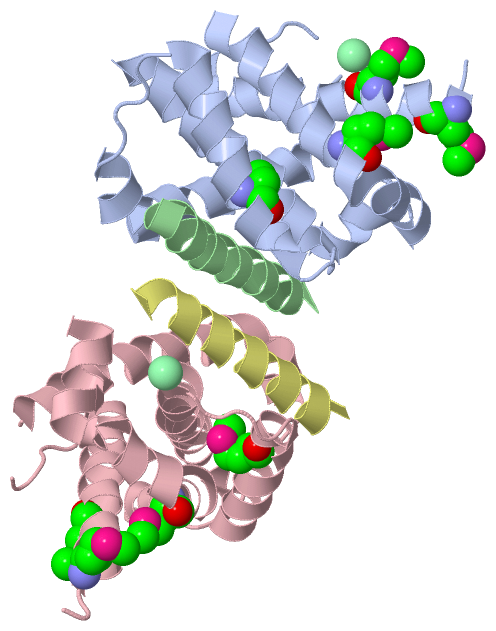
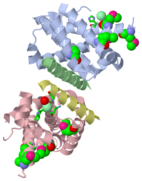
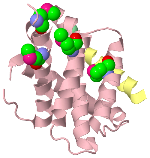
 Visualization

(-) Interactive Views

Asymmetric Unit
  Complete Structure
    Jena3D(integrated viewing of ligand, site, SAP, PROSITE, SCOP information)
    WebMol | AstexViewer[tm]@PDBe
(Java Applets, require no local installation except for Java; loading may be slow)
    STRAP
(Java WebStart application, automatic local installation, requires Java; full application with system access!)
    RasMol
(require local installation)
    Molscript (VRML)
(requires installation of a VRML viewer; select preferred view via VRML and generate a mono or stereo PDF format file)
 
  Ligands, Modified Residues, Ions
    CL  [ RasMol | Jena3D ]  +environment [ RasMol | Jena3D ]
    MSE  [ RasMol | Jena3D ]  +environment [ RasMol | Jena3D ]
 
  Sites
    AC1  [ RasMol ]  +environment [ RasMol ]
    AC2  [ RasMol ]  +environment [ RasMol ]
 
  Cis Peptide Bonds
(no "Cis Peptide Bonds" information available for 2vof)
 
Biological Units
  Complete Structure
    Biological Unit 1  [ Jena3D ]
    Biological Unit 2  [ Jena3D ]
    Biological Unit 3  [ Jena3D ]

(-) Still Images

Jmol
  protein: cartoon or spacefill or dots and stick; nucleic acid: cartoon and stick; ligands: spacefill; active site: stick
Molscript
  protein, nucleic acid: cartoon; ligands: spacefill; active site: ball and stick

 Databases and Analysis Tools

(-) Databases

Access by PDB/NDB ID
  2vof
    Family and Domain InformationProDom | SYSTERS
    General Structural InformationGlycoscienceDB | MMDB | NDB | OCA | PDB | PDBe | PDBj | PDBsum | PDBWiki | PQS | PROTEOPEDIA
    Orientation in MembranesOPM
    Protein SurfaceSURFACE
    Secondary StructureDSSP (structure derived) | HSSP (homology derived)
    Structural GenomicsGeneCensus
    Structural NeighboursCE | VAST
    Structure ClassificationCATH | Dali | SCOP
    Validation and Original DataBMRB Data View | BMRB Restraints Grid | EDS | PROCHECK | RECOORD | WHAT_CHECK
 
Access by UniProt ID/Accession number
  B2LA1_MOUSE | Q07440
    Comparative Protein Structure ModelsModBase
    Genomic InformationEnsembl
    Protein-protein InteractionDIP
    Sequence, Family and Domain InformationInterPro | Pfam | SMART | UniProtKB/SwissProt
  BBC3_MOUSE | Q99ML1
    Comparative Protein Structure ModelsModBase
    Genomic InformationEnsembl
    Protein-protein InteractionDIP
    Sequence, Family and Domain InformationInterPro | Pfam | SMART | UniProtKB/SwissProt
 
Access by Enzyme Classificator   (EC Number)
  (no 'Enzyme Classificator' available)
    General Enzyme InformationBRENDA | EC-PDB | Enzyme | IntEnz
    PathwayKEGG | MetaCyc
 
Access by Disease Identifier   (MIM ID)
  (no 'MIM ID' available)
    Disease InformationOMIM
 
Access by GenAge ID
  (no 'GenAge ID' available)
    Age Related InformationGenAge

(-) Analysis Tools

Access by PDB/NDB ID
    Domain InformationXDom
    Interatomic Contacts of Structural UnitsCSU
    Ligand-protein ContactsLPC
    Protein CavitiescastP
    Sequence and Secondary StructurePDBCartoon
    Structure AlignmentSTRAP(Java WebStart application, automatic local installation, requires Java; full application with system access!)
    Structure and Sequence BrowserSTING
 
Access by UniProt ID/Accession number
  B2LA1_MOUSE | Q07440
    Protein Disorder PredictionDisEMBL | FoldIndex | GLOBPLOT (for more information see DisProt)
  BBC3_MOUSE | Q99ML1
    Protein Disorder PredictionDisEMBL | FoldIndex | GLOBPLOT (for more information see DisProt)

 Related Entries

(-) Entries Sharing at Least One Protein Chain (UniProt ID)

UniProtKB/Swiss-Prot
        B2LA1_MOUSE | Q074402vog 2voh 2voi
        BBC3_MOUSE | Q99ML12roc

(-) Related Entries Specified in the PDB File

2vog STRUCTURE OF MOUSE A1 BOUND TO THE BMF BH3-DOMAIN RELATED ID: 2VOH RELATED DB: PDB STRUCTURE OF MOUSE A1 BOUND TO THE BAK BH3-DOMAIN RELATED ID: 2VOI RELATED DB: PDB STRUCTURE OF MOUSE A1 BOUND TO THE BID BH3-DOMAIN