|
|
|
|
 Description
|
|
Compounds
|
||||||||||||||||||||||||||||||||||||||||||||||||||||
Chains, Units
Summary Information (see also Sequences/Alignments below) |
Ligands, Modified Residues, Ions (3, 5)| Asymmetric/Biological Unit (3, 5) |
Sites (5, 5)
Asymmetric Unit (5, 5)
|
SS Bonds (1, 1)
Asymmetric/Biological Unit
|
||||||||
Cis Peptide Bonds (2, 2)
Asymmetric/Biological Unit
|
||||||||||||
SAPs(SNPs)/Variants (0, 0)| (no "SAP(SNP)/Variant" information available for 2VK3) |
PROSITE Motifs (1, 1)| Asymmetric/Biological Unit (1, 1) |
Exons (0, 0)| (no "Exon" information available for 2VK3) |
Sequences/Alignments
Asymmetric/Biological UnitChain A from PDB Type:PROTEIN Length:142 aligned with PROF2_RAT | Q9EPC6 from UniProtKB/Swiss-Prot Length:140 Alignment length:142 1 | 8 18 28 38 48 58 68 78 88 98 108 118 128 138 PROF2_RAT - --MAGWQSYVDNLMCDGCCQEAAIVGYCDAKYVWAATAGGVFQSITPAEIDVIIGKDREGFFTNGLTLGGKKCSVIRDSLYVDSDCTMDIRTKSQGGEPTYNVAVGRAGRVLVFVMGKEGVHGGGLNKKAYSMAKYLRDSGF 140 SCOP domains d2vk3a_ A: automated matches SCOP domains CATH domains 2vk3A00 A:-2-139 Dynein light chain 2a, cytoplasmic CATH domains Pfam domains ----Profilin-2vk3A01 A:2-134 ----- Pfam domains SAPs(SNPs) ---------------------------------------------------------------------------------------------------------------------------------------------- SAPs(SNPs) PROSITE --PROFILIN ----------------------------------------------------------------------------------------------------------------------------------- PROSITE Transcript ---------------------------------------------------------------------------------------------------------------------------------------------- Transcript 2vk3 A -2 GSMAGWQSYVDNLMCDGCCQEAAIVGYCDAKYVWAATAGGVFQSITPAEIDVIIGKDREGFFTNGLTLGGKKCSVIRDSLYVDSDCTMDIRTKSQGGEPTYNVAVGRAGRVLVFVMGKEGVHGGGLNKKAYSMAKYLRDSGF 139 7 17 27 37 47 57 67 77 87 97 107 117 127 137
|
||||||||||||||||||||
SCOP Domains (1, 1)
Asymmetric/Biological Unit
 |
CATH Domains (1, 1)
Asymmetric/Biological Unit
|
Pfam Domains (1, 1)
Asymmetric/Biological Unit
|
Gene Ontology (6, 6)|
Asymmetric/Biological Unit(hide GO term definitions) Chain A (PROF2_RAT | Q9EPC6)
|
||||||||||||||||||||||||||||||||||||||||||||||||||||||
Interactive Views
|
||||||||||||||||||||||||||||||||||||||||||||||||||||||||||||||||||||||||||||||||||||||||||||||||||||||||||||||||||||||||||||||||||||||||||||||||||||||||||||||||||||||||
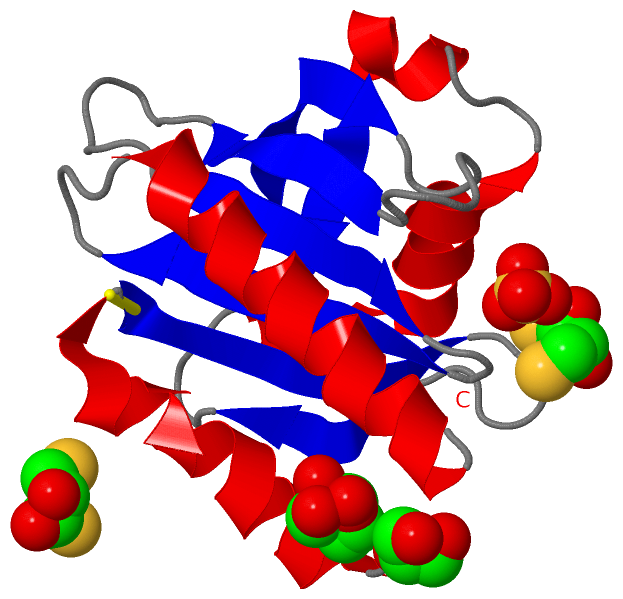
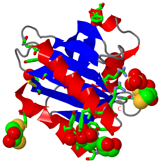
Still Images
|
||||||||||||||||
Databases
|
||||||||||||||||||||||||||||||||||||||||||||||||||||||||||||||||||||||||||||||||||||||||||||||||||||||||||||||||||||||||||||||||||||||||||||||||||||||||||||||||
Analysis Tools
|
|||||||||||||||||||||||||||||||||||||||||||||||||||||||||||||
Entries Sharing at Least One Protein Chain (UniProt ID)
Related Entries Specified in the PDB File
|
|