Show PDB file:   
         Plain Text   HTML   (compressed file size)
QuickSearch:   
by PDB,NDB,UniProt,PROSITE Code or Search Term(s)  
(-)Biol.Unit 1 - manually
(-)Asymmetric Unit
(-)Asym. Unit - sites
(-)Biological Unit 1
(-)Biol. Unit 1 - sites
collapse expand < >
Image Biol.Unit 1 - manually
Biol.Unit 1 - manually  (Jmol Viewer)
Image Asymmetric Unit
Asymmetric Unit  (Jmol Viewer)
Image Asym. Unit - sites
Asym. Unit - sites  (Jmol Viewer)
Image Biological Unit 1
Biological Unit 1  (Jmol Viewer)
Image Biol. Unit 1 - sites
Biol. Unit 1 - sites  (Jmol Viewer)

(-) Description

Title :  DHFR R67 COMPLEXED WITH NADP AND DIHYDROFOLATE
 
Authors :  J. M. Krahn, R. E. London
Date :  16 Oct 07  (Deposition) - 03 Jun 08  (Release) - 24 Feb 09  (Revision)
Method :  X-RAY DIFFRACTION
Resolution :  1.26
Chains :  Asym. Unit :  A
Biol. Unit 1:  A  (4x)
Keywords :  Oxidoreductase, Nadp, Dihydrofolate, Asymmetric Ligand Binding, Antibiotic Resistance, Methotrexate Resistance, One-Carbon Metabolism, Plasmid, Trimethoprim Resistance (Keyword Search: [Gene Ontology, PubMed, Web (Google))
 
Reference :  J. M. Krahn, M. R. Jackson, E. F. Derose, E. E. Howell, R. E. London
Crystal Structure Of A Type Ii Dihydrofolate Reductase Catalytic Ternary Complex.
Biochemistry V. 46 14878 2007
PubMed-ID: 18052202  |  Reference-DOI: 10.1021/BI701532R
(for further references see the PDB file header)

(-) Compounds

Molecule 1 - DIHYDROFOLATE REDUCTASE TYPE 2
    ChainsA
    EC Number1.5.1.3
    Organism ScientificESCHERICHIA COLI
    StrainTMP-RESISTANT, CONTAINING R67 DHFR OVERPRODUCING PLASMID PLZ1
    SynonymDIHYDROFOLATE REDUCTASE TYPE II

 Structural Features

(-) Chains, Units

  1
Asymmetric Unit A
Biological Unit 1 (4x)A

Summary Information (see also Sequences/Alignments below)

(-) Ligands, Modified Residues, Ions  (3, 5)

Asymmetric Unit (3, 5)
No.NameCountTypeFull Name
1DHF1Ligand/IonDIHYDROFOLIC ACID
2MRD3Ligand/Ion(4R)-2-METHYLPENTANE-2,4-DIOL
3NAP1Ligand/IonNADP NICOTINAMIDE-ADENINE-DINUCLEOTIDE PHOSPHATE
Biological Unit 1 (3, 20)
No.NameCountTypeFull Name
1DHF4Ligand/IonDIHYDROFOLIC ACID
2MRD12Ligand/Ion(4R)-2-METHYLPENTANE-2,4-DIOL
3NAP4Ligand/IonNADP NICOTINAMIDE-ADENINE-DINUCLEOTIDE PHOSPHATE

(-) Sites  (5, 5)

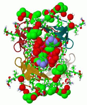
Asymmetric Unit (5, 5)
No.NameEvidenceResiduesDescription
1AC1SOFTWAREGLN A:67 , ILE A:68 , TYR A:69 , HOH A:547 , HOH A:550 , HOH A:575 , HOH A:581 , HOH A:596 , HOH A:603 , HOH A:605 , HOH A:608BINDING SITE FOR RESIDUE DHF A 0
2AC2SOFTWARELYS A:32 , SER A:34 , GLY A:35 , ALA A:36 , LEU A:50 , GLY A:64 , SER A:65 , GLN A:67 , ILE A:68 , TYR A:69 , PRO A:70 , ALA A:72 , ALA A:73 , HOH A:503 , HOH A:504 , HOH A:506 , HOH A:508 , HOH A:509 , HOH A:510 , HOH A:527 , HOH A:538 , HOH A:547 , HOH A:550 , HOH A:565 , HOH A:567 , HOH A:575 , HOH A:578 , HOH A:581 , HOH A:590 , HOH A:591 , HOH A:592 , HOH A:596 , HOH A:597 , HOH A:605 , HOH A:608BINDING SITE FOR RESIDUE NAP A 1
3AC3SOFTWAREALA A:22 , GLY A:25 , ARG A:76 , ILE A:77 , ASN A:78 , HOH A:559 , HOH A:580 , HOH A:595 , HOH A:600BINDING SITE FOR RESIDUE MRD A 500
4AC4SOFTWAREGLY A:35 , ASN A:49 , LEU A:50 , PRO A:52 , HOH A:570BINDING SITE FOR RESIDUE MRD A 501
5AC5SOFTWAREPHE A:24 , GLY A:25 , TRP A:45 , HOH A:600BINDING SITE FOR RESIDUE MRD A 502

(-) SS Bonds  (0, 0)

(no "SS Bond" information available for 2RK1)

(-) Cis Peptide Bonds  (0, 0)

(no "Cis Peptide Bond" information available for 2RK1)

 Sequence-Structure Mapping

(-) SAPs(SNPs)/Variants  (0, 0)

(no "SAP(SNP)/Variant" information available for 2RK1)

(-) PROSITE Motifs  (0, 0)

(no "PROSITE Motif" information available for 2RK1)

(-) Exons   (0, 0)

(no "Exon" information available for 2RK1)

(-) Sequences/Alignments

Asymmetric Unit
   Reformat: Number of residues per line =  ('0' or empty: single-line sequence representation)
  Number of residues per labelling interval =   
  UniProt sequence: complete  aligned part    
   Show mapping: SCOP domains CATH domains Pfam domains Secondary structure (by author)
SAPs(SNPs) PROSITE motifs Exons
(details for a mapped element are shown in a popup box when the mouse pointer rests over it)
Chain A from PDB  Type:PROTEIN  Length:58
 aligned with DYR21_ECOLX | P00383 from UniProtKB/Swiss-Prot  Length:78

    Alignment length:58
                                    30        40        50        60        70        
           DYR21_ECOLX   21 NATFGMGDRVRKKSGAAWQGQIVGWYCTNLTPEGYAVESEAHPGSVQIYPVAALERIN 78
               SCOP domains d2rk1a_ A: automated matches                               SCOP domains
               CATH domains 2rk1A00 A:21-78  [code=2.30.30.60, no name defined]        CATH domains
               Pfam domains DHFR_2-2rk1A01 A:21-78                                     Pfam domains
         Sec.struct. author ........eeee......eeeeeeee.......eeeeee......eeeee...eee.. Sec.struct. author
                 SAPs(SNPs) ---------------------------------------------------------- SAPs(SNPs)
                    PROSITE ---------------------------------------------------------- PROSITE
                 Transcript ---------------------------------------------------------- Transcript
                  2rk1 A 21 NATFGMGDRVRKKSGAAWQGQIVGWYCTNLTPEGYAVESEAHPGSVQIYPVAALERIN 78
                                    30        40        50        60        70        

   Legend:   → Mismatch (orange background)
  - → Gap (green background, '-', border residues have a numbering label)
    → Modified Residue (blue background, lower-case, 'x' indicates undefined single-letter code, labelled with number + name)
  x → Chemical Group (purple background, 'x', labelled with number + name, e.g. ACE or NH2)
  extra numbering lines below/above indicate numbering irregularities and modified residue names etc., number ends below/above '|'

 Classification and Annotation

(-) SCOP Domains  (1, 1)

Asymmetric Unit

(-) CATH Domains  (1, 1)

Asymmetric Unit
(-)
Class: Mainly Beta (13760)

(-) Pfam Domains  (1, 1)

Asymmetric Unit

(-) Gene Ontology  (8, 8)

Asymmetric Unit(hide GO term definitions)
Chain A   (DYR21_ECOLX | P00383)
molecular function
    GO:0004146    dihydrofolate reductase activity    Catalysis of the reaction: 5,6,7,8-tetrahydrofolate + NADP+ = 7,8-dihydrofolate + NADPH + H+.
    GO:0016491    oxidoreductase activity    Catalysis of an oxidation-reduction (redox) reaction, a reversible chemical reaction in which the oxidation state of an atom or atoms within a molecule is altered. One substrate acts as a hydrogen or electron donor and becomes oxidized, while the other acts as hydrogen or electron acceptor and becomes reduced.
biological process
    GO:0006730    one-carbon metabolic process    The chemical reactions and pathways involving the transfer of one-carbon units in various oxidation states.
    GO:0055114    oxidation-reduction process    A metabolic process that results in the removal or addition of one or more electrons to or from a substance, with or without the concomitant removal or addition of a proton or protons.
    GO:0046677    response to antibiotic    Any process that results in a change in state or activity of a cell or an organism (in terms of movement, secretion, enzyme production, gene expression, etc.) as a result of an antibiotic stimulus. An antibiotic is a chemical substance produced by a microorganism which has the capacity to inhibit the growth of or to kill other microorganisms.
    GO:0042493    response to drug    Any process that results in a change in state or activity of a cell or an organism (in terms of movement, secretion, enzyme production, gene expression, etc.) as a result of a drug stimulus. A drug is a substance used in the diagnosis, treatment or prevention of a disease.
    GO:0031427    response to methotrexate    Any process that results in a change in state or activity of a cell or an organism (in terms of movement, secretion, enzyme production, gene expression, etc.) as a result of a methotrexate stimulus. Methotrexate is 4-amino-10-methylformic acid, a folic acid analogue that is a potent competitive inhibitor of dihydrofolate reductase.
    GO:0046654    tetrahydrofolate biosynthetic process    The chemical reactions and pathways resulting in the formation of tetrahydrofolate, 5,6,7,8-tetrahydrofolic acid, a folate derivative bearing additional hydrogens on the pterin group.

 Visualization

(-) Interactive Views

Asymmetric Unit
  Complete Structure
    Jena3D(integrated viewing of ligand, site, SAP, PROSITE, SCOP information)
    WebMol | AstexViewer[tm]@PDBe
(Java Applets, require no local installation except for Java; loading may be slow)
    STRAP
(Java WebStart application, automatic local installation, requires Java; full application with system access!)
    RasMol
(require local installation)
    Molscript (VRML)
(requires installation of a VRML viewer; select preferred view via VRML and generate a mono or stereo PDF format file)
 
  Ligands, Modified Residues, Ions
    DHF  [ RasMol | Jena3D ]  +environment [ RasMol | Jena3D ]
    MRD  [ RasMol | Jena3D ]  +environment [ RasMol | Jena3D ]
    NAP  [ RasMol | Jena3D ]  +environment [ RasMol | Jena3D ]
 
  Sites
    AC1  [ RasMol ]  +environment [ RasMol ]
    AC2  [ RasMol ]  +environment [ RasMol ]
    AC3  [ RasMol ]  +environment [ RasMol ]
    AC4  [ RasMol ]  +environment [ RasMol ]
    AC5  [ RasMol ]  +environment [ RasMol ]
 
  Cis Peptide Bonds
(no "Cis Peptide Bonds" information available for 2rk1)
 
Biological Unit
  Complete Structure
    Biological Unit 1  [ Jena3D ]

(-) Still Images

Jmol
  protein: cartoon or spacefill or dots and stick; nucleic acid: cartoon and stick; ligands: spacefill; active site: stick
Molscript
  protein, nucleic acid: cartoon; ligands: spacefill; active site: ball and stick

 Databases and Analysis Tools

(-) Databases

Access by PDB/NDB ID
  2rk1
    Family and Domain InformationProDom | SYSTERS
    General Structural InformationGlycoscienceDB | MMDB | NDB | OCA | PDB | PDBe | PDBj | PDBsum | PDBWiki | PQS | PROTEOPEDIA
    Orientation in MembranesOPM
    Protein SurfaceSURFACE
    Secondary StructureDSSP (structure derived) | HSSP (homology derived)
    Structural GenomicsGeneCensus
    Structural NeighboursCE | VAST
    Structure ClassificationCATH | Dali | SCOP
    Validation and Original DataBMRB Data View | BMRB Restraints Grid | EDS | PROCHECK | RECOORD | WHAT_CHECK
 
Access by UniProt ID/Accession number
  DYR21_ECOLX | P00383
    Comparative Protein Structure ModelsModBase
    Genomic InformationEnsembl
    Protein-protein InteractionDIP
    Sequence, Family and Domain InformationInterPro | Pfam | SMART | UniProtKB/SwissProt
 
Access by Enzyme Classificator   (EC Number)
  1.5.1.3
    General Enzyme InformationBRENDA | EC-PDB | Enzyme | IntEnz
    PathwayKEGG | MetaCyc
 
Access by Disease Identifier   (MIM ID)
  (no 'MIM ID' available)
    Disease InformationOMIM
 
Access by GenAge ID
  (no 'GenAge ID' available)
    Age Related InformationGenAge

(-) Analysis Tools

Access by PDB/NDB ID
    Domain InformationXDom
    Interatomic Contacts of Structural UnitsCSU
    Ligand-protein ContactsLPC
    Protein CavitiescastP
    Sequence and Secondary StructurePDBCartoon
    Structure AlignmentSTRAP(Java WebStart application, automatic local installation, requires Java; full application with system access!)
    Structure and Sequence BrowserSTING
 
Access by UniProt ID/Accession number
  DYR21_ECOLX | P00383
    Protein Disorder PredictionDisEMBL | FoldIndex | GLOBPLOT (for more information see DisProt)

 Related Entries

(-) Entries Sharing at Least One Protein Chain (UniProt ID)

UniProtKB/Swiss-Prot
        DYR21_ECOLX | P003831vie 1vif 2gqv 2p4t 2rh2 2rk2 3sfm

(-) Related Entries Specified in the PDB File

2p4t A MUTANT FORM OF THE SAME PROTEIN (Q67H) COMPLEXED WITH NADP. THE MUTANT HAS A HIGHER AFFINITY FOR NADP, BUT APPEARS TO BIND DIFFERENTLY.
2rh2 HIGH RESOLUTION STRUCTURE OF THE SAME PROTEIN WITH NO LIGANDS, USED AS A STARTING MODEL FOR THIS STRUCTURE.