Show PDB file:   
         Plain Text   HTML   (compressed file size)
QuickSearch:   
by PDB,NDB,UniProt,PROSITE Code or Search Term(s)  
(-)Biol.Unit 1 - manually
(-)Asymmetric Unit
(-)Asym. Unit - sites
(-)Biological Unit 1
(-)Biol. Unit 1 - sites
collapse expand < >
Image Biol.Unit 1 - manually
Biol.Unit 1 - manually  (Jmol Viewer)
Image Asymmetric Unit
Asymmetric Unit  (Jmol Viewer)
Image Asym. Unit - sites
Asym. Unit - sites  (Jmol Viewer)
Image Biological Unit 1
Biological Unit 1  (Jmol Viewer)
Image Biol. Unit 1 - sites
Biol. Unit 1 - sites  (Jmol Viewer)

(-) Description

Title :  CRYSTAL STRUCTURE OF HUMAN GLYCOLATE OXIDASE IN COMPLEX WITH GLYOXYLATE
 
Authors :  M. S. Murray, R. P. Holmes, W. T. Lowther
Date :  25 Sep 07  (Deposition) - 26 Feb 08  (Release) - 13 Jul 11  (Revision)
Method :  X-RAY DIFFRACTION
Resolution :  1.65
Chains :  Asym. Unit :  A
Biol. Unit 1:  A  (4x)
Keywords :  Gox, Haox1, Glycolate Oxidase, Hydroxy Acid Oxidase, Glyoxylate, Glyoxylic Acid, Flavoprotein, Fmn, Glycolate Pathway, Oxidoreductase, Peroxisome (Keyword Search: [Gene Ontology, PubMed, Web (Google))
 
Reference :  M. S. Murray, R. P. Holmes, W. T. Lowther
Active Site And Loop 4 Movements Within Human Glycolate Oxidase: Implications For Substrate Specificity And Drug Design.
Biochemistry V. 47 2439 2008
PubMed-ID: 18215067  |  Reference-DOI: 10.1021/BI701710R

(-) Compounds

Molecule 1 - HYDROXYACID OXIDASE 1
    ChainsA
    EC Number1.1.3.15
    EngineeredYES
    Expression SystemESCHERICHIA COLI
    Expression System PlasmidPET28A
    Expression System StrainC41(DE3)
    Expression System Taxid562
    Expression System Vector TypePLASMID
    GeneHAO1, GOX1, HAOX1
    Organism CommonHUMAN
    Organism ScientificHOMO SAPIENS
    Organism Taxid9606
    SynonymHAOX1, GLYCOLATE OXIDASE, GOX

 Structural Features

(-) Chains, Units

  1
Asymmetric Unit A
Biological Unit 1 (4x)A

Summary Information (see also Sequences/Alignments below)

(-) Ligands, Modified Residues, Ions  (2, 2)

Asymmetric Unit (2, 2)
No.NameCountTypeFull Name
1FMN1Ligand/IonFLAVIN MONONUCLEOTIDE
2GLV1Ligand/IonGLYOXYLIC ACID
Biological Unit 1 (2, 8)
No.NameCountTypeFull Name
1FMN4Ligand/IonFLAVIN MONONUCLEOTIDE
2GLV4Ligand/IonGLYOXYLIC ACID

(-) Sites  (2, 2)

Asymmetric Unit (2, 2)
No.NameEvidenceResiduesDescription
1AC1SOFTWARETYR A:26 , TYR A:27 , ALA A:79 , THR A:80 , ALA A:81 , SER A:108 , GLN A:130 , TYR A:132 , THR A:158 , LYS A:236 , SER A:258 , HIS A:260 , GLY A:261 , ARG A:263 , ASP A:291 , GLY A:292 , GLY A:293 , ARG A:295 , GLY A:314 , ARG A:315 , HOH A:380 , HOH A:389 , HOH A:399BINDING SITE FOR RESIDUE FMN A 371
2AC2SOFTWARETYR A:26 , TRP A:110 , TYR A:132 , ARG A:167 , LEU A:205 , HIS A:260 , ARG A:263BINDING SITE FOR RESIDUE GLV A 372

(-) SS Bonds  (0, 0)

(no "SS Bond" information available for 2RDU)

(-) Cis Peptide Bonds  (0, 0)

(no "Cis Peptide Bond" information available for 2RDU)

 Sequence-Structure Mapping

(-) SAPs(SNPs)/Variants  (0, 0)

(no "SAP(SNP)/Variant" information available for 2RDU)

(-) PROSITE Motifs  (1, 1)

Asymmetric Unit (1, 1)
 PROSITEUniProtKBPDB
No.IDACDescriptionIDLocationCountLocation
1FMN_HYDROXY_ACID_DH_1PS00557 FMN-dependent alpha-hydroxy acid dehydrogenases active site.HAOX1_HUMAN258-264  1A:258-264
Biological Unit 1 (1, 4)
 PROSITEUniProtKBPDB
No.IDACDescriptionIDLocationCountLocation
1FMN_HYDROXY_ACID_DH_1PS00557 FMN-dependent alpha-hydroxy acid dehydrogenases active site.HAOX1_HUMAN258-264  4A:258-264

(-) Exons   (8, 8)

Asymmetric Unit (8, 8)
 ENSEMBLUniProtKBPDB
No.Transcript IDExonExon IDGenome LocationLengthIDLocationLengthCountLocationLength
1.1ENST000003787891ENSE00001478813chr20:7921121-7920933189HAOX1_HUMAN1-46461A:3-4644
1.2ENST000003787892ENSE00000658559chr20:7915282-7915131152HAOX1_HUMAN46-97521A:46-9752
1.3ENST000003787893ENSE00000658558chr20:7895066-7894811256HAOX1_HUMAN97-182861A:97-18286
1.4ENST000003787894ENSE00000658557chr20:7886976-7886801176HAOX1_HUMAN182-241601A:182-24160
1.5ENST000003787895ENSE00000658556chr20:7875871-787578092HAOX1_HUMAN241-271311A:241-27131
1.6ENST000003787896ENSE00000658555chr20:7866511-7866353159HAOX1_HUMAN272-324531A:272-32453
1.7ENST000003787897ENSE00000658554chr20:7866237-786616870HAOX1_HUMAN325-348241A:325-34824
1.8ENST000003787898ENSE00001478762chr20:7864310-7863628683HAOX1_HUMAN348-370231A:348-36215

(-) Sequences/Alignments

Asymmetric Unit
   Reformat: Number of residues per line =  ('0' or empty: single-line sequence representation)
  Number of residues per labelling interval =   
  UniProt sequence: complete  aligned part    
   Show mapping: SCOP domains CATH domains Pfam domains Secondary structure (by author)
SAPs(SNPs) PROSITE motifs Exons
(details for a mapped element are shown in a popup box when the mouse pointer rests over it)
Chain A from PDB  Type:PROTEIN  Length:360
 aligned with HAOX1_HUMAN | Q9UJM8 from UniProtKB/Swiss-Prot  Length:370

    Alignment length:360
                                    12        22        32        42        52        62        72        82        92       102       112       122       132       142       152       162       172       182       192       202       212       222       232       242       252       262       272       282       292       302       312       322       332       342       352       362
          HAOX1_HUMAN     3 PRLICINDYEQHAKSVLPKSIYDYYRSGANDEETLADNIAAFSRWKLYPRMLRNVAETDLSTSVLGQRVSMPICVGATAMQRMAHVDGELATVRACQSLGTGMMLSSWATSSIEEVAEAGPEALRWLQLYIYKDREVTKKLVRQAEKMGYKAIFVTVDTPYLGNRLDDVRNRFKLPPQLRMKNFETSTLSFSPEENFGDDSGLAAYVAKAIDPSISWEDIKWLRRLTSLPIVAKGILRGDDAREAVKHGLNGILVSNHGARQLDGVPATIDVLPEIVEAVEGKVEVFLDGGVRKGTDVLKALALGAKAVFVGRPIVWGLAFQGEKGVQDVLEILKEEFRLAMALSGCQNVKVIDKTLVRK 362
               SCOP domains d2rdua_ A: automated matches                                                                                                                                                                                                                                                                                                                                             SCOP domains
               CATH domains 2rduA00 A:3-362 Aldolase class I                                                                                                                                                                                                                                                                                                                                         CATH domains
               Pfam domains ------------FMN_dh-2rduA01 A:15-362                                                                                                                                                                                                                                                                                                                                      Pfam domains
         Sec.struct. author ....hhhhhhhhhhhhhhhhhhhhhhh....hhhhhhhhhhhhhhee..............eee..eee...eee....hhhhh..hhhhhhhhhhhhhh.eeee......hhhhhhhhh...eeeeee....hhhhhhhhhhhhhhh....eeee........hhhhhhhh............................hhhhhhhhhhh....hhhhhhhhhhhh...eeeeee.hhhhhhhhhhh...eeee............hhhhhhhhhhhhhh...eeeee....hhhhhhhhhhh...eeeehhhhhhhhhhhhhhhhhhhhhhhhhhhhhhhhhhh..hhhhhhhh.ee. Sec.struct. author
                 SAPs(SNPs) ------------------------------------------------------------------------------------------------------------------------------------------------------------------------------------------------------------------------------------------------------------------------------------------------------------------------------------------------------------------------ SAPs(SNPs)
                    PROSITE ---------------------------------------------------------------------------------------------------------------------------------------------------------------------------------------------------------------------------------------------------------------FMN_HYD-------------------------------------------------------------------------------------------------- PROSITE
           Transcript 1 (1) Exon 1.1  PDB: A:3-46 UniProt: 1-46         --------------------------------------------------Exon 1.3  PDB: A:97-182 UniProt: 97-182                                               ----------------------------------------------------------Exon 1.5  PDB: A:241-271       Exon 1.6  PDB: A:272-324 UniProt: 272-324            Exon 1.7  PDB: A:325-348-------------- Transcript 1 (1)
           Transcript 1 (2) -------------------------------------------Exon 1.2  PDB: A:46-97 UniProt: 46-97               ------------------------------------------------------------------------------------Exon 1.4  PDB: A:182-241 UniProt: 182-241                   ----------------------------------------------------------------------------------------------------------Exon 1.8        Transcript 1 (2)
                 2rdu A   3 PRLICINDYEQHAKSVLPKSIYDYYRSGANDEETLADNIAAFSRWKLYPRMLRNVAETDLSTSVLGQRVSMPICVGATAMQRMAHVDGELATVRACQSLGTGMMLSSWATSSIEEVAEAGPEALRWLQLYIYKDREVTKKLVRQAEKMGYKAIFVTVDTPYLGNRLDDVRNRFKLPPQLRMKNFETSTLSFSPEENFGDDSGLAAYVAKAIDPSISWEDIKWLRRLTSLPIVAKGILRGDDAREAVKHGLNGILVSNHGARQLDGVPATIDVLPEIVEAVEGKVEVFLDGGVRKGTDVLKALALGAKAVFVGRPIVWGLAFQGEKGVQDVLEILKEEFRLAMALSGCQNVKVIDKTLVRK 362
                                    12        22        32        42        52        62        72        82        92       102       112       122       132       142       152       162       172       182       192       202       212       222       232       242       252       262       272       282       292       302       312       322       332       342       352       362

   Legend:   → Mismatch (orange background)
  - → Gap (green background, '-', border residues have a numbering label)
    → Modified Residue (blue background, lower-case, 'x' indicates undefined single-letter code, labelled with number + name)
  x → Chemical Group (purple background, 'x', labelled with number + name, e.g. ACE or NH2)
  extra numbering lines below/above indicate numbering irregularities and modified residue names etc., number ends below/above '|'

 Classification and Annotation

(-) SCOP Domains  (1, 1)

Asymmetric Unit

(-) CATH Domains  (1, 1)

Asymmetric Unit
(-)
Class: Alpha Beta (26913)

(-) Pfam Domains  (1, 1)

Asymmetric Unit

(-) Gene Ontology  (17, 17)

Asymmetric Unit(hide GO term definitions)
Chain A   (HAOX1_HUMAN | Q9UJM8)
molecular function
    GO:0003973    (S)-2-hydroxy-acid oxidase activity    Catalysis of the reaction: (S)-2-hydroxy-acid + O2 = 2-oxo acid + hydrogen peroxide.
    GO:0010181    FMN binding    Interacting selectively and non-covalently with flavin mono nucleotide. Flavin mono nucleotide (FMN) is the coenzyme or the prosthetic group of various flavoprotein oxidoreductase enzymes.
    GO:0003824    catalytic activity    Catalysis of a biochemical reaction at physiological temperatures. In biologically catalyzed reactions, the reactants are known as substrates, and the catalysts are naturally occurring macromolecular substances known as enzymes. Enzymes possess specific binding sites for substrates, and are usually composed wholly or largely of protein, but RNA that has catalytic activity (ribozyme) is often also regarded as enzymatic.
    GO:0008891    glycolate oxidase activity    Catalysis of the reaction: glycolate + O2 = glyoxylate + hydrogen peroxide.
    GO:0047969    glyoxylate oxidase activity    Catalysis of the reaction: glyoxylate + H(2)O + O(2) = H(2)O(2) + H(+) + oxalate.
    GO:0052853    long-chain-(S)-2-hydroxy-long-chain-acid oxidase activity    Catalysis of the reaction: long-chain-(S)-2-hydroxy-acid + O2 = long-chain-2-oxo acid + hydrogen peroxide. Long chain refers to a chain length of 14 to 18 carbons.
    GO:0052854    medium-chain-(S)-2-hydroxy-acid oxidase activity    Catalysis of the reaction: medium-chain-(S)-2-hydroxy-acid + O2 = medium-chain-2-oxo acid + hydrogen peroxide. Medium chain refers to a chain length of between 8 and 12 carbons.
    GO:0016491    oxidoreductase activity    Catalysis of an oxidation-reduction (redox) reaction, a reversible chemical reaction in which the oxidation state of an atom or atoms within a molecule is altered. One substrate acts as a hydrogen or electron donor and becomes oxidized, while the other acts as hydrogen or electron acceptor and becomes reduced.
    GO:0005102    receptor binding    Interacting selectively and non-covalently with one or more specific sites on a receptor molecule, a macromolecule that undergoes combination with a hormone, neurotransmitter, drug or intracellular messenger to initiate a change in cell function.
    GO:0052852    very-long-chain-(S)-2-hydroxy-acid oxidase activity    Catalysis of the reaction: very-long-chain (S)-2-hydroxy-acid + O2 = very-long-chain 2-oxo acid + hydrogen peroxide. Very long chain refers to a chain length of greater than 18 carbons.
biological process
    GO:0001561    fatty acid alpha-oxidation    A metabolic pathway by which 3-methyl branched fatty acids are degraded. These compounds are not degraded by the normal peroxisomal beta-oxidation pathway, because the 3-methyl blocks the dehydrogenation of the hydroxyl group by hydroxyacyl-CoA dehydrogenase. The 3-methyl branched fatty acid is converted in several steps to pristenic acid, which can then feed into the beta-oxidative pathway.
    GO:0046296    glycolate catabolic process    The chemical reactions and pathways resulting in the breakdown of glycolate, the anion of hydroxyethanoic acid (glycolic acid).
    GO:0046487    glyoxylate metabolic process    The chemical reactions and pathways involving glyoxylate, the anion of glyoxylic acid, HOC-COOH.
    GO:0055114    oxidation-reduction process    A metabolic process that results in the removal or addition of one or more electrons to or from a substance, with or without the concomitant removal or addition of a proton or protons.
    GO:0006979    response to oxidative stress    Any process that results in a change in state or activity of a cell or an organism (in terms of movement, secretion, enzyme production, gene expression, etc.) as a result of oxidative stress, a state often resulting from exposure to high levels of reactive oxygen species, e.g. superoxide anions, hydrogen peroxide (H2O2), and hydroxyl radicals.
cellular component
    GO:0005782    peroxisomal matrix    The volume contained within the membranes of a peroxisome; in many cells the matrix contains a crystalloid core largely composed of urate oxidase.
    GO:0005777    peroxisome    A small organelle enclosed by a single membrane, and found in most eukaryotic cells. Contains peroxidases and other enzymes involved in a variety of metabolic processes including free radical detoxification, lipid catabolism and biosynthesis, and hydrogen peroxide metabolism.
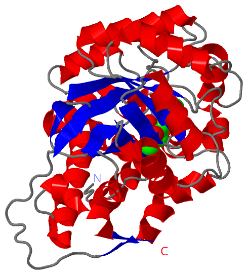
 Visualization

(-) Interactive Views

Asymmetric Unit
  Complete Structure
    Jena3D(integrated viewing of ligand, site, SAP, PROSITE, SCOP information)
    WebMol | AstexViewer[tm]@PDBe
(Java Applets, require no local installation except for Java; loading may be slow)
    STRAP
(Java WebStart application, automatic local installation, requires Java; full application with system access!)
    RasMol
(require local installation)
    Molscript (VRML)
(requires installation of a VRML viewer; select preferred view via VRML and generate a mono or stereo PDF format file)
 
  Ligands, Modified Residues, Ions
    FMN  [ RasMol | Jena3D ]  +environment [ RasMol | Jena3D ]
    GLV  [ RasMol | Jena3D ]  +environment [ RasMol | Jena3D ]
 
  Sites
    AC1  [ RasMol ]  +environment [ RasMol ]
    AC2  [ RasMol ]  +environment [ RasMol ]
 
  Cis Peptide Bonds
(no "Cis Peptide Bonds" information available for 2rdu)
 
Biological Unit
  Complete Structure
    Biological Unit 1  [ Jena3D ]

(-) Still Images

Jmol
  protein: cartoon or spacefill or dots and stick; nucleic acid: cartoon and stick; ligands: spacefill; active site: stick
Molscript
  protein, nucleic acid: cartoon; ligands: spacefill; active site: ball and stick

 Databases and Analysis Tools

(-) Databases

Access by PDB/NDB ID
  2rdu
    Family and Domain InformationProDom | SYSTERS
    General Structural InformationGlycoscienceDB | MMDB | NDB | OCA | PDB | PDBe | PDBj | PDBsum | PDBWiki | PQS | PROTEOPEDIA
    Orientation in MembranesOPM
    Protein SurfaceSURFACE
    Secondary StructureDSSP (structure derived) | HSSP (homology derived)
    Structural GenomicsGeneCensus
    Structural NeighboursCE | VAST
    Structure ClassificationCATH | Dali | SCOP
    Validation and Original DataBMRB Data View | BMRB Restraints Grid | EDS | PROCHECK | RECOORD | WHAT_CHECK
 
Access by UniProt ID/Accession number
  HAOX1_HUMAN | Q9UJM8
    Comparative Protein Structure ModelsModBase
    Genomic InformationEnsembl
    Protein-protein InteractionDIP
    Sequence, Family and Domain InformationInterPro | Pfam | SMART | UniProtKB/SwissProt
 
Access by Enzyme Classificator   (EC Number)
  1.1.3.15
    General Enzyme InformationBRENDA | EC-PDB | Enzyme | IntEnz
    PathwayKEGG | MetaCyc
 
Access by Disease Identifier   (MIM ID)
  (no 'MIM ID' available)
    Disease InformationOMIM
 
Access by GenAge ID
  (no 'GenAge ID' available)
    Age Related InformationGenAge

(-) Analysis Tools

Access by PDB/NDB ID
    Domain InformationXDom
    Interatomic Contacts of Structural UnitsCSU
    Ligand-protein ContactsLPC
    Protein CavitiescastP
    Sequence and Secondary StructurePDBCartoon
    Structure AlignmentSTRAP(Java WebStart application, automatic local installation, requires Java; full application with system access!)
    Structure and Sequence BrowserSTING
 
Access by UniProt ID/Accession number
  HAOX1_HUMAN | Q9UJM8
    Protein Disorder PredictionDisEMBL | FoldIndex | GLOBPLOT (for more information see DisProt)

 Related Entries

(-) Entries Sharing at Least One Protein Chain (UniProt ID)

UniProtKB/Swiss-Prot
        HAOX1_HUMAN | Q9UJM82nzl 2rdt 2rdw 2w0u

(-) Related Entries Specified in the PDB File

1gox SPINACH GLYCOLATE OXIDASE
2rdt HUMAN GLYCOLATE OXIDASE IN COMPLEX WITH CDST
2rdw HUMAN GLYCOLATE OXIDASE IN COMPLEX WITH SULFATE