Show PDB file:   
         Plain Text   HTML   (compressed file size)
QuickSearch:   
by PDB,NDB,UniProt,PROSITE Code or Search Term(s)  
(-)Asym.Unit - manually
(-)Asymmetric Unit
(-)Asym. Unit - sites
(-)Biological Unit 1
(-)Biol. Unit 1 - sites
(-)Biological Unit 2
collapse expand < >
Image Asym.Unit - manually
Asym.Unit - manually  (Jmol Viewer)
Image Asymmetric Unit
Asymmetric Unit  (Jmol Viewer)
Image Asym. Unit - sites
Asym. Unit - sites  (Jmol Viewer)
Image Biological Unit 1
Biological Unit 1  (Jmol Viewer)
Image Biol. Unit 1 - sites
Biol. Unit 1 - sites  (Jmol Viewer)
Image Biological Unit 2
Biological Unit 2  (Jmol Viewer)

(-) Description

Title :  STRUCTURE OF THE SAM DOMAIN OF HUMAN EPHRIN TYPE-B RECEPTOR 4
 
Authors :  J. R. Walker, D. Cuerrier, C. Butler-Cole, J. Weigelt, C. H. Arrowsmith, A. M. Edwards, A. Bochkarev, S. Dhe-Paganon, Structural Genomics Consortium (Sgc)
Date :  11 Jul 07  (Deposition) - 24 Jul 07  (Release) - 24 Feb 09  (Revision)
Method :  X-RAY DIFFRACTION
Resolution :  2.10
Chains :  Asym. Unit :  A,B
Biol. Unit 1:  A  (1x)
Biol. Unit 2:  B  (1x)
Keywords :  Atp-Binding, Glycoprotein, Kinase, Membrane, Nucleotide- Binding, Phosphorylation, Receptor, Repeat, Signal, Transferase, Transmembrane, Tyrosine-Protein Kinase, Structural Genomics, Structural Genomics Consortium, Sgc (Keyword Search: [Gene Ontology, PubMed, Web (Google))
 
Reference :  J. R. Walker, D. Cuerrier, C. Butler-Cole, J. Weigelt, C. H. Arrowsmith, A. M. Edwards, A. Bochkarev, S. Dhe-Paganon
Sam Domain Of Human Ephrin Type-B Receptor 4 (Ephb4)
To Be Published
PubMed: search
(for further references see the PDB file header)

(-) Compounds

Molecule 1 - EPHRIN TYPE-B RECEPTOR 4
    ChainsA, B
    EC Number2.7.10.1
    EngineeredYES
    Expression SystemESCHERICHIA COLI BL21(DE3)
    Expression System PlasmidPET28-MHL
    Expression System StrainBL21 (DE3)
    Expression System Taxid469008
    Expression System Vector TypePLASMID
    FragmentSAM (STERILE ALPHA MOTIF) DOMAIN
    GeneEPHB4, HTK, TYRO11
    Organism CommonHUMAN
    Organism ScientificHOMO SAPIENS
    Organism Taxid9606
    SynonymTYROSINE-PROTEIN KINASE RECEPTOR HTK, TYROSINE- PROTEIN KINASE TYRO11

 Structural Features

(-) Chains, Units

  12
Asymmetric Unit AB
Biological Unit 1 (1x)A 
Biological Unit 2 (1x) B

Summary Information (see also Sequences/Alignments below)

(-) Ligands, Modified Residues, Ions  (1, 2)

Asymmetric Unit (1, 2)
No.NameCountTypeFull Name
1CL2Ligand/IonCHLORIDE ION
Biological Unit 1 (0, 0)
No.NameCountTypeFull Name
1CL-1Ligand/IonCHLORIDE ION
Biological Unit 2 (0, 0)
No.NameCountTypeFull Name
1CL-1Ligand/IonCHLORIDE ION

(-) Sites  (1, 1)

Asymmetric Unit (1, 1)
No.NameEvidenceResiduesDescription
1AC1SOFTWAREARG A:917BINDING SITE FOR RESIDUE CL A 2

(-) SS Bonds  (0, 0)

(no "SS Bond" information available for 2QKQ)

(-) Cis Peptide Bonds  (0, 0)

(no "Cis Peptide Bond" information available for 2QKQ)

 Sequence-Structure Mapping

(-) SAPs(SNPs)/Variants  (0, 0)

(no "SAP(SNP)/Variant" information available for 2QKQ)

(-) PROSITE Motifs  (0, 0)

(no "PROSITE Motif" information available for 2QKQ)

(-) Exons   (2, 4)

Asymmetric Unit (2, 4)
 ENSEMBLUniProtKBPDB
No.Transcript IDExonExon IDGenome LocationLengthIDLocationLengthCountLocationLength
1.1bENST000003581731bENSE00001868923chr7:100425121-100424601521EPHB4_HUMAN1-18180--
1.2bENST000003581732bENSE00000825052chr7:100421895-10042182571EPHB4_HUMAN18-41240--
1.3ENST000003581733ENSE00000825051chr7:100421553-100421266288EPHB4_HUMAN42-137960--
1.4ENST000003581734ENSE00000825050chr7:100420289-100419893397EPHB4_HUMAN138-2701330--
1.5bENST000003581735bENSE00000825049chr7:100417918-100417763156EPHB4_HUMAN270-322530--
1.6ENST000003581736ENSE00000825048chr7:100417511-100417179333EPHB4_HUMAN322-4331120--
1.7bENST000003581737bENSE00000710240chr7:100416266-100416142125EPHB4_HUMAN433-474420--
1.8cENST000003581738cENSE00001706820chr7:100414979-100414814166EPHB4_HUMAN475-530560--
1.9cENST000003581739cENSE00001665919chr7:100411643-100411541103EPHB4_HUMAN530-564350--
1.10ENST0000035817310ENSE00001787983chr7:100411338-10041127465EPHB4_HUMAN564-586230--
1.11bENST0000035817311bENSE00001754468chr7:100410830-100410717114EPHB4_HUMAN586-624390--
1.12ENST0000035817312ENSE00001631272chr7:100410616-100410369248EPHB4_HUMAN624-706830--
1.13aENST0000035817313aENSE00001753966chr7:100405202-100404987216EPHB4_HUMAN707-778720--
1.14aENST0000035817314aENSE00001623534chr7:100404191-100404042150EPHB4_HUMAN779-828500--
1.15ENST0000035817315ENSE00001783205chr7:100403316-100403123194EPHB4_HUMAN829-893650--
1.16ENST0000035817316ENSE00001649970chr7:100402943-100402788156EPHB4_HUMAN893-945532A:908-945
B:908-945
38
38
1.17bENST0000035817317bENSE00001639386chr7:100401212-1004001871026EPHB4_HUMAN945-987432A:945-968
B:945-968
24
24

(-) Sequences/Alignments

Asymmetric Unit
   Reformat: Number of residues per line =  ('0' or empty: single-line sequence representation)
  Number of residues per labelling interval =   
  UniProt sequence: complete  aligned part    
   Show mapping: SCOP domains CATH domains Pfam domains Secondary structure (by author)
SAPs(SNPs) PROSITE motifs Exons
(details for a mapped element are shown in a popup box when the mouse pointer rests over it)
Chain A from PDB  Type:PROTEIN  Length:61
 aligned with EPHB4_HUMAN | P54760 from UniProtKB/Swiss-Prot  Length:987

    Alignment length:61
                                   917       927       937       947       957       967 
          EPHB4_HUMAN   908 AFGSVGEWLRAIKMGRYEESFAAAGFGSFELVSQISAEDLLRIGVTLAGHQKKILASVQHM 968
               SCOP domains d2qkqa_ A: automated matches                                  SCOP domains
               CATH domains 2qkqA00 A:908-968 Transcription Factor, Ets-1                 CATH domains
               Pfam domains ------------------------------------------------------------- Pfam domains
         Sec.struct. author ...hhhhhhhhh.hhhhhhhhhhh...hhhhhh..hhhhhhhhh..hhhhhhhhhhhhh.. Sec.struct. author
                 SAPs(SNPs) ------------------------------------------------------------- SAPs(SNPs)
                    PROSITE ------------------------------------------------------------- PROSITE
           Transcript 1 (1) Exon 1.16  PDB: A:908-945 [INCOMPLETE]----------------------- Transcript 1 (1)
           Transcript 1 (2) -------------------------------------Exon 1.17b [INCOMPLETE]  Transcript 1 (2)
                 2qkq A 908 AFGSVGEWLRAIKMGRYEESFAAAGFGSFELVSQISAEDLLRIGVTLAGHQKKILASVQHM 968
                                   917       927       937       947       957       967 

Chain B from PDB  Type:PROTEIN  Length:61
 aligned with EPHB4_HUMAN | P54760 from UniProtKB/Swiss-Prot  Length:987

    Alignment length:61
                                   917       927       937       947       957       967 
          EPHB4_HUMAN   908 AFGSVGEWLRAIKMGRYEESFAAAGFGSFELVSQISAEDLLRIGVTLAGHQKKILASVQHM 968
               SCOP domains d2qkqb_ B: automated matches                                  SCOP domains
               CATH domains 2qkqB00 B:908-968 Transcription Factor, Ets-1                 CATH domains
           Pfam domains (1) SAM_1-2qkqB01 B:908-968                                       Pfam domains (1)
           Pfam domains (2) SAM_1-2qkqB02 B:908-968                                       Pfam domains (2)
         Sec.struct. author ...hhhhhhhhh.hhhhhhhhhhhh..hhhhhh..hhhhhhhhh..hhhhhhhhhhhhh.. Sec.struct. author
                 SAPs(SNPs) ------------------------------------------------------------- SAPs(SNPs)
                    PROSITE ------------------------------------------------------------- PROSITE
           Transcript 1 (1) Exon 1.16  PDB: B:908-945 [INCOMPLETE]----------------------- Transcript 1 (1)
           Transcript 1 (2) -------------------------------------Exon 1.17b [INCOMPLETE]  Transcript 1 (2)
                 2qkq B 908 AFGSVGEWLRAIKMGRYEESFAAAGFGSFELVSQISAEDLLRIGVTLAGHQKKILASVQHM 968
                                   917       927       937       947       957       967 

   Legend:   → Mismatch (orange background)
  - → Gap (green background, '-', border residues have a numbering label)
    → Modified Residue (blue background, lower-case, 'x' indicates undefined single-letter code, labelled with number + name)
  x → Chemical Group (purple background, 'x', labelled with number + name, e.g. ACE or NH2)
  extra numbering lines below/above indicate numbering irregularities and modified residue names etc., number ends below/above '|'

 Classification and Annotation

(-) SCOP Domains  (1, 2)

Asymmetric Unit

(-) CATH Domains  (1, 2)

Asymmetric Unit

(-) Pfam Domains  (1, 2)

Asymmetric Unit
(-)
Clan: SAM (26)

(-) Gene Ontology  (27, 27)

Asymmetric Unit(hide GO term definitions)
Chain A,B   (EPHB4_HUMAN | P54760)
molecular function
    GO:0005524    ATP binding    Interacting selectively and non-covalently with ATP, adenosine 5'-triphosphate, a universally important coenzyme and enzyme regulator.
    GO:0005003    ephrin receptor activity    Combining with an ephrin to initiate a change in cell activity.
    GO:0016301    kinase activity    Catalysis of the transfer of a phosphate group, usually from ATP, to a substrate molecule.
    GO:0000166    nucleotide binding    Interacting selectively and non-covalently with a nucleotide, any compound consisting of a nucleoside that is esterified with (ortho)phosphate or an oligophosphate at any hydroxyl group on the ribose or deoxyribose.
    GO:0005515    protein binding    Interacting selectively and non-covalently with any protein or protein complex (a complex of two or more proteins that may include other nonprotein molecules).
    GO:0004672    protein kinase activity    Catalysis of the phosphorylation of an amino acid residue in a protein, usually according to the reaction: a protein + ATP = a phosphoprotein + ADP.
    GO:0004713    protein tyrosine kinase activity    Catalysis of the reaction: ATP + a protein tyrosine = ADP + protein tyrosine phosphate.
    GO:0016740    transferase activity    Catalysis of the transfer of a group, e.g. a methyl group, glycosyl group, acyl group, phosphorus-containing, or other groups, from one compound (generally regarded as the donor) to another compound (generally regarded as the acceptor). Transferase is the systematic name for any enzyme of EC class 2.
    GO:0004714    transmembrane receptor protein tyrosine kinase activity    Combining with a signal and transmitting the signal from one side of the membrane to the other to initiate a change in cell activity by catalysis of the reaction: ATP + a protein-L-tyrosine = ADP + a protein-L-tyrosine phosphate.
biological process
    GO:0001525    angiogenesis    Blood vessel formation when new vessels emerge from the proliferation of pre-existing blood vessels.
    GO:0007155    cell adhesion    The attachment of a cell, either to another cell or to an underlying substrate such as the extracellular matrix, via cell adhesion molecules.
    GO:0002042    cell migration involved in sprouting angiogenesis    The orderly movement of endothelial cells into the extracellular matrix in order to form new blood vessels involved in sprouting angiogenesis.
    GO:0048013    ephrin receptor signaling pathway    The series of molecular signals generated as a consequence of an ephrin receptor binding to an ephrin.
    GO:0003007    heart morphogenesis    The developmental process in which the heart is generated and organized. The heart is a hollow, muscular organ, which, by contracting rhythmically, keeps up the circulation of the blood.
    GO:0007275    multicellular organism development    The biological process whose specific outcome is the progression of a multicellular organism over time from an initial condition (e.g. a zygote or a young adult) to a later condition (e.g. a multicellular animal or an aged adult).
    GO:0018108    peptidyl-tyrosine phosphorylation    The phosphorylation of peptidyl-tyrosine to form peptidyl-O4'-phospho-L-tyrosine.
    GO:0016310    phosphorylation    The process of introducing a phosphate group into a molecule, usually with the formation of a phosphoric ester, a phosphoric anhydride or a phosphoric amide.
    GO:0046777    protein autophosphorylation    The phosphorylation by a protein of one or more of its own amino acid residues (cis-autophosphorylation), or residues on an identical protein (trans-autophosphorylation).
    GO:0006468    protein phosphorylation    The process of introducing a phosphate group on to a protein.
    GO:0007169    transmembrane receptor protein tyrosine kinase signaling pathway    A series of molecular signals initiated by the binding of an extracellular ligand to a receptor on the surface of the target cell where the receptor possesses tyrosine kinase activity, and ending with regulation of a downstream cellular process, e.g. transcription.
cellular component
    GO:0005829    cytosol    The part of the cytoplasm that does not contain organelles but which does contain other particulate matter, such as protein complexes.
    GO:0070062    extracellular exosome    A vesicle that is released into the extracellular region by fusion of the limiting endosomal membrane of a multivesicular body with the plasma membrane. Extracellular exosomes, also simply called exosomes, have a diameter of about 40-100 nm.
    GO:0005576    extracellular region    The space external to the outermost structure of a cell. For cells without external protective or external encapsulating structures this refers to space outside of the plasma membrane. This term covers the host cell environment outside an intracellular parasite.
    GO:0016021    integral component of membrane    The component of a membrane consisting of the gene products and protein complexes having at least some part of their peptide sequence embedded in the hydrophobic region of the membrane.
    GO:0005887    integral component of plasma membrane    The component of the plasma membrane consisting of the gene products and protein complexes having at least some part of their peptide sequence embedded in the hydrophobic region of the membrane.
    GO:0016020    membrane    A lipid bilayer along with all the proteins and protein complexes embedded in it an attached to it.
    GO:0005886    plasma membrane    The membrane surrounding a cell that separates the cell from its external environment. It consists of a phospholipid bilayer and associated proteins.
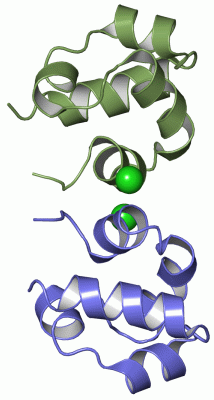
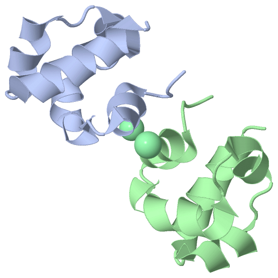
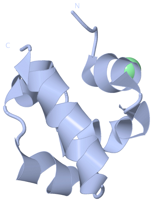
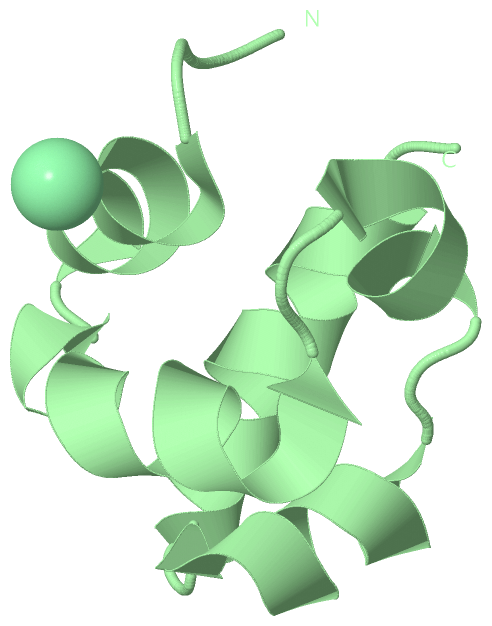
 Visualization

(-) Interactive Views

Asymmetric Unit
  Complete Structure
    Jena3D(integrated viewing of ligand, site, SAP, PROSITE, SCOP information)
    WebMol | AstexViewer[tm]@PDBe
(Java Applets, require no local installation except for Java; loading may be slow)
    STRAP
(Java WebStart application, automatic local installation, requires Java; full application with system access!)
    RasMol
(require local installation)
    Molscript (VRML)
(requires installation of a VRML viewer; select preferred view via VRML and generate a mono or stereo PDF format file)
 
  Ligands, Modified Residues, Ions
    CL  [ RasMol | Jena3D ]  +environment [ RasMol | Jena3D ]
 
  Sites
    AC1  [ RasMol ]  +environment [ RasMol ]
 
  Cis Peptide Bonds
(no "Cis Peptide Bonds" information available for 2qkq)
 
Biological Units
  Complete Structure
    Biological Unit 1  [ Jena3D ]
    Biological Unit 2  [ Jena3D ]

(-) Still Images

Jmol
  protein: cartoon or spacefill or dots and stick; nucleic acid: cartoon and stick; ligands: spacefill; active site: stick
Molscript
  protein, nucleic acid: cartoon; ligands: spacefill; active site: ball and stick

 Databases and Analysis Tools

(-) Databases

Access by PDB/NDB ID
  2qkq
    Family and Domain InformationProDom | SYSTERS
    General Structural InformationGlycoscienceDB | MMDB | NDB | OCA | PDB | PDBe | PDBj | PDBsum | PDBWiki | PQS | PROTEOPEDIA
    Orientation in MembranesOPM
    Protein SurfaceSURFACE
    Secondary StructureDSSP (structure derived) | HSSP (homology derived)
    Structural GenomicsGeneCensus
    Structural NeighboursCE | VAST
    Structure ClassificationCATH | Dali | SCOP
    Validation and Original DataBMRB Data View | BMRB Restraints Grid | EDS | PROCHECK | RECOORD | WHAT_CHECK
 
Access by UniProt ID/Accession number
  EPHB4_HUMAN | P54760
    Comparative Protein Structure ModelsModBase
    Genomic InformationEnsembl
    Protein-protein InteractionDIP
    Sequence, Family and Domain InformationInterPro | Pfam | SMART | UniProtKB/SwissProt
 
Access by Enzyme Classificator   (EC Number)
  2.7.10.1
    General Enzyme InformationBRENDA | EC-PDB | Enzyme | IntEnz
    PathwayKEGG | MetaCyc
 
Access by Disease Identifier   (MIM ID)
  (no 'MIM ID' available)
    Disease InformationOMIM
 
Access by GenAge ID
  (no 'GenAge ID' available)
    Age Related InformationGenAge

(-) Analysis Tools

Access by PDB/NDB ID
    Domain InformationXDom
    Interatomic Contacts of Structural UnitsCSU
    Ligand-protein ContactsLPC
    Protein CavitiescastP
    Sequence and Secondary StructurePDBCartoon
    Structure AlignmentSTRAP(Java WebStart application, automatic local installation, requires Java; full application with system access!)
    Structure and Sequence BrowserSTING
 
Access by UniProt ID/Accession number
  EPHB4_HUMAN | P54760
    Protein Disorder PredictionDisEMBL | FoldIndex | GLOBPLOT (for more information see DisProt)

 Related Entries

(-) Entries Sharing at Least One Protein Chain (UniProt ID)

UniProtKB/Swiss-Prot
        EPHB4_HUMAN | P547602bba 2e7h 2hle 2vwu 2vwv 2vww 2vwx 2vwy 2vwz 2vx0 2vx1 2x9f 2xvd 2yn8 3zew 4aw5 4bb4

(-) Related Entries Specified in the PDB File

(no "Related Entries Specified in the PDB File" available for 2QKQ)