|
|
|
|
 Description
|
|
Compounds
|
||||||||||||||||||||||||
Chains, Units
Summary Information (see also Sequences/Alignments below) |
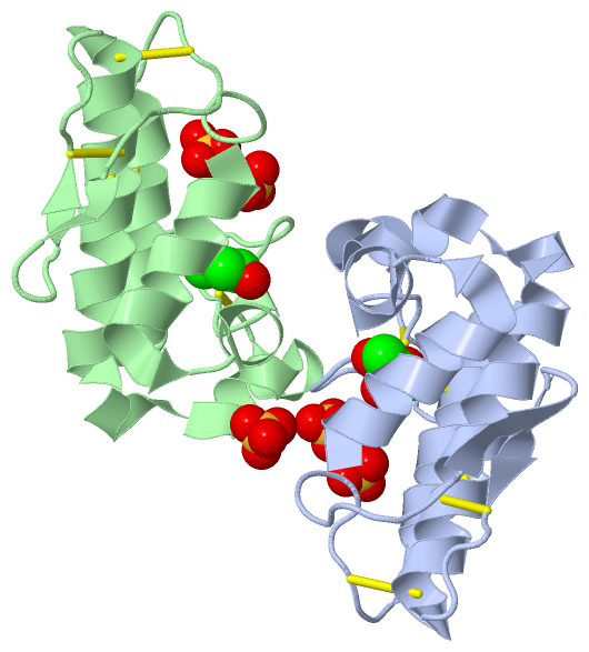
Ligands, Modified Residues, Ions (2, 7)| Asymmetric Unit (2, 7) Biological Unit 1 (2, 7) Biological Unit 2 (2, 14) |
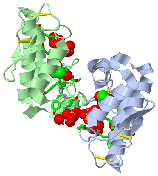
Sites (7, 7)
Asymmetric Unit (7, 7)
|
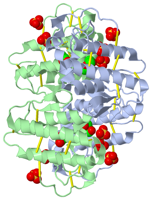
SS Bonds (14, 14)
Asymmetric Unit
|
||||||||||||||||||||||||||||||||||||||||||||||||||||||||||||
Cis Peptide Bonds (0, 0)| (no "Cis Peptide Bond" information available for 2PH4) |
SAPs(SNPs)/Variants (0, 0)| (no "SAP(SNP)/Variant" information available for 2PH4) |
PROSITE Motifs (2, 4)
Asymmetric Unit (2, 4)
|
||||||||||||||||||||||||||||||||||||||||||||||||||||||||||||||||||||||||||||||||||||||||||||||||
Exons (0, 0)| (no "Exon" information available for 2PH4) |
Sequences/Alignments
Asymmetric UnitChain A from PDB Type:PROTEIN Length:121 aligned with PA2H_PROMB | P84776 from UniProtKB/Swiss-Prot Length:121 Alignment length:121 10 20 30 40 50 60 70 80 90 100 110 120 PA2H_PROMB 1 SLIELTKMVFQETGKNPVTYYTLYGCNCGVGRRGKPKDATDRCCFVHRCCYKKLTGCDPKKDRYSYSWENKAIVCGEKNPCLKELCECDKAVAICLRKNLGTYDKNYRFTMKFLCDKPEKC 121 SCOP domains d2ph4a_ A: automated matches SCOP domains CATH domains 2ph4A00 A:1-133 Phospholipase A2 CATH domains Pfam domains ------------------------------------------------------------------------------------------------------------------------- Pfam domains SAPs(SNPs) ------------------------------------------------------------------------------------------------------------------------- SAPs(SNPs) PROSITE ------------------------------------------PA2_HIS ----------------------------------PA2_ASP -------------------------- PROSITE Transcript ------------------------------------------------------------------------------------------------------------------------- Transcript 2ph4 A 1 SLIELTKMVFQETGKNPVTYYTLYGCNCGVGRRGKPKDATDRCCFVHRCCYKKLTGCDPKKDRYSYSWENKAIVCGEKNPCLKELCECDKAVAICLRKNLGTYDKNYRFTMKFLCDKPEKC 133 10 || 21 31 41 51 || ||69 79 90 100 110 120 || ||132 13| 53| 61| 88| 123| || 15 57 67 90 125 || 127| 129 Chain B from PDB Type:PROTEIN Length:121 aligned with PA2H_PROMB | P84776 from UniProtKB/Swiss-Prot Length:121 Alignment length:121 10 20 30 40 50 60 70 80 90 100 110 120 PA2H_PROMB 1 SLIELTKMVFQETGKNPVTYYTLYGCNCGVGRRGKPKDATDRCCFVHRCCYKKLTGCDPKKDRYSYSWENKAIVCGEKNPCLKELCECDKAVAICLRKNLGTYDKNYRFTMKFLCDKPEKC 121 SCOP domains d2ph4b_ B: automated matches SCOP domains CATH domains 2ph4B00 B:1-133 Phospholipase A2 CATH domains Pfam domains ------------------------------------------------------------------------------------------------------------------------- Pfam domains SAPs(SNPs) ------------------------------------------------------------------------------------------------------------------------- SAPs(SNPs) PROSITE ------------------------------------------PA2_HIS ----------------------------------PA2_ASP -------------------------- PROSITE Transcript ------------------------------------------------------------------------------------------------------------------------- Transcript 2ph4 B 1 SLIELTKMVFQETGKNPVTYYTLYGCNCGVGRRGKPKDATDRCCFVHRCCYKKLTGCDPKKDRYSYSWENKAIVCGEKNPCLKELCECDKAVAICLRKNLGTYDKNYRFTMKFLCDKPEKC 133 10 || 21 31 41 51 || ||69 79 90 100 110 120 || ||132 13| 53| 61| 88| 123| || 15 57 67 90 125 || 127| 129
|
||||||||||||||||||||
SCOP Domains (1, 2)
Asymmetric Unit
 |
CATH Domains (1, 2)
Asymmetric Unit
|
Pfam Domains (0, 0)| (no "Pfam Domain" information available for 2PH4) |
Gene Ontology (4, 4)|
Asymmetric Unit(hide GO term definitions) Chain A,B (PA2H_PROMB | P84776)
|
||||||||||||||||||||||||||||||||||||||||||
Interactive Views
|
||||||||||||||||||||||||||||||||||||||||||||||||||||||||||||||||||||||||||||||||||||||||||||||||||||||||||||||||||||||||||||||||||||||||||||||||||||||||||||||||||||||||||||||||||||||||||||||
Still Images
|
||||||||||||||||
Databases
|
||||||||||||||||||||||||||||||||||||||||||||||||||||||||||||||||||||||||||||||||||||||||||||||||||||||||||||||||||||||||||||||||||||||||||||||||||||||||||||||||
Analysis Tools
|
|||||||||||||||||||||||||||||||||||||||||||||||||||||||||||||
Entries Sharing at Least One Protein Chain (UniProt ID)
Related Entries Specified in the PDB File
|
|